Embed Size (px)
Citation preview
Acido-basicity of Mg-Ni-Al Mixed Oxides from LDH Precursors: A FTIR and XPS Study
O. Cairon,* ,† E. Dumitriu, ‡ and C. Guimon†
Institut Pluridiciplinaire de Recherche sur l’EnVironnement et les Mate´riauxsEquipe de Chimie Physique(IPREMsECP)sUMR (CNRS) 5254, 2 aV. P. Angot, 64053 Pau Cedex 9, France, and Laboratory ofCatalysis, Technical UniVersity of Iasi, 71 D. Mangeron, Iasi, Romania
ReceiVed: NoVember 6, 2006; In Final Form: March 8, 2007
The acido-basicity of Mg-Ni-Al mixed oxides generated from the calcination of layered double hydroxides(with the atomic ratios Mg/Ni variable and Mg+ Ni/Al equal to 2) has been investigated by adsorptionexperiments, using FTIR with pyrrole and pyridine as probe molecules and XPS with ammonia and sulfurdioxide. The spectroscopic data were compared with microcalorimetric data and with the results of catalytictests (cyclohexanol conversion). The acidity, comparable to that of alumina, is mainly of Lewis type. It increaseswith the nickel content. The FTIR spectra of chemisorbed pyrrole indicate the presence of strong, mediumand weak basic sites. The bulk basicity of trimetallic oxides is stronger than that of the bimetallic ones. Theeffective basicity of the bimetallic Mg-Al oxide is minimized by the presence of numerous remaining carbonateions. The XPS analyses show that the surface basicity increases with the Mg content, as indicated by thereaction test. Moreover it appears that the majority of the basic surface sites are hydroxyl OH- groups.
1. Introduction
Ni-based catalysts (with or without other metals or promoters)are widely used in many processes such as selective hydro-dehydrogenation,1,2 methane reforming or cracking,3,4 andproduction of synthesis gas.5. The activity of the catalysts in allthese processes depends mainly on the dispersion, the reduc-ibility of the metal, and the resistance to deactivation. Theseproperties depend on the composition, the method of preparation,and the conditions of activation. Generally, these oxides alsohave acid/and base properties, useful in the field of the acid orbasic catalysis. This applies to the mixed oxides generated bycalcination from layered double hydroxide precursors.
Layered double hydroxidessLDHssbelong to a large classof natural and synthetic anionic clays.6 LDH compounds havethe general formula [M1-x
2+Mx3+(OH)2]x+[Ax/n
n-]‚mH2O, whereM2+ ) Mg2+, Ni2+, Zn2+..., M3+ ) Al3+, Fe3+..., and A )anion. Their structure is similar to that of brucite Mg(OH)2
where octahedra of Mg2+ (6-fold coordinated to OH-) shareedges to form infinite sheets. These octahedra are stacked ontop of each other and are held together by hydrogen bonding.When Mg2+ is substituted by a trivalent cation (with a radiusclose to that of Mg2+), a formal positive charge appears inhydroxyl layers. Electroneutrality and the stability of the systemare ensured by anions An- (most frequently carbonates) andwater, located in the interlayer region. Typically,x is in the0.2-0.33 range (M2+/M3+ between 2 and 4).
Calcination of LDH induces dehydration, dehydroxylation andloss of compensating anions, and leads to acidic and basic mixedoxides with a high surface area, homogeneous interdispersionof the metals and better resistance to sintering than correspond-ing supported catalysts. The activity of these catalysts relatedto their acid-base properties depends on their composition, thepreparation method, and the treatment conditions (calcination
temperature). Thus, it is very important to characterize theseacid-base properties, i.e., the nature (Bro¨nsted or Lewis), thestrength, and the density of sites.
Besides catalytic reactions, this characterization is usuallycarried out with the help of thermal or spectroscopic methodsbased on gas-phase adsorption of basic or acid probes. The mostimportant and widely used techniques are microcalorimetry,7,8
differential scanning calorimetry (DSC),9 temperature-pro-grammed desorption (TPD),10,11 thermogravimetry (TGA),12
Fourier-transform IR spectroscopy (FTIR)10,13-15 and X-rayphotoelectron spectroscopy (XPS).16,17Different gaseous probemolecules can be used with the latter two techniques. Besidescarbon monoxide, most of the basic probe molecules used arenitrogen-containing bases such as ammonia, pyridine and itsmethylated derivatives (lutidines), pyrrole, acetonitrile...Acidprobes are less numerous, the most frequently used being carbonor sulfur dioxide and sometimes pyrrole.
In a previous paper,18 we reported the thermal behavior of aseries of Mg-Ni-Al LDHs and the adsorption microcalorimetrydata (NH3, SO2)obtained on mixed oxides resulting fromcalcination at 723 K. These data gave information on thestrength and the concentration of the acid and basic sites. Theaim of this work is to specify these properties by determiningthe nature and the distribution (bulk, surface) of the sites. Forthis purpose, we use the two spectrometric techniques mentionedabove, FTIR (with adsorption of pyrrole and pyridine) and XPS(with adsorption of ammonia and sulfur dioxide). Their datawill then be compared to the results of the catalytic conversionof cyclohexanol. This reaction leads indeed to cyclohexene whenacid sites are present and to cyclohexanone with acid-baseassistance.
2. Experimental
2.1. Materials. LDHs compounds with different Mg/Ni/Almolar ratios were elaborated by coprecipitation at lowsupersaturation,6,19-21 with both the precipitants (NaOH and Na2-CO3) and the corresponding metal nitrates being added slowly,
* Corresponding author. E-mail: [email protected].† IPREMsECP.‡ Technical University of Iasi.
8015J. Phys. Chem. C2007,111,8015-8023
10.1021/jp0673011 CCC: $37.00 © 2007 American Chemical SocietyPublished on Web 05/12/2007
while the pH was held constant. The precipitate was hydro-thermally treated in a Teflon-coated autoclave at 453 K for12 h. The resulting solid was isolated by filtration, washed withdeionized water and dried overnight at 353 K.
The samples are named according to their formal composi-tion: Mg/Ni/Al 2/0/1, 1/1/1, 0.33/1.67/1, and 0/2/1. The realcompositions determined by ICP AES (Table 1) are very closeto these values. A very slight excess of carbonate ions can beobserved, which is often the case in this type of material.
Calcination was carried out at 723 K (1 K‚min-1) for 2 hbefore adsorption, analysis and reaction test.
2.2. Characterization.For the FT-IR measurements, powderswere pressed into self-supporting pellets and placed inside anIR cell in which in situ activation and gas dosage could bedone.22 Spectra were collected at a resolution of 2 cm-1 on aNicolet Magna 560 spectrometer. The oxide form was obtainedby in situ activation of the self-supported wafer of LDH at720 K under reduced pressure (10-3 Pa) for 5 h. After activation,the background spectrum of the oxide was recorded at roomtemperature. Freshly distilled pyrrole was introduced progres-sively at room temperature by doses up to an equilibriumpressure of 80 Pa. The IR spectra were recorded after ca. 10-15 min equilibration with each successive pyrrole doses. In theFigures 1-4, the background spectrum was subtracted from eachof the spectra recorded after pyrrole introduction, except whena new reference specified in the legend was used.
XPS analyses were performed at room temperature with anSSI spectrometer using monochromatic and focused (spotdiameter of 600µm, 100 W) Al KR radiation (1486.6 eV) at aresidual pressure of 10-7 Pa. The introduction chamber of thespectrometer was coupled with a glovebox for protecting thesamples from the atmosphere between adsorption and theanalysis. Charge effects were compensated by using a flood gun(5 eV). The hemispherical analyzer ran with a constant passenergy of 50 eV for high-resolution spectra. The experimentalbands were fitted with theoretical bands (80% Gaussian, 20%Lorentzian) with a least-squares algorithm using a nonlinearbaseline. Before analysis, the adsorption of NH3 or SO2 isperformed at atmospheric pressure and 353 K under helium flow(20 cm3 min-1).
After a first adsorption at 353 K until an equilibrium pressureof about 67 Pa, the samples were outgassed at 10-4 Pa for 30min at the same temperature and a second adsorption wasperformed on each sample until an equilibrium pressure of about27 Pa was attained, in order to calculate the amount ofirreversibly chemisorbed NH3 (or SO2) at this pressure, whichprovides an estimate of the number of strong acidic (or basic)sites.
The catalytic reactions were run in a pulse type microreactor(stainless steel, inner diameter 3.5 mm, and length 80 mm)containing 20 mg of catalyst with particle size of 0.25-0.43mm. A 0.1 µL sample of cyclohexanol was injected undernitrogen flow (20 cm3 min-1, 250 kPa) at a constant temperature(648 K). The reaction products were analyzed with an on-lineGC (Gas chromatograph HP 5890, SUPELCOWAX-10 fused
silica capillary column, 30 m× 0.2 mm × 0.2 µm filmthickness) equipped with FID or TCD and computing integrator.
3. Results and Discussion
3.1. FTIR Study. Most of the probes usually employed forFT-IR analyses characterize either the acidic or basic properties.Pyridine is the most widely used for the study of the acidity ofsolids while carbon dioxide allows the characterization of thebasicity. Unfortunately, it was not possible to use CO2 becauseof the presence of some residual carbonate ions in our samplesafter the calcination procedure.18 Because of its weak acidity,pyrrole (C4H4NH) PYH, is reported as a useful probe for thebasicity either of alkaline exchanged zeolites or metal oxides.23-26
The H-donor pyrrole properties allow C4H4NH...O bridges toform with basic oxygens. This formation results in the batho-chromic shift of the broadν(NH) vibration compared to theposition at 3497 cm-1 for the “free” molecule in CCl4 whichshifts to ca. 3400 cm-1 in the liquid due to intermolecularH-bonding. The shift observed increases with the basic forceof the sites probed and reflects the O2- basicity strength.23,24
Furthermore, pyrrole has been reported to chemisorb disso-ciatively over strong basic metal oxides and deprotonation ofthe pyrrole on the strongest basic oxygens leads to theproduction of pyrrolate anionssPY-sstabilized by the cationsof surface or by the acid sites.26 In Pyrrolate anions, a∼70 cm-1 downshift of theν(B15) ring-stretching comparedto the fundamental vibration of pyrrole (∼1522 cm-1) isnoticed.27 Moreover, the dissociation of pyrrole leads to adecrease inν(CΗ) frequency and two bands at∼3100 and∼3050 cm-1 appear. As far as pyrrole adsorption is either non-dissociative or dissociative, we report in Table 2 some of theobserved vibrational frequencies of pyrrole (C2V symmetry) ingaseous, liquid and solid states and of the solid pyrrolate anionfor comparison. Only the vibrations from literature valuesdiscussed in this work are reported here.
Τhe π-electrons of pyrrole also act as an H-acceptor, wheninteracting with superficial free hydroxyl groups. The H-bondformation between the surface hydroxyl groups and the probeprovokes the decrease in the intensity of the OH bands and thesimultaneous appearance of a broad band at lower frequency.Scokart reported a broad band at 3550 cm-1 for hydroxyl groupsof pure silica gel.23 A 200 cm-1 downshift was measured inthe case of acid solids such as silica-alumina or alumina. Theamphoteric properties of pyrrole allow acidity measurementsto be made. Thus, the choice of this probe appeared appropriatefor the study of our materials although the pyrrole makes theinterpretation and assignments of IR spectra quite complex sinceit reacts with both acid and basic sites. To clarify the presentationof our results, we first report a short overview of the results
TABLE 1: Elemental Composition (ICP-AES) of the LDHPrecursors
usual name formulaBET area(m2 g-1)
2/0/1 [Mg0.67Al 0.33(OH)2](CO32-)0.19‚0.65H2O 190
1/1/1 [Mg0.35Ni0.34Al 0.31(OH)2](CO32-)0.21‚0.93H2O 280
0.33/1.67/1 [Mg0.12Ni0.55Al 0.32(OH)2](CO32-)0.19‚0.96H2O 270
0/2/1 [Ni0.66Al 0.33(OH)2](CO32-)0.20‚0.77H2O 250
TABLE 2: Some of Experimental Frequencies (cm-1) ofPyrrole in Gaseous, Liquid, and Solid States and of the SolidPyrrolate Anion13,23,26,27
C4H4NH/(PYH) C4H4N-/(PY-)
assignment symmetry gas solid liquid solid
ν(NH) A1,9 3527 3400ν(CH) A1,8 3148 3129 3133 3074
B1,16 3140 3123 3108 3090A1,7 3125 3113 3108 3051B1,17 3116 3105 3133 3051
νR (cycle)ν(CdC) or B1,15 1546 1522 1531 1453ν(C-C) or A1,6 1472 1472 1472 1442ν(C-N) B1,14 1424 1432 1418 1361
A1,5 1391 1379 1384 1344
8016 J. Phys. Chem. C, Vol. 111, No. 22, 2007 Cairon et al.
obtained over a mixed oxide taken as an example (Mg-Ni-Al0.33/1.67/1) and then we will present the detailed study ofpyrrole adsorption, considering the different steps in theadsorption progress. Then we shall report the comparative studyof the adsorption on the series of oxides and the main resultsobtained.
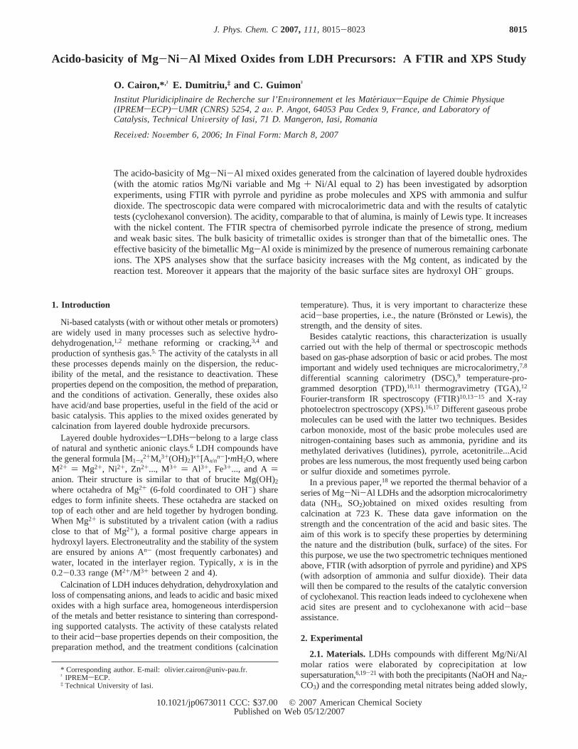
3.1.1. Adsorption of Pyrrole on Mg-Ni-Al 0.33/1.67/1.Figure 1 reports pyrrole adsorption presented in the two spectralregions: A, 4000-2500 cm-1; and B, 1800-1200 cm-1, forsuccessive doses of pyrrole up to saturation, corresponding toa vapor pressureP ) 80 Pa in the cell. The background spectrumof the original outgassed oxide at 720 K is subtracted. The mainfeatures observed in these IR spectra containing adsorbed pyrroleare, for the A region, a strong, broad band between∼3400 and∼3200 cm-1, ν(NH), with two series of narrow bands between3200 and 3000 cm-1, ν(CH), and 3000-2800 cm-1. Severalbands detected on this last range are not clearly attributed butdescribed as combination bands observed only in the presenceof basic sites.24,25In the B region, the ring-stretching vibrations
of adsorbed pyrrole show the complexity of the recorded IRspectra. Nevertheless, among theνR vibrations, a very intenseband at∼1450 cm-1 is detected on introduction of ever lowquantities of pyrrole. This band is characteristic of the formationof the pyrrolate species (see Table 2 for comparison).
As more pyrrole was introduced, i.e., increasing vaporpressure, some minor bands around this position appeared. Torefine the interpretation of IR spectra in the two A and B regions,three steps of pyrrole coverage should be considered:- low surface coverage corresponding to low quantities of pyrroleintroduce up to a vapor pressure Pe 0.4 Pa in the cell (Figure2),- medium surface coverage corresponding to a vapor pressurein the range of 0.4-10 Pa (Figure 3),- and finally high surface coverage corresponding to a vaporpressure in the range of 10-80 Pa (Figure 4).
Note that all the pyrrole introduced was not totally adsorbedduring these three selected steps, this is why the vapor pressuresare only for guidance but they had been pointed out becausethey corresponded to main modifications on the IR spectraindicating the end of formation of some entities or to new onesemerging.
Figure 1. FTIR difference spectra of Mg-Ni-Al (0.33/1.67/1) afteradsorption of pyrrole,P ) 0.5, 0.1, 0.2, 0.3, 0.4, 0.6, 0.9, 2, 3, 5, 10,20, 40, and 80 Pa (equilibration pressure in the cell), activated oxidespectrum subtracted. 4000-2500 (A) and 1800-1200 cm-1 (B) ranges.
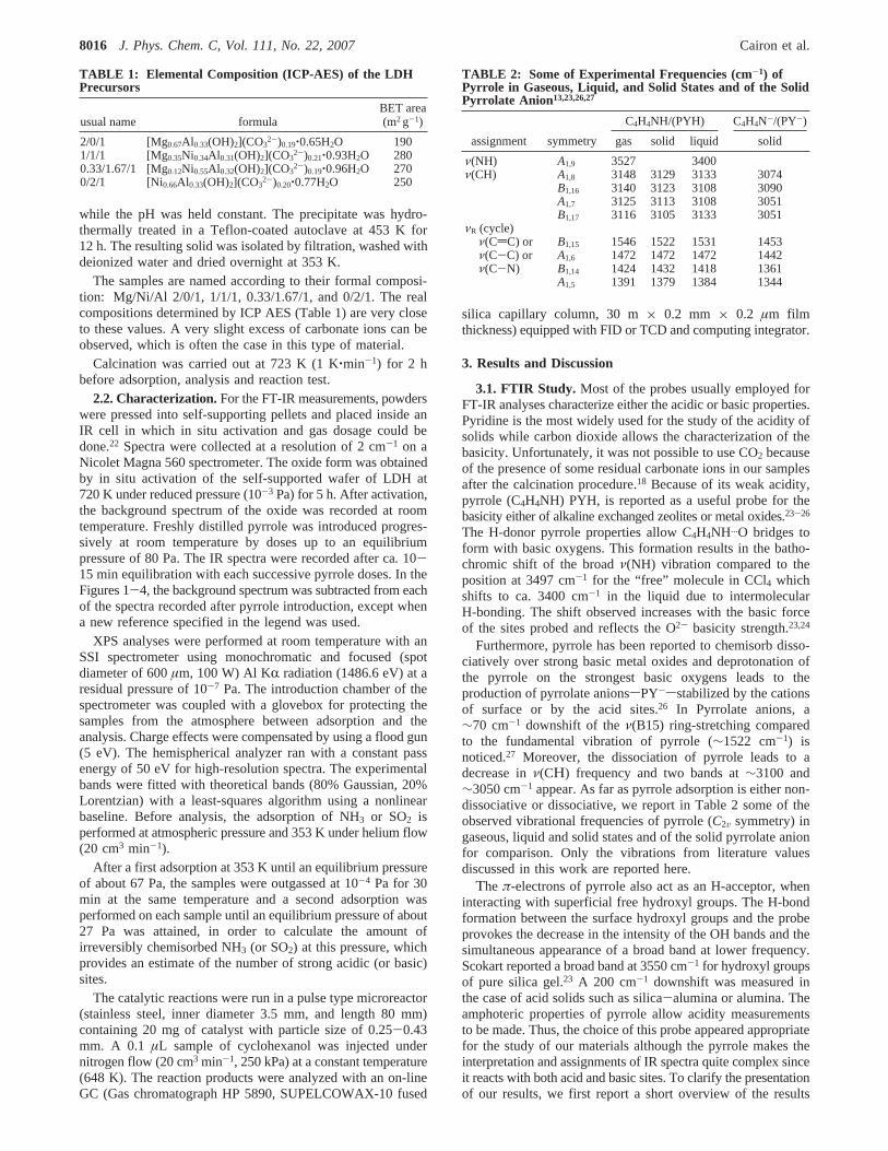
Figure 2. FTIR spectra of Mg-Ni-Al (0.33/1.67/1) after adsorptionof pyrrole (P e 0.4 Pa), activated oxide spectrum subtracted.
Acido-Basicity of Mg-Ni-Al Mixed Oxides J. Phys. Chem. C, Vol. 111, No. 22, 20078017
Adsorption of pyrrole at low surface coverage (Figure 2). Atthe beginning of the adsorption, the appearance in the B regionof the ring-stretching vibrations of adsorbed pyrrole, theν(B15)band at∼1452 cm-1, reveals the formation of pyrrolate species(see Table 2 for comparison). It is accompanied by theν(CH)band at 3102 cm-1 in the A high-frequency region as previouslymentioned.26,27 Dissociation of pyrrole on the very basic O2-
sites is confirmed by the simultaneous appearance of a barelydetectableν(OH) shoulder at 3650 cm-1 (Figure 2A).22 Thespectra after first doses of pyrrole show that in the two regionsA and B, many other bands simultaneously appear and increasedepending upon surface coverage. This feature is indicative thatnot only the strongest basic centers are probed.
In A region, the 3318 cm-1 band, progressively increasingin intensity and moderately shifting to 3327 cm-1, correspondsto the NH-stretching frequency of pyrrole bound to basic O2-
by a hydrogen bond. Theν(NH) stretching frequency may becompared to the∼3497 cm-1 for the “free” molecule in CCl4and the downshift is similar to the one observed for magnesiumoxide, indicating an equivalent basic strength.23 Theν(CH) bandat 3120 cm-1 and the ring stretching vibrations at 1530 and
1418 cm-1 confirm the presence of non-dissociated pyrrole H-bound to basic O2-. In the 3000-2800 cm-1 range, severalbands are detected but not clearly attributed, and are describedas combination bands observed only in the presence of basicsites.24,25
In addition to bands due to the above characterized O2- basicsites, the IR spectra show “negative” bands at ca. 3750 and3705 cm-1 corresponding to the interaction via theπ-systembetween the probe and the surface hydroxyl groups initiallydetectable on the background spectrum of the oxide. They maybe related to the broad and growing band at∼3545 cm-1, andreveal the presence of H-bond formation. The measured shift(about 200 cm-1) would indicate that the acidity of the probedhydroxyl groups is close to that of alumina and silica.23
Unfortunately the lack of structure of the broad band does notallow us to discriminate the acidity strength of these two OHgroups. In the B region, the bands at 1590 and 1541 cm-1 aresimilar to those observed in the case of alumina and can beattributed to the ring stretching vibrations of pyrrole in interac-tion with acid sites.23,24
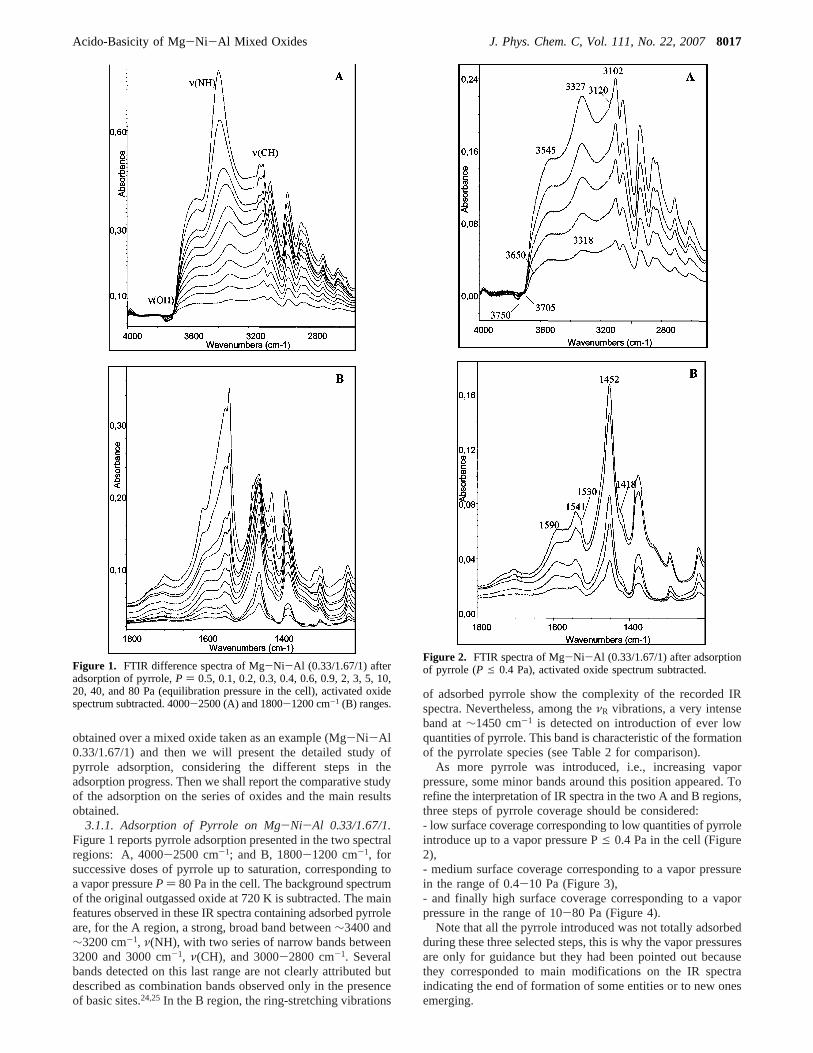
Figure 3. FTIR spectra of Mg-Ni-Al (0.33/1.67/1) after adsorptionof pyrrole (0.4< P e 10 Pa),P ) 0.4 Pa corresponding spectrumsubtracted.
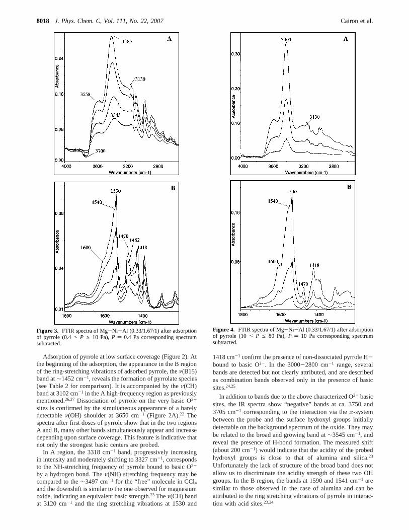
Figure 4. FTIR spectra of Mg-Ni-Al (0.33/1.67/1) after adsorptionof pyrrole (10 < P e 80 Pa),P ) 10 Pa corresponding spectrumsubtracted.
8018 J. Phys. Chem. C, Vol. 111, No. 22, 2007 Cairon et al.
As already noted, the interpretation of the IR spectra isdifficult because of the numerous adsorbed species formed.However, we can clearly highlight the formation of pyrrolates,which indicates the existence of very strong basic sitescharacterizing the first stage of adsorption.
Adsorption of pyrrole at medium surface coverage (Figure3). Figure 3 reports spectra recorded for the next doses of pyrrolewith a vapor pressure in the range of 0.4-10 Pa. The lastspectrum showed in Figure 2 was selected as the new back-ground and subtracted because it corresponds to the end of thepyrrolate formation, i.e., the 1452 cm-1 band does not increaseany further and its maximum in intensity has been reached. Thuswe can assess and refine the bands around this position,emerging during this second step, in order to identify theadsorbed species. Particular attention was paid to checking thatsuch background subtractions do not involve any artifact.28
As expected, the 1452 cm-1, ν(B15), and the corresponding3100 cm-1, ν(CH), bands are missing, indicating that there isno further formation of pyrrolates, i.e., that the very basicoxygens are not probed during this second step. The adsorptionof pyrrole continues on the basic oxygen as observed by theν(NH) band which position shifts gradually from 3345 to 3385cm-1. This shifting probably results from the progressivecontribution of the 3400 cm-1 band due to a pyrrole liquid-likephase (Table 2),13,23 rather than from the strength of the basicsites heterogeneity. The 1470 cm-1 and the 3130 cm-1, ν(CH),bands are consistent with the presence of pyrrole in the liquid-like state resulting from the increase of the surface coverage(multilayers formation).29
The interaction of pyrrole with the OH groups continues asindicated by the disappearance of ill-defined hydroxyl groupsaround 3700 cm-1 and could be accompanied by the broad bandtoward the low wavenumbers with the maximum emerging at∼3558 cm-1. The∼150 cm-1 downshift indicates the weaknessof the acidity of the hydroxyl groups involved in question andalso show that is very difficult to get any precise informationabout the strength of acidic sites involved. The interaction ofthe pyrrole with the acid sites continues as indicated by thering bands at∼1600 and∼1540 cm-1, as previously observed(Figure 2).
Looking closely at the 1470 cm-1 shape band, a below peakat ∼1462 cm-1 is perceptible for the first probe doses duringthis second adsorption step. This trend does not resulting fromour subtracting procedure, using the saturation of pyrrolatesformation as the blank spectrum to avoid their main contribution.As was suggested by Binet et al.26 the interaction between thepyrrole probe and the oxygen anions could lead to a polarizablehydrogen bridge linking O2- and PYH for less basic O2- sites.The H-bridged complex so formed, [PY‚‚‚H‚‚‚O]-, is stabilizedby cations or by acidic hydroxyl groups. Its formation, observedon highly dehydroxylated alumina, results in the appearance oftwo narrow bands at 1461, 1380 cm-1 assigned to theν(B15)andν(B14) ring stretching respectively. The 60 cm-1 downshiftof ν(B15) is consistent with the intermediate basic strengthclaimed by these authors compared to the 70 cm-1 downshiftwhen pyrrolate formation occurs. The shoulder appearing at1462 cm-1 near the 1470 cm-1 band in our spectra, maytherefore be assigned to theν(B15) ring vibration of pyrroleadsorbed on O2- with an intermediate basic strength. Moreover,this attribution is consistent with the observation of the broadcontinuum in the region 3600-3000 cm-1, due to the polarizablehydrogen bridge of the [PY‚‚‚H‚‚‚O]- complex linking the O2-.
Adsorption of pyrrole at high surface coverage (Figure 4).Figure 4 reports spectra recorded for the highest doses of pyrrole
with a vapor pressure 10e P e 80 Pa. The last spectrumshowed in Figure 3 was selected as new background andsubtracted.
In the ν(NH) region, the intense band at∼3400 cm-1 isassigned to the NH-stretching of pyrrole liquid-like phase.13,23,25
The constant position around this wavenumber indicates thatthere is no other significant contribution of pyrrole adsorbedon O2- basic centers. The high coverage step spectra(10 e P e 80 Pa) confirm our previous interpretation: thenarrow peak at 1470 cm-1 and the 3130 cm-1 band are due toliquid-like pyrrole.13,23The narrow shape of the 1470 cm-1 band,without any surrounding bands like in Figure 3, confirms theexistence of the 1462 cm-1 band resulting from theν(Β15) ringvibration observed at medium coverage in this figure andassigned to the adsorption on intermediate basic site. Moreoverthe 1530 and 1418 cm-1 bands are still observed while nointeraction of pyrrole with weak basic sites occurs (no NH-stretching shift). This feature indicates that these two bandsappear twice, i.e., for non-dissociated pyrrole and for pyrrolein the liquid-like state as previously mentioned.24 The 1600 and1542 cm-1 bands which does not correspond to liquid pyrrole,reveal that the adsorption of pyrrole on acid sites continues.Concerning the acid properties, complementary adsorption ofpyridine will be performed.
To summarize this section, pyrrole adsorbed in our mixedoxide reveals three basic strength centers:- the very basic ones induce dissociative pyrrole adsorption(PY-) and are the most numerous,- the intermediates are hydrogen bridged linking the O2- andallow the [PY...H...O]- complex to be formed,- the “normal” basic ones cause pyrrole molecule to be non-dissociatively H-bonded.
3.1.2. ComparatiVe Study of the Pyrrole Adsorption on theMg-Ni-Al Mixed Oxides.The goal of this study is to determinethe relative basicity of the Mg-Ni-Al mixed oxides. Quantita-tive conclusions will not be proposed because of the lack ofknowledge of integrated molar absorption coefficients valuesfor the ν(NH) or the ring vibration bands of adsorbed pyrrole.However, since the BET surface areas are in comparable range(Table 1) and the intensity of a specific IR band is a functionof the amount of pyrrole adsorbed, it should reflect the numberof probed sites.28,29So, on this assumption and in order to makesome valid comparisons, we carried out the adsorption of pyrrolein the same conditions (same coverage rate, 0< P e 40 Pa),all the spectra being normalized at 11.5 mg pellets. For the sakeof clarity, we will not present all the spectra and the study isfocused on the three basic strength sites revealed by thecorresponding ring vibrations, i.e., 1452, 1462, and 1530 cm-1
for the strong, medium and weak basic sites respectively.The acidic sites were estimated from the 1593, 1542, and
1490 cm-1 bands,23 after pyrrole adsorption and these resultswere supplemented by the adsorption of pyridine.
Basic sites. The recorded spectra, obtained by subtracting theoriginal spectra of activated oxide, are presented in Figure 5,for both the A (4000-2500 cm-1) and the B (1800-1200 cm-1)regions. In A, all the oxides, except MgNiAl 2/0/1, present thebands observed and discussed previously. The absence ofvibration bands for the oxide MgNiAl 2/0/1 is explained bythe saturation of the IR signal. This saturation is due to thenumerous residual carbonate species as shown in Figure 6.However, the low intensities of theν(CH) andν(NH) bands(Figure 5) show that pyrrole is very slightly adsorbed on weakbasic oxygens and moreover, the low intensity of theν(CH)band (3100 cm-1) is in agreement with the very few strong basic
Acido-Basicity of Mg-Ni-Al Mixed Oxides J. Phys. Chem. C, Vol. 111, No. 22, 20078019

sites of this oxide. Theν(NH) stretching frequency is relatedwith the strength of the basic oxygens.24,25 This frequencydecreases slightly (from 3330 to 3310 cm-1) when the Ni/Mgratio increases, showing a slight increase of the strength of theLewis sites with increasing Ni content.
For the other oxides, the comparison of the relative intensitiesof the ring bands allows us to determine the relative proportionsof the basic sites for at least two distinct strength. Thus, thestrongest basic oxygens, involving the formation of pyrrolates,PY- at ca.1450 cm-1, are the most prevalent in the mixed oxideMgNiAl 0.33/1.67/1 then decrease in MgNiAl 1/1/1 andMgNiAl 0/2/1. This order is the same than that of the amountsof irreversibly chemisorbed SO2 (Table 3) which correspondto the concentration of the strong and medium basic sites.
Regarding the oxygens with intermediate basic strength,forming the H-bridged complex [PY...H...O]-, the width of theband near 1460 cm-1 indicates a slightly higher contributionfor the oxide MgNiAl 0/2/1 than for the two others,MgNiAl 0.33/1.67/1 and MgNiAl 1/1/1.
The number of weak basic oxygen revealed by the non-dissociative adsorption of pyrrole can be estimated, starting from
the intensity of theν(NH) vibrations (Figure 5). For the firstpyrrole doses, theν(NH) position is roughly the same for allthe samples at∼3320 cm-1. At higher surface coverage, itremains in the same order in our experimental conditions(P < 40 Pa) in spite of the contribution due to the liquid-likepyrrole phase. It also appears that the number of basic sitesimplied in non-dissociative adsorption decreases slightly fromoxides MgNiAl 0/2/1 to MgNiAl 0.33/1.67/1 then toMgNiAl 1/1/1, and remains very low for the oxide MgNiAl2/0/1 (Figure 5A). This result is confirmed by the reduction ofthe ring bands at 1530 and 1418 cm-1 due to the no-dissociatedpyrrole (Figure 5B).
Acid sites. The acid character of our oxides can be highlightedby the vibration bands at 1590, 1540 and 1490 cm-1. Thesebands have variable intensities according to the oxide studied.
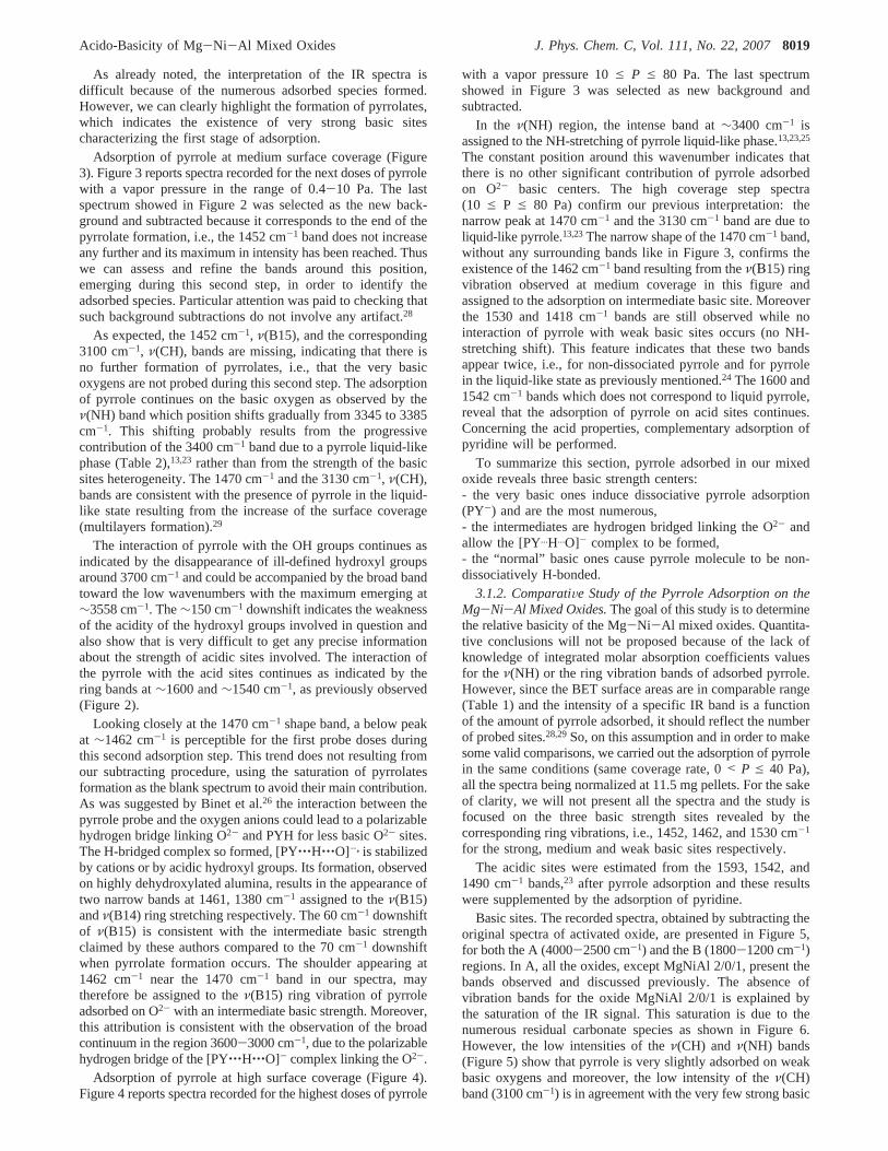
It appears that the acidity of the three MgNiAl oxides isapproximately identical for the oxides MgNiAl 0/2/1 and 0.33/1.67/1 and slightly lower for the 1/1/1 oxide (Figure 5B). Thisresult is confirmed by the adsorption of pyridine (Figure 7).The 1445 cm-1 band (ν19b, νCN) characteristic of coordinatedpyridine, Lpy species, indicates that the strength of the Lewisacid sites is quite weak since it vanishes after a brief pumpingat 473 K. Moreover, no pyridinium species, pyH+ at 1545 cm-1
(ν19b, νCN) are detected. This last feature indicates that theBronsted acid sites are very weak, which is in agreement withthe 200 cm-1 ∆ν(OH) measured downshift (Figure 2).
To conclude this FT-IR comparative study, the basicity ofthe oxides, measured as the propensity to give pyrrolates anions,are in the following order: MgNiAl 0.33/1.67/1> MgNiAl1/1/1> MgNiAl 0/2/1 . MgNiAl 2/0/1. As shown by the rateof residual carbonate species (Figure 6), the thermal stabilityof these ions is related with the composition of oxides: thecarbonate or polycarbonate content increases with the Mg/Niratio as previously observed.18 All the mixed oxides have a verylow Bronsted acidity, close to that of silica or alumina. The
Figure 5. FTIR spectra of Mg-Ni-Al mixed oxides after adsorptionof pyrrole up to 40 Pa: Mg-Ni-Al 0/2/1 (a), Mg-Ni-Al 0.33/1.67/1(b), Mg-Ni-Al 1/1/1 (c), and Mg-Ni-Al 2/0/1 (d).
Figure 6. FTIR spectra (1200-1800 cm-1) of calcined (723 K) Mg-Ni-Al mixed oxides: Mg-Ni-Al 0/2/1 (a), Mg-Ni-Al 0.33/1.67/1(b), Mg-Ni-Al 1/1/1 (c), and Mg-Ni-Al 2/0/1 (d).
TABLE 3: Amounts of Irreversibly Chemisorbed NH 3 andSO2 (µmol g-1)
Mg-Ni-Al 2/0/1 1/1/1 0.33/1.67/1 0/2/1
NH3 54.5 200 278.5 343SO2 120 543 619 490
8020 J. Phys. Chem. C, Vol. 111, No. 22, 2007 Cairon et al.
acidity is essentially of Lewis type with a moderate strength.The density of the acid Lewis sites is increasing slightly withNi/Mg ratio in agreement with the volumetric data (Table 3).
4. XPS Study
XPS is a specific spectrometry for surface studies (the depthof the layer analyzed is less than 10 nm). This technique iscomplementary to FTIR and microcalorimetry. As noted above,the potential of XPS in the study of catalytic surfaces is wellrecognized. Besides the classical analysis of the superficialchemical composition, this technique can be applied to studythe surface reactivity of solids by using the adsorption of probemolecules in the same manner as for FTIR and microcalorim-etry. The method proposed by Defosse and Canesson for acidsolids30 has been applied by several authors to the study of theacidity and the basicity of various families of zeolites16,17,31orother solids.17,32-35
Because of the weakness of the interaction between thepyrrole and most acid and basic sites of oxides, this moleculeis not suitable for an XPS study since the analyses are performedin UHV conditions (10-7 Pa). A recording at liquid-nitrogentemperature could avoid the desorption of chemisorbed pyrrole.31
So, as in the previous microcalorimetry study,18 we chose sulfurdioxide and ammonia which are probes with stronger acidityand basicity.
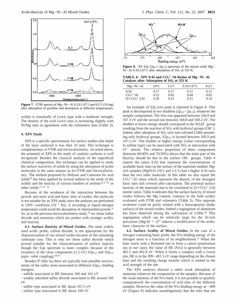
4.1. Surface Basicity of Mixed Oxides.The most widelyused acidic probe, carbon dioxide, is not appropriate for thecharacterization of our mixed oxides because of the remainingcarbonate ions. This is why we chose sulfur dioxide whichproved suitable for the characterization of surface basicitythough the S2p spectrum is more complex because of theexistence of the close components (1.2 eV) S2p3/2 and S2p1/2
(spin-orbit coupling).34,35
Besides S0 (like S8) there are typically four possible environ-ments of the sulfur atom associated to different S2p3/2 bindingenergies:- sulfide associated to BE between 160 and 163 eV- weakly adsorbed sulfur dioxide associated to BE around 166eV- sulfite type associated to BE about 167.5 eV- sulfate type associated to BE about 169 eV
An example of S2p core peak is reported in Figure 8. Thispeak is decomposed in two doublets (2p3/2-2p1/2), whatever thesample composition. The first one appeared between 166.8 and167.3 eV and the second one between 168.8 and 169.2 eV. Thedoublet at lower energy should correspond to the SO3H- groupresulting from the reaction of SO2 with hydroxyl groups (OH-).Indeed, after adsoption of SO2 over non-calcined LDHs present-ing only hydroxyl groups, S2p3/2 is located between 166.9 and167.2 eV. The doublet at higher energy (value correspondingto sulfate type) can be associated with SO2 in interaction withO2- anions. The relative proportion of these components(between 60/40% and 70/30%) shows that the main part of thebasicity should be due to the surface OH- groups. Table 4reports the ratios S/Al that represent the concentrations ofavailable basic sites on the surface of the materials studied. Mg-rich samples (MgNiAl 2/0/1 and 1/1/1) had a higher S/Al ratiothan the two other materials. In this table we also report theCO3
2-/Al ratios which represent the density of the strongestbasic sites still covered after calcination. The potential surfacebasicity of the materials has to be correlated to [S+CO3
2-]/Alatomic ratios. Table 4 indicates that the surface basicity of mixedoxides follows the Mg content, contrary to the bulk acidityevaluated with FTIR and volumetry (Table 3). This oppositeevolution could be partly related with a heterogeneity (bulk/surface) of the mixed oxides. Indeed a segregation of aluminumhas been observed during the calcination of LDHs.36 Thissegregation which can be relatively large for the Al-richprecursors (Mg/Al< 1)37 induces a modification of the acido-basic character of the surface.
4.2. Surface Acidity of Mixed Oxides. In the case of anitrogen containing basic probe, the N1s binding energy of thenitrogen atom is a function of its neighborhood.17 When thebase reacts with a Bro¨nsted site to form a cation (ammoniumion in our case), the value of BE (N1s) is generally between401.5 and 402.8 eV. When it forms a complex with a Lewissite, BE is in the 399-401.5 eV range depending on the chosenbase and the resulting charge transfer which is related to theacid strength of the site.
The XPS analyses showed a rather weak adsorption ofammonia whatever the composition of the samples. Because ofthe weak intensity of the N1s band, it is not possible to quantifycomparatively the concentration of acid sites of the differentsamples. However the value of the N1s binding energy at∼400eV (Figure 9) indicates unambiguously that the sites that are
Figure 7. FTIR spectra of Mg-Ni-Al 0.33/1.67/1 and 0/2/1 (10 mg)after adsorption of pyridine and desorption at different temperatures.
Figure 8. XP S2p (2p3/2-2p1/2) spectrum of the mixed oxide Mg-Ni-Al 0.33/1.67/1 after adsorption of SO2 at 353 K.
TABLE 4: XPS S/Al and CO32-/Al Ratios of Mg-Ni-Al
Catalysts after Adsorption of SO2 at 353 K
Mg-Ni-Al 2/0/1 1/1/1 0.33/1.67/1 0/2/1
S/Al 0.17 0.17 0.11 0.12CO3
2-/Al 0.12 0.05 0.04 0.02[S+CO3
2-]/Al 0.29 0.22 0.15 0.14
Acido-Basicity of Mg-Ni-Al Mixed Oxides J. Phys. Chem. C, Vol. 111, No. 22, 20078021
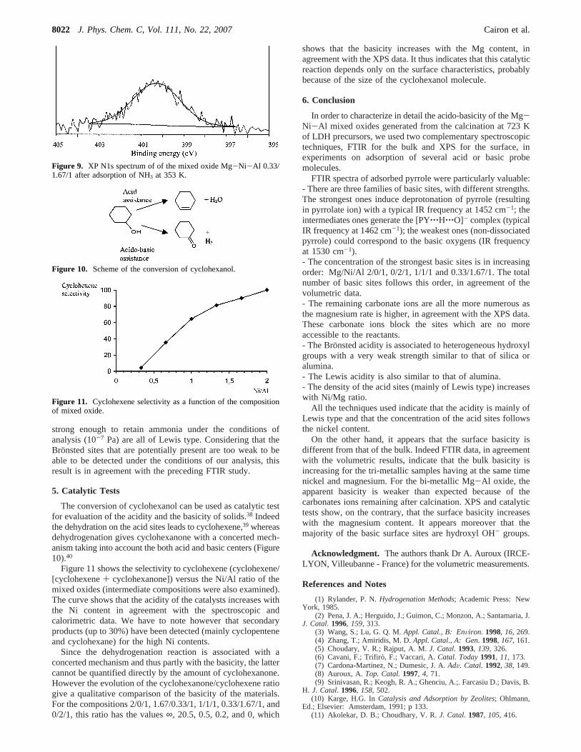
strong enough to retain ammonia under the conditions ofanalysis (10-7 Pa) are all of Lewis type. Considering that theBronsted sites that are potentially present are too weak to beable to be detected under the conditions of our analysis, thisresult is in agreement with the preceding FTIR study.
5. Catalytic Tests
The conversion of cyclohexanol can be used as catalytic testfor evaluation of the acidity and the basicity of solids.38 Indeedthe dehydration on the acid sites leads to cyclohexene,39 whereasdehydrogenation gives cyclohexanone with a concerted mech-anism taking into account the both acid and basic centers (Figure10).40
Figure 11 shows the selectivity to cyclohexene (cyclohexene/[cyclohexene+ cyclohexanone]) versus the Ni/Al ratio of themixed oxides (intermediate compositions were also examined).The curve shows that the acidity of the catalysts increases withthe Ni content in agreement with the spectroscopic andcalorimetric data. We have to note however that secondaryproducts (up to 30%) have been detected (mainly cyclopenteneand cyclohexane) for the high Ni contents.
Since the dehydrogenation reaction is associated with aconcerted mechanism and thus partly with the basicity, the lattercannot be quantified directly by the amount of cyclohexanone.However the evolution of the cyclohexanone/cyclohexene ratiogive a qualitative comparison of the basicity of the materials.For the compositions 2/0/1, 1.67/0.33/1, 1/1/1, 0.33/1.67/1, and0/2/1, this ratio has the values∞, 20.5, 0.5, 0.2, and 0, which
shows that the basicity increases with the Mg content, inagreement with the XPS data. It thus indicates that this catalyticreaction depends only on the surface characteristics, probablybecause of the size of the cyclohexanol molecule.
6. Conclusion
In order to characterize in detail the acido-basicity of the Mg-Ni-Al mixed oxides generated from the calcination at 723 Kof LDH precursors, we used two complementary spectroscopictechniques, FTIR for the bulk and XPS for the surface, inexperiments on adsorption of several acid or basic probemolecules.
FTIR spectra of adsorbed pyrrole were particularly valuable:- There are three families of basic sites, with different strengths.The strongest ones induce deprotonation of pyrrole (resultingin pyrrolate ion) with a typical IR frequency at 1452 cm-1; theintermediates ones generate the [PY‚‚‚H‚‚‚O]- complex (typicalIR frequency at 1462 cm-1); the weakest ones (non-dissociatedpyrrole) could correspond to the basic oxygens (IR frequencyat 1530 cm-1).- The concentration of the strongest basic sites is in increasingorder: Mg/Ni/Al 2/0/1, 0/2/1, 1/1/1 and 0.33/1.67/1. The totalnumber of basic sites follows this order, in agreement of thevolumetric data.- The remaining carbonate ions are all the more numerous asthe magnesium rate is higher, in agreement with the XPS data.These carbonate ions block the sites which are no moreaccessible to the reactants.- The Bronsted acidity is associated to heterogeneous hydroxylgroups with a very weak strength similar to that of silica oralumina.- The Lewis acidity is also similar to that of alumina.- The density of the acid sites (mainly of Lewis type) increaseswith Ni/Mg ratio.
All the techniques used indicate that the acidity is mainly ofLewis type and that the concentration of the acid sites followsthe nickel content.
On the other hand, it appears that the surface basicity isdifferent from that of the bulk. Indeed FTIR data, in agreementwith the volumetric results, indicate that the bulk basicity isincreasing for the tri-metallic samples having at the same timenickel and magnesium. For the bi-metallic Mg-Al oxide, theapparent basicity is weaker than expected because of thecarbonates ions remaining after calcination. XPS and catalytictests show, on the contrary, that the surface basicity increaseswith the magnesium content. It appears moreover that themajority of the basic surface sites are hydroxyl OH- groups.
Acknowledgment. The authors thank Dr A. Auroux (IRCE-LYON, Villeubanne - France) for the volumetric measurements.
References and Notes
(1) Rylander, P. N.Hydrogenation Methods; Academic Press: NewYork, 1985.
(2) Pena, J. A.; Herguido, J.; Guimon, C.; Monzon, A.; Santamaria, J.J. Catal. 1996, 159, 313.
(3) Wang, S.; Lu, G. Q. M.Appl. Catal., B: EnViron. 1998, 16, 269.(4) Zhang, T.; Amiridis, M. D.Appl. Catal., A: Gen.1998, 167, 161.(5) Choudary, V. R.; Rajput, A. M.J. Catal. 1993, 139, 326.(6) Cavani, F.; Trifiro, F.; Vaccari, A.Catal. Today1991, 11, 173.(7) Cardona-Martinez, N.; Dumesic, J. A.AdV. Catal. 1992, 38, 149.(8) Auroux, A. Top. Catal.1997, 4, 71.(9) Srinivasan, R.; Keogh, R. A.; Ghenciu, A.;. Farcasiu D.; Davis, B.
H. J. Catal. 1996, 158, 502.(10) Karge, H.G. InCatalysis and Adsorption by Zeolites; Ohlmann,
Ed.; Elsevier: Amsterdam, 1991; p 133.(11) Akolekar, D. B.; Choudhary, V. R.J. Catal. 1987, 105, 416.
Figure 9. XP N1s spectrum of of the mixed oxide Mg-Ni-Al 0.33/1.67/1 after adsorption of NH3 at 353 K.
Figure 10. Scheme of the conversion of cyclohexanol.
Figure 11. Cyclohexene selectivity as a function of the compositionof mixed oxide.
8022 J. Phys. Chem. C, Vol. 111, No. 22, 2007 Cairon et al.
(12) Biaglow, A. I.; Parrillo, D. J.; Gorte, R. J.J. Catal. 1993, 144,193.
(13) Lavalley, J. C.Catal. Today1996, 27, 377.(14) Kustov, L. M.Top. Catal.1997, 4, 131.(15) Knozinger, H. InHandbook of Heterogeneous Catalysis; Ertl, G.,
Knozinger, H., Weitkamp, J., Eds.; Wiley-VCH: Weinheim, Germany,1997; Vol. 2, p 707.
(16) Kaliaguine, S.Stud. Surf. Sci. Catal.1996, 102, 191 and referencestherein.
(17) Guimon, C.; Martinez, H. InRecent Research DeVelopments inCatalysis; Pandalai, S. G., Ed.; Research Signpost: Kerala, India, 2003;Vol. 2, p 99.
(18) Casenave, S.; Martinez, H.; Guimon, C.; Auroux, A.; Hulea, V.;Cordoneanu, A.; Dumitriu, E.Thermochim. Acta2001, 379, 85.
(19) Brocker, F. J.; Dethlefsen, W.; Kaempfer, K. K.; Marosi, L.;Scwarzmann, M.; Triebskorn, B.; Zirker, G. (BASF AG). German Patent2,255,909, 1972.
(20) Woltermann, G. M. (Ashland Oil). US Patent 4,454,244, 1984.(21) Vaccari, A.Catal. Today1998, 41, 53.(22) Cairon, O.FTIR Study of Modified EMT and Y Zeolites and
Catalysis Aspects. PhD Thesis, Caen University, France, 1996.(23) Scokart, P. O.; Rouxhet, P.J. Chem. Soc. Faraday Trans. 1980,
76, 1476.(24) Huang, M.; Kaliaguine, S.J. Chem. Soc. Faraday Trans. 1992,
88, 751.(25) Murphy, D.; Massiani, P.; Franck, R.; Barthomeuf, D.J. Phys.
Chem. 1996, 100, 6731.
(26) Binet, C.; Jadi, A.; Lamotte, J.; Lavalley, J. C.J. Chem. Soc.,Faraday Trans. 1996, 92, 123.
(27) Lautie, A.; Novak, A.C. R. Acad. Sci. Paris, B1973, 276, 27.(28) Cairon, O.; Thomas, K.; Chevreau, T.Microporous Mesoporous
Mater. 2001, 46, 327.(29) Cairon O.; Chevreau, T.J. Chem. Soc. Faraday Trans. 1998, 94,
323.(30) Defosse, C.; Canesson, P.J. Chem. Soc. Faraday Trans. 1976, 11,
2565.(31) Huang, M.; Adnot, A.; Kaliaguine, S.J. Catal. 1992, 137,
322.(32) Johansson, M.; Klier, K.Top. Catal.1997, 4, 99.(33) Auroux, A.; Gervasini, A.; Guimon, C.J. Phys. Chem. B1999,
103, 7195.(34) Gergely, B.; Guimon, C.; Gervasini, A.; Auroux, A.Surf. Interface
Anal. 2000, 30, 61.(35) Guimon, C.; Gervasini, A.; Auroux, A.J. Phys. Chem. B2001,
105, 10316.(36) Hibino, T.; Tsunashima, A.Chem. Mater.1998, 10, 4055.(37) Diez, V. K.; Apesteguia, C. R.; Di, Cosimo, J. I.J. Catal.2003,
215, 220.(38) Casenave, S.; Martinez, H.; Guimon, C.; Auroux, A.; Hulea, V.;
Dumitriu, E. J. Therm. Anal. Cal. 2003, 72, 191.(39) Ai, M. J. Catal. 1975, 40, 318.(40) Begouhanova, C. P.; Al-Zihari, M. A.Catal. Lett.1991, 11, 245.
Acido-Basicity of Mg-Ni-Al Mixed Oxides J. Phys. Chem. C, Vol. 111, No. 22, 20078023


![Transcriptions pour Piano - Sheet music...JÏAN DB плп. L'époux!que je choisis, duo LES SHOÎÎHBES. Ait! faut-l] à mon âge, ftir. LS TOILE BUIC, romance .., BiL'BUS. Valet d'un](https://img.pdfslide.fr/doc/110x75/5eb4bc430ea4590e7f63975a/transcriptions-pour-piano-sheet-music-jan-db-lpouxque-je-choisis.jpg)