Embed Size (px)
Citation preview
R
aplgrptnp(Lê©
M
A
ohSS2mi
0d
Annales d’Endocrinologie 71 (2010) 291–296
Original article
Antioxidant protection of Malaysian tualang honey in pancreas of normaland streptozotocin-induced diabetic rats
Action protectrice anti-oxydante du miel malésien Tualang sur le pancréas de rats normaux oudiabétiques induits par la streptozotocine
O.O. Erejuwa a,∗, S.A. Sulaiman a, M.S. Wahab a,K.N.S. Sirajudeen b, M.S. MD. Salleh c, S. Gurtu d
a Department of Pharmacology, School of Medical Sciences, Universiti Sains Malaysia, 16150 Kubang Kerian, Kelantan, Malaysiab Department of Chemical Pathology, School of Medical Sciences, Universiti Sains Malaysia, 16150 Kubang Kerian, Kelantan, Malaysia
c Department of Pathology, School of Medical Sciences, Universiti Sains Malaysia, 16150 Kubang Kerian, Kelantan, Malaysiad School of Medicine and Health Sciences, Monash University Sunway Campus, Jalan Lagoon Selatan, 46150, Bandar Sunway, Selangor, Malaysia
Available online 15 April 2010
ésumé
La glucotoxicité contribue à la dysfonction � cellulaire par le biais du stress oxydatif. Notre étude précédente avait démontré que le miel Tualangméliorait le stress oxydatif rénal et induisait un effet hypoglycémiant chez des rats présentant un diabète induit par la streptozotocine (STZ). Larésente étude avait pour but d’évaluer si l’effet hypoglycémiant du miel Tualang pouvait en partie être lié à son action protectrice anti-oxydante sure pancréas. Un diabète a été induit par une seule dose de STZ (60 mg/kg ; voie intrapéritonéale). Les rats diabétiques étaient randomisés en deuxroupes dont l’un recevait de l’eau distillée 0,5 ml/j et l’autre du miel Tualang (1 g/kg par jour). En parallèle, deux groupes de rats non diabétiquesecevaient de l’eau distillée (0,5 ml/j) ou du miel Tualang (1 g/kg par jour). Les animaux étaient traités oralement pendant 28 jours. À la fin de laériode de traitement, les rats traités par le miel avaient une glycémie significativement plus basse (p < 0,05) lorsqu’ils étaient comparés aux ratsémoins diabétiques [8,8 (5,8) mmol/L versus 17,9 (2,6) mmol/L ; médiane (interquartile)]. Le pancréas des rats diabétiques témoins contenait desiveaux significativement plus élevés de malondialdéhyde (MDA) ainsi qu’une ascension de l’activité superoxyde dismutase (SOD) et glutathionéroxidase (GPx). L’activité Catalase (CAT) était significativement réduite tandis que la glutathion-S-transférase (GST) et la glutathion réductaseGR) étaient inchangées dans le pancréas des rats diabétiques. Le miel Tualang réduisait significativement les niveaux élevés de MDA (p < 0,05).e traitement par le miel restaurait également des activités SOD et CAT. Ces résultats suggèrent que l’effet hypoglycémiant du miel Tualang peuttre attribué à ses effets anti-oxydants sur le pancréas.
2010 Elsevier Masson SAS. Tous droits réservés.
ots clés : Stress oxydatif ; Diabète sucré ; Glucotoxicité ; Streptozotocine ; Miel Tualang ; Pancréas
bstract
Glucotoxicity contributes to �-cell dysfunction through oxidative stress. Our previous study demonstrated that tualang honey ameliorated renalxidative stress and produced hypoglycemic effect in streptozotocin (STZ)-induced diabetic rats. This present study investigated the hypothesis thatypoglycemic effect of tualang honey might partly be due to protection of pancreas against oxidative stress. Diabetes was induced by a single dose ofTZ (60 mg/kg; ip). Diabetic rats were randomly divided into two groups and administered distilled water (0.5 ml/d) and tualang honey (1.0 g/kg/d).
imilarly, two groups of non-diabetic rats received distilled water (0.5 ml/d) and tualang honey (1.0 g/kg/d). The animals were treated orally for8 days. At the end of the treatment period, the honey-treated diabetic rats had significantly (p < 0.05) reduced blood glucose levels [8.8 (5.8) mmol/L;edian (interquartile range)] compared with the diabetic control rats [17.9 (2.6) mmol/L]. The pancreas of diabetic control rats showed significantlyncreased levels of malondialdehyde (MDA) and up-regulation of superoxide dismutase (SOD) and glutathione peroxidase (GPx) activities. Catalase
∗ Corresponding author. Tel.: +609 7666877; fax: +609 7653370.E-mail address: [email protected] (O.O. Erejuwa).
003-4266/$ – see front matter © 2010 Elsevier Masson SAS. All rights reserved.oi:10.1016/j.ando.2010.03.003
2
(ta©
K
1
oatboCstoe[soosavti[
atalGfpcaiptrr[gptfbaaerr(
92 O.O. Erejuwa et al. / Annales d’Endocrinologie 71 (2010) 291–296
CAT) activity was significantly reduced while glutathione-S-transferase (GST) and glutathione reductase (GR) activities remained unchanged inhe pancreas of diabetic rats. Tualang honey significantly (p < 0.05) reduced elevated MDA levels. Honey treatment also restored SOD and CATctivities. These results suggest that hypoglycemic effect of tualang honey might be attributed to its antioxidative effect on the pancreas.
2010 Elsevier Masson SAS. All rights reserved.
Tualan
2
2
tgcdwku
2
FMl((w
2
haiRKtMISao
2
oibb
eywords: Oxidative stress; Diabetes mellitus; Glucotoxicity; Streptozotocin;
. Introduction
Reactive oxygen species (ROS) is implicated in the etiol-gy of diabetes induced by chemical agents such as alloxannd streptozotocin (STZ) in experimental animals [1]. Besideshe pathogenesis of diabetes, ROS is associated with dia-etic status and this condition has been proposed as onef the pathogenic mechanisms of diabetic complications [2].hronic hyperglycemia impairs �-cell function and insulin
ensitivity, a phenomenon known as glucotoxicity [3]. Gluco-oxicity is believed to contribute to �-cell dysfunction throughxidative stress, a consequence of increased mitochondrial gen-ration of ROS that follows excessive glucose metabolism4]. The pancreatic �-cells are highly susceptible to oxidativetress because they have very low expressions and activitiesf anti-oxidative enzymes [5]. The role of oxidative stressn pancreatic �-cells is further reinforced by studies whichhowed that alloxan generates ROS in the pancreas and thatntioxidant drug, n-acetylcysteine (NAC) inhibits NF-�B acti-ation and reduces hyperglycemia [6]. Studies have shownhat over-expression of antioxidant enzymes protects againstncreased levels of free radicals in �-cells and its micro milieu7,8].
Honey is a supersaturated sugar solution of which fructosend glucose are the predominant constituents [9]. In additiono carbohydrates, honey contains protein including enzymes,mino acids, vitamins and minerals, antioxidants such as cata-ase, peroxidase, alkaloids, polyphenols and flavonoids [10–14].enerally, honey consists of variable compositions. These dif-
erences depend on floral sources, geographical origin, totalhenolic content, water proportion and color [15,16]. Theseompositional variations have been reported to influence thentioxidant properties and other therapeutic effects of honeyn both in vitro and in vivo studies [17,18]. Tualang honey isroduced by Apis dorsata, the bees which build their hives onualang tree (Koompasia excelsa). In our previous study, we haveeported that tualang honey reduced hyperglycemia and amelio-ated oxidative stress in kidney of STZ-induced diabetic rats19]. Based on our results, we have hypothesized that hypo-lycemic effect of tualang honey might have been mediatedartly through ameliorating oxidative stress in the pancreas. Inhe literature, interest on the role and use of natural antioxidantsor prevention of oxidative stress and free radical damage in dia-etes has recently increased. So far, there are no available databout the effect of honey on pancreas in STZ-induced diabeticnimals. Therefore, this study was carried out to investigate the
ffect of chronic hyperglycemia on lipid peroxidation and freeadical scavenging enzymes in pancreas of normal and diabeticats supplemented and not supplemented with tualang honeyAgroMas®, Malaysia).eGcd
g honey; Pancreas
. Materials and methods
.1. Chemicals
STZ, tris(hydroxymethyl)aminomethane-HCl (Tris-HCl),hiobarbituric acid (TBA), reduced glutathione (GSH), oxidizedlutathione (GSSG) and glutathione reductase (GR) were pur-hased from Sigma–Aldrich (St. Louis, MO, USA). Superoxideismutase (SOD) and Glutathione peroxidase (GPx) assay kitsere purchased from Cayman (MI, USA). Bio-Rad protein assayit was purchased from Bio-Rad (USA). All other chemicalssed were of analytical grade.
.2. Composition and preparation of tualang honey
Tualang honey (AgroMas®, Malaysia) was supplied byederal Agricultural Marketing Authority (FAMA), Kedah,alaysia. The composition of tualang honey is presented as fol-
ows: total reducing sugar (67.5%) [fructose (29.6%), glucose30.0%), maltose (7.9%); fructose/glucose ratio (0.99)], sucrose0.6%) and water (20.0%). It was diluted with 0.5 mL of distilledater and prepared freshly each time it was administered.
.3. Experimental animals
Twenty-four male Sprague-Dawley rats weighing 250–300 g,oused in a well ventilated animal room at ambient temper-ture (25 ± 2 ◦C) with 12-h light and dark cycles, were usedn this study. The animals were bred in Laboratory Animalesearch Unit of Universiti Sains Malaysia, Health Campus,elantan, Malaysia. An ethical approval was obtained from
he Animal Ethics Committee of Universiti Sains Malaysia,alaysia. The care and handling of our animals followed the
nstitutional Guidelines for the Care and Use of Animals forcientific Purposes from Helsinki Declaration. Rats had freeccess to standard chow and drinking water ad libitum, unlesstherwise stated.
.4. Induction of diabetes and treatment
After an overnight fast, diabetes was induced by a single dosef STZ (60 mg/kg body weight) administered intraperitoneallyn citrate buffer (0.1 mol/L, pH 4.5). Control rats received citrateuffer alone without STZ. Two days after STZ injection, fastinglood samples were collected from the tail vein and used for the
stimation of blood glucose concentrations using an Accu-Cheklucometer (Roche, Germany). Animals with blood glucoseoncentrations equal to 12 mmol/L or greater with symptoms ofiabetes mellitus such as polyuria, polydipsia, polyphagia and
’Endo
wTo
••
••
4wodc−
2
mhtctp(
2
2
mhc2owmwaaww
2
CTsanSa
2
CTwpcNOc
2
[tpenmta1
2
t2wtsuot
2
a0Ccwtrm3ep
2
O.O. Erejuwa et al. / Annales d
eight loss were considered diabetic and included in the study.he animals were randomly divided into four groups consistingf six animals and treated as follows:
Group 1: normal rats given distilled water (0.5 mL);Group 2: normal rats administered tualang honey (1.0 g/kgbody weight);Group 3: diabetic rats administered distilled water (0.5 mL);Group 4: diabetic rats treated with tualang honey (1.0 g/kgbody weight).
The animals were treated by oral gavage once daily forweeks. Fasting blood glucose and body weight were measuredeekly. After 4 weeks of treatment, the animals were fastedvernight and sacrificed by decapitation. Pancreata were rapidlyissected and washed in ice-cold normal saline (0.9% NaCl),lots washed off, then frozen in liquid nitrogen and stored at80 ◦C till use.
.5. Preparation of homogenates
Ten percent (w/v) homogenation of pancreatic tissues wereade in Tris-HCl (0.1 M, pH 7.4) using an ice-chilled glass
omogenizing vessel in a homogenizer fitted with Teflon pes-le (Glas-Col, USA) at 900 rpm. The suspended mixture wasentrifuged at 1000 × g for 10 min at 4 ◦C in a refrigerated cen-rifuge. The resulting supernatant was used for the assay of totalrotein, activities of antioxidant enzymes and malondialdehydeMDA) concentrations.
.6. Sample assays
.6.1. Lipid peroxidation assayLipid peroxidation was determined as MDA according to the
ethod of Ohkawa et al. [20]. Briefly, 100 �L of pancreaticomogenates or MDA standards were pipetted into test tubesontaining 1.5 mL of 20% (w/v) glacial acetic acid (pH 3.5),00 �L of 8.1% (w/v) sodium dodecyl sulphate (SDS), 1.5 mLf 0.8% (w/v) thiobarbituric acid (TBA) and 700 �L of distilledater. The test tubes were incubated at 95 ◦C for 60 min with aarble on top of each test tube. After incubation, the test tubesere cooled and then centrifuged at 3000 × g for 10 min. The
mount of MDA formed was measured spectrophotometricallyt 532 nm. 1,1,3,3-Tetraethoxypropane (TEP), a form of MDA,as used as standard in this assay. The concentration of MDAas expressed as nmol of MDA per mg protein.
.6.2. Superoxide dismutase (SOD) assaySuperoxide dismutase (SOD) activity was measured using
ayman assay kit according to the manufacturer’s instructions.his assay kit utilizes a tetrazolium salt for the detection of
uperoxide radicals generated by xanthine oxidase and hypox-nthine. One unit of SOD was defined as the amount of enzymeeeded to exhibit 50% dismutation of superoxide radical. TheOD assay measures all the three types of SOD (Cu/Zn, Mn,nd FeSOD).aadt
crinologie 71 (2010) 291–296 293
.6.3. Glutathione peroxidase (GPx) assayGlutathione peroxidase (GPx) activity was measured using
ayman assay kits according to the manufacturer’s instructions.his kit measures GPx activity indirectly by a coupled reactionith glutathione reductase (GR). Oxidized glutathione (GSSG),roduced upon reduction of hydroperoxide by GPx, is recy-led to its reduced state by GR and NADPH. The oxidation ofADPH is accompanied by a decrease in absorbance at 340 nm.ne unit of GPx was defined as the amount of enzyme that
atalyzes the oxidation of 1 nmol of NADPH per minute at 25 ◦C.
.6.4. Catalase (CAT) assayCAT activity was measured according to the method of Gott
21]. Briefly, this assay involves the incubation of sample testube containing 0.5 mL of hydrogen peroxide and 0.1 mL ofancreatic homogenate. After incubation at 37 ◦C for 60 s, thenzymatic reaction was stopped by addition of 0.5 mL of ammo-ium molybdate solution. The yellow complex of ammoniumolybdate and hydrogen peroxide was then measured spec-
rophotometrically at 405 nm. One unit of CAT was defineds the amount of enzyme that catalyzes the decomposition of�mol of hydrogen peroxide per minute.
.6.5. Glutathione reductase (GR) assayGlutathione reductase (GR) activity was assayed according
o the method of Goldberg and Spooner [22]. Briefly, 1 mL of.728 mM GSSG solution and 40 �L of pancreatic homogenateere incubated for 5 min at 37 ◦C. After incubation, the reac-
ion was initiated by addition of 200 �L of 1.054 mM NADPHolution. The decrease in absorbance was measured at 340 nmsing spectrophotometer and recorded every 30 s over a periodf 5 min. One unit of GR was defined as the amount of enzymehat catalyzes the oxidation of 1 nmol of NADPH per minute.
.6.6. Glutathione-S-transferase (GST) assayGlutathione-S-transferase (GST) activity was assayed
ccording to the method of Habig et al. [23]. Briefly, 2 mL of.3 M potassium phosphate buffer (pH 6.35), 75 �L of 30 mMDNB solution, 725 �L of distilled water and 0.1 mL of pan-reatic homogenate were pipetted into a test tube. The test tubeas vortexed and incubated at 37 ◦C for 10 min. After incuba-
ion, the reaction was initiated by addition of 100 �L of 30 mMeduced glutathione solution. The decrease in absorbance waseasured spectrophotometrically at 340 nm and recorded every
0 s for 4 min. One unit of GST was defined as the amount ofnzyme that catalyzes the conjugation of 1 nmol of GSH-CDNBer minute.
.6.7. Protein assayProtein concentration was estimated using Bio-Rad protein
ssay kit based on the method of Bradford [24]. The assay isdye-binding assay in which a differential color change of a
ye, with maximum absorbance at 595 nm, occurs in responseo various concentrations of protein.
294 O.O. Erejuwa et al. / Annales d’Endocrinologie 71 (2010) 291–296
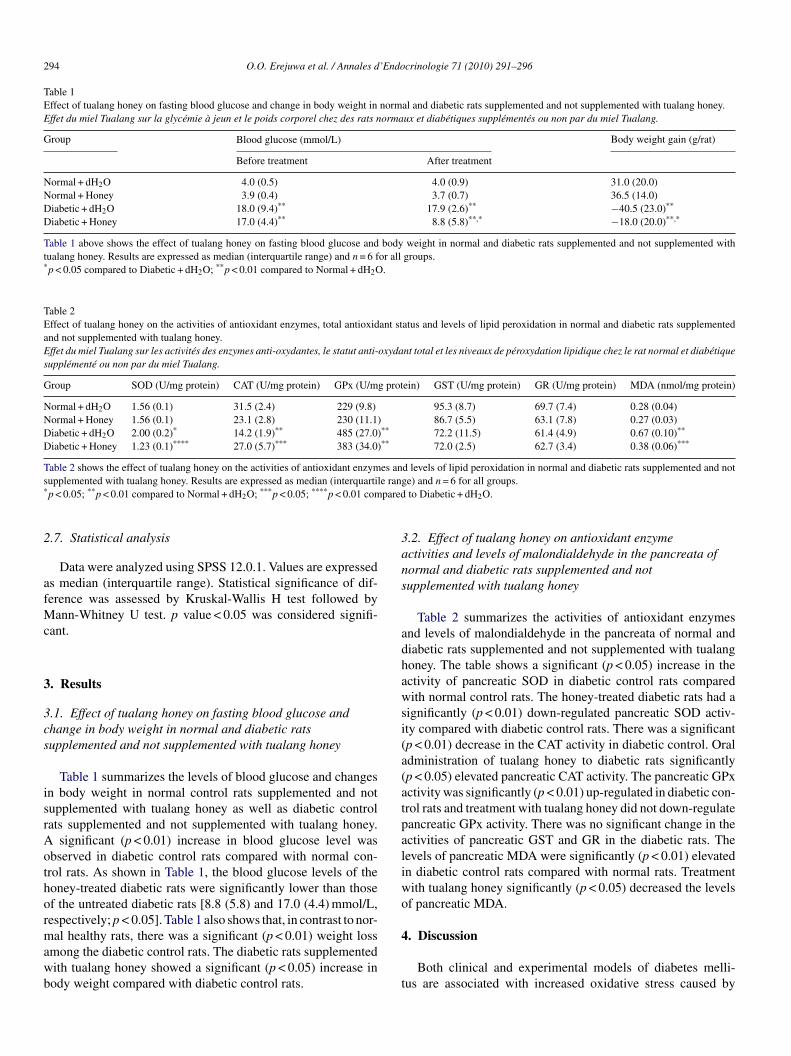
Table 1Effect of tualang honey on fasting blood glucose and change in body weight in normal and diabetic rats supplemented and not supplemented with tualang honey.Effet du miel Tualang sur la glycémie à jeun et le poids corporel chez des rats normaux et diabétiques supplémentés ou non par du miel Tualang.
Group Blood glucose (mmol/L) Body weight gain (g/rat)
Before treatment After treatment
Normal + dH2O 4.0 (0.5) 4.0 (0.9) 31.0 (20.0)Normal + Honey 3.9 (0.4) 3.7 (0.7) 36.5 (14.0)Diabetic + dH2O 18.0 (9.4)** 17.9 (2.6)** −40.5 (23.0)**
Diabetic + Honey 17.0 (4.4)** 8.8 (5.8)**,* −18.0 (20.0)**,*
Table 1 above shows the effect of tualang honey on fasting blood glucose and body weight in normal and diabetic rats supplemented and not supplemented withtualang honey. Results are expressed as median (interquartile range) and n = 6 for all groups.*p < 0.05 compared to Diabetic + dH2O; **p < 0.01 compared to Normal + dH2O.
Table 2Effect of tualang honey on the activities of antioxidant enzymes, total antioxidant status and levels of lipid peroxidation in normal and diabetic rats supplementedand not supplemented with tualang honey.Effet du miel Tualang sur les activités des enzymes anti-oxydantes, le statut anti-oxydant total et les niveaux de péroxydation lipidique chez le rat normal et diabétiquesupplémenté ou non par du miel Tualang.
Group SOD (U/mg protein) CAT (U/mg protein) GPx (U/mg protein) GST (U/mg protein) GR (U/mg protein) MDA (nmol/mg protein)
Normal + dH2O 1.56 (0.1) 31.5 (2.4) 229 (9.8) 95.3 (8.7) 69.7 (7.4) 0.28 (0.04)Normal + Honey 1.56 (0.1) 23.1 (2.8) 230 (11.1) 86.7 (5.5) 63.1 (7.8) 0.27 (0.03)Diabetic + dH2O 2.00 (0.2)* 14.2 (1.9)** 485 (27.0)** 72.2 (11.5) 61.4 (4.9) 0.67 (0.10)**
Diabetic + Honey 1.23 (0.1)**** 27.0 (5.7)*** 383 (34.0)** 72.0 (2.5) 62.7 (3.4) 0.38 (0.06)***
Table 2 shows the effect of tualang honey on the activities of antioxidant enzymes and levels of lipid peroxidation in normal and diabetic rats supplemented and nots e rang* pared
2
afMc
3
3cs
isrAothormawb
3ans
adhawsi(a(atpaliw
upplemented with tualang honey. Results are expressed as median (interquartilp < 0.05; **p < 0.01 compared to Normal + dH2O; ***p < 0.05; ****p < 0.01 com
.7. Statistical analysis
Data were analyzed using SPSS 12.0.1. Values are expresseds median (interquartile range). Statistical significance of dif-erence was assessed by Kruskal-Wallis H test followed by
ann-Whitney U test. p value < 0.05 was considered signifi-ant.
. Results
.1. Effect of tualang honey on fasting blood glucose andhange in body weight in normal and diabetic ratsupplemented and not supplemented with tualang honey
Table 1 summarizes the levels of blood glucose and changesn body weight in normal control rats supplemented and notupplemented with tualang honey as well as diabetic controlats supplemented and not supplemented with tualang honey.
significant (p < 0.01) increase in blood glucose level wasbserved in diabetic control rats compared with normal con-rol rats. As shown in Table 1, the blood glucose levels of theoney-treated diabetic rats were significantly lower than those
f the untreated diabetic rats [8.8 (5.8) and 17.0 (4.4) mmol/L,espectively; p < 0.05]. Table 1 also shows that, in contrast to nor-al healthy rats, there was a significant (p < 0.01) weight lossmong the diabetic control rats. The diabetic rats supplementedith tualang honey showed a significant (p < 0.05) increase inody weight compared with diabetic control rats.
o
4
t
e) and n = 6 for all groups.to Diabetic + dH2O.
.2. Effect of tualang honey on antioxidant enzymectivities and levels of malondialdehyde in the pancreata oformal and diabetic rats supplemented and notupplemented with tualang honey
Table 2 summarizes the activities of antioxidant enzymesnd levels of malondialdehyde in the pancreata of normal andiabetic rats supplemented and not supplemented with tualangoney. The table shows a significant (p < 0.05) increase in thectivity of pancreatic SOD in diabetic control rats comparedith normal control rats. The honey-treated diabetic rats had a
ignificantly (p < 0.01) down-regulated pancreatic SOD activ-ty compared with diabetic control rats. There was a significantp < 0.01) decrease in the CAT activity in diabetic control. Oraldministration of tualang honey to diabetic rats significantlyp < 0.05) elevated pancreatic CAT activity. The pancreatic GPxctivity was significantly (p < 0.01) up-regulated in diabetic con-rol rats and treatment with tualang honey did not down-regulateancreatic GPx activity. There was no significant change in thectivities of pancreatic GST and GR in the diabetic rats. Theevels of pancreatic MDA were significantly (p < 0.01) elevatedn diabetic control rats compared with normal rats. Treatmentith tualang honey significantly (p < 0.05) decreased the levelsf pancreatic MDA.
. Discussion
Both clinical and experimental models of diabetes melli-us are associated with increased oxidative stress caused by
’Endo
codoSiRwps1edectbic
soameaelcwmMfhf
wriaetivemEhcdcIimiHm
TMb(miattmss
oTpeTcTchpost
C
A
aUcs
R
O.O. Erejuwa et al. / Annales d
hronic hyperglycemia [25]. Oxidative stress is postulated asne of the mechanisms by which glucotoxicity produces itseleterious effects [26]. STZ has a selective cytotoxic actionn pancreatic �-cells. Although the exact mechanism by whichTZ produces its �-cell cytotoxic effect is not fully understood,
t is believed to be mediated by release of nitric oxide (NO) andOS resulting in alkylation of DNA [27]. In the present study,e examined the possible protective effect of tualang honey onancreatic tissue in STZ-induced diabetic rats. In our previoustudy, we have demonstrated that tualang honey at a dose of.0 g/kg improved body weight, decreased blood glucose lev-ls and ameliorated oxidative stress in kidneys of STZ-inducediabetic rats [19]. We had postulated that the antihyperglycemicffect of tualang honey might partly be due to protection of pan-reatic �-cells against STZ-induced diabetic oxidative stress. Inhe present study, tualang honey treatment at a dose of 1.0 g/kgody weight decreased elevated blood glucose and moderatelyncreased body weight in STZ-induced diabetic rats. This isonsistent with our previous findings [19].
ROS attack membrane phospholipids and cause the conver-ion of unsaturated fatty acids to lipid peroxides. Peroxidationf fatty acids containing three or more double bonds gener-tes MDA. These lipid peroxides are highly toxic products thatay bring about inactivation and damage of membrane bound
nzymes, proteins and cell membranes either through directttack by free radicals or through chemical modification by itsnd products, such as MDA [28]. In our study, pancreatic MDAevels were significantly increased in diabetic control rats. This isonsistent with other studies showing that diabetes is associatedith elevated levels of lipid peroxides [29]. Tualang honey treat-ent decreased the elevated MDA. Increased levels of pancreaticDA in diabetic rats might be a consequence of increased ROS
ormation and decreased antioxidants caused by STZ as well asyperglycemia, which in view of augmented effect of oxygenree radicals might have led to accumulation of MDA [4,27,29].
Our data showed that superoxide dismutase (SOD) activityas up-regulated while catalase (CAT) activity was down-
egulated in the pancreas of diabetic control rats. Othernvestigators also reported similar findings [30,31]. Increasedctivity of SOD could be due to its induction by increased gen-ration of superoxide anions. This is often a defense mechanismo protect the pancreatic �-cells from the detrimental effect ofncreased superoxide production [7,32]. Although SOD con-erts superoxide radicals to hydrogen peroxide, experimentalvidence show that over-expression of SOD activity may pro-ote toxicity of superoxide-radical-forming compounds [33].nhanced SOD activity might lead to an increased turnover ofydrogen peroxide generation. Catalase (CAT) catalyzes theonversion of hydrogen peroxide to water and oxygen. Theown-regulation of CAT activity might be a consequence of gly-ation [34], and inhibition by elevated levels of superoxide [35].n line with these data, enhanced SOD and reduced CAT activ-ties might result in accumulation of hydrogen peroxide which
ight undergo Fenton reaction in the presence of transition metalons such as copper and iron to generate hydroxyl radicals [36].ydroxyl radical is a highly reactive species proposed as theain initiator of most free radical induced tissue damage [37].
crinologie 71 (2010) 291–296 295
his could have contributed to the significantly elevated levels ofDA observed in the diabetic control rats. The pancreas of dia-
etic rats showed increased activity of glutathione peroxidaseGPx). The elevated GPx activity in the diabetic rat pancreasight be a compensatory mechanism to detoxify organic and
norganic peroxides including excess hydrogen peroxide gener-ted by increased SOD activity [38,39]. Our results showed thathe activities of glutathione reductase (GR) and glutathione-S-ransferase (GST) did not change in diabetic rat pancreas. This
ay indicate that these enzymes (GR and GST) do not playignificant roles in protecting the pancreas against oxidativetress.
In conclusion, our results suggest that pancreatic tissuesf diabetic rats are exposed to an increased oxidative stress.ualang honey supplementation showed protective effects on theancreas against STZ-induced diabetic oxidative stress. This isvident by the reduced levels of lipid peroxidation marker, MDA.ualang honey administration also restored pancreatic SOD andatalase activities, while GPx activity remained up-regulated.hese protective effects of tualang honey on the diabetic pan-reas against oxidative stress might be responsible for itsypoglycemic effect. Our data showed that tualang honey did notroduce any effect on blood glucose, antioxidant enzymes andther oxidative stress markers in non-diabetic pancreas. Furthertudies are required to elucidate the exact mechanism by whichualang honey protects the pancreas against oxidative stress.
onflict of interest statement
The authors declare that there is no conflict of interest.
cknowledgements
This study was supported by Universiti Sains Malaysia. Welso acknowledge Fellowship awarded to the first author byniversiti Sains Malaysia. We are very grateful to Federal Agri-
ultural Marketing Authority (FAMA), Kedah, Malaysia forupplying the tualang honey.
eferences
[1] Oberley LW. Free radicals and diabetes. Free Radic Biol Med1988;5:113–24.
[2] Giugliano D, Ceriello A, Paolisso G. Oxidative stress and diabeticcomplications (Review). Diabetes Care 1996;19:257–67.
[3] Kaiser N, Leibowitz G, Nesher R. Glucotoxicity and beta-cell failure intype 2 diabetes mellitus. J Pediatr Endocrinol Metab 2003;16:5–22.
[4] Ihara Y, Toyokuni S, Uchida K, Odaka H, Tanaka T, Ikeda H, et al. Hyper-glycemia causes oxidative stress in pancreatic �-cells of GK rats, a modelof type 2 diabetes. Diabetes 1999;48:927–32.
[5] Grankvist K, Marklund SL, Taljedal IB. CuZn-superoxide dismutase, Mn-superoxide dismutase, catalase and glutathione peroxidase in pancreaticislets and other tissues in the mouse. Biochem J 1981;199:393–8.
[6] Ho E, Chen G, Bray TM. Supplementation of N-acetylcysteine inhibits
NFkappaB activation and protects against alloxan-induced diabetes in CD-1 mice. FASEB J 1999;13:1845–54.[7] Kubisch HM, Wang J, Bray TM, Phillips JP. Targeted over-expression ofCu/Zn superoxide dismutase protects pancreatic �-cells against oxidativestress. Diabetes 1997;46:1563–6.
2 ’Endo
[
[
[
[
[
[
[
[
[
[
[
[
[
[
[
[
[
[
[
[
[
[
[
[
[
[
[
[
[
96 O.O. Erejuwa et al. / Annales d
[8] Benhamou PY, Moriscot C, Richard MJ, Beatrix O, Badet L, PattouF, et al. Adenovirus-mediated catalase gene transfer reduces oxidantstress in human, porcine and rat pancreatic islets. Diabetologia 1998;41:1093–100.
[9] White JW. Composition of honey. In: Carne E, editor. Honey: A compre-hensive survey. London: Heinemann; 1979. p. 157–207.
10] Gheldof N, Wang XH, Engeseth NJ. Identification and quantification ofantioxidant components of honeys from various floral sources. J AgricFood Chem 2002;50:5870–7.
11] Schepartz AI. Honey catalase: occurrence and some kinetic properties. JApic Res 1966;5:167–76.
12] Crane E. Honey: a comprehensive survey. New York, USA: Crane russakand company; 1975.
13] Aljadi AM, Kamaruddin MY. Evaluation of the phenolic contents andantioxidant capacities of two Malaysian floral honeys. Food Chem2004;85:513–8.
14] Rosenblat G, Angonnet S, Goroshit A, Tabak M, Neeman I. Antioxidantproperties of honey produced by bees fed with medical plant extracts. In: In“Proceedings of the International Conference on Bee Products: Properties,Applications and Apitherapy” held in Tel Aviv, Israel, May 26–30. NewYork: Plenum Press; 1996. p. 49–55.
15] Mohamed M, Sirajudeen KNS, Swamy M, Yaacob NS, Sulaiman SA. Stud-ies on the antioxidant properties of tualang honey of Malaysia. Afr J TradCAM 2010;7(1):59–63.
16] Frankel S, Robinson GE, Berenbaum MR. Antioxidant capacity and cor-related characteristics of 14 unifloral honeys. J Apic Res 1998;37(1):27–31.
17] Nagai T, Inoue R, Kanamori N, Suzuki N, Nagashima T. Characterizationof honey from different floral sources. Its functional properties and effectsof honey species on storage of meat. Food Chem 2006;97:256–62.
18] Gheldof N, Engeseth NJ. Antioxidant capacity of honeys from various floralsources based on the determination of oxygen radical absorbance capacityand inhibition of in vitro lipoprotein oxidation in human serum samples. JAgric Food Chem 2002;50:3050–5.
19] Erejuwa OO, Sulaiman SA, Ab Wahab MS, Sirajudeen KNS, SalzihanMS. Effects of Malaysian tualang honey supplementation on glycemia,free radical scavenging enzymes and markers of oxidative stress in kid-neys of normal and streptozotocin-induced diabetic rats. Int J Cardiol2009;137:S45.
20] Ohkawa H, Ohishi N, Yagi K. Assay for lipid peroxides in animal tissuesby thiobarbituric acid reaction. Anal Biochem 1979;95:351–8.
21] Gott L. A simple method for determination of serum catalase activ-
ity and revision of reference range. Clin Chim Acta 1991;196(2–3):143–51.22] Goldberg DM, Spooner RJ. Assay of glutathione reductase. In: BergmeyenHV, editor. Methods of enzymatic analysis. FL: Verlag Chemie DeerfieldBeach; 1983. p. 258–65.
[
crinologie 71 (2010) 291–296
23] Habig WH, Pabst MJ, Jakoby WB. Glutathione-S-transferases. Thefirst enzymatic step in mercapturic acid formation. J Biol Chem1974;249:7130–9.
24] Bradford M. A rapid and sensitive method for the quantitation of microgramquantities of protein utilizing the principle of protein-dye binding. AnalBiochem 1976;72:248–54.
25] Baynes JW, Thorpe SR. The role of oxidative stress in diabeticcomplications. Curr Opin Endocrinol 1999;3:277–84.
26] Kaneto H, Kajimoto Y, Miyagawa J, Matsuoka T, Fujitani Y, UmayaharaY. Beneficial effects of antioxidants in diabetes: possible protection ofpancreatic beta-cells against glucose toxicity. Diabetes 1999;48:2398–406.
27] Haluzik M, Nedvidkova J. The role of nitric oxide in the development ofstreptozotocin-induced diabetes mellitus: experimental and clinical impli-cation. Physiol Res 2000;49:37–42.
28] Hennig B, Chow CK. Lipid peroxidation and endothelial cell injury: impli-cations in atherosclerosis. Free Radic Biol Med 1988;4:99–106.
29] Yagi K. Lipid peroxides and human diseases. Chem Phys Lipids1987;45:337–51.
30] Razieh Y, Amin A, Shirin J. Experimental diabetes treated with Achilleasantolina: Effect on pancreatic oxidative parameters. J Ethnopharmacol2007;112:13–8.
31] Kakkar R, Mantha SV, Radhi J, Prasad K, Kalra J. Increased oxidativestress in rat liver and pancreas during progression of streptozotocin-induceddiabetes. Clin Sci 1998;94:623–32.
32] Tiedge M, Lortz S, Munday R, Lenzen S. Protection against theco-operative toxicity of nitric oxide and oxygen free radicals by over-expression of antioxidant enzymes in bioengineered insulin-producingRINm5F cells. Diabetologia 1999;42:849–55.
33] Lortz S, Gurgul-Convey E, Lenzen S, Tiedge M. Importance of mito-chondrial superoxide dismutase expression in insulin-producing cells forthe toxicity of reactive oxygen species and pro-inflammatory cytokines.Diabetologia 2005;48:1541–8.
34] Wolff SP, Dean RT. Glucose autoxidation and protein modification: Thepotential role of ‘autoxidative glycosylation’ in diabetes. Biochem J1987;245:243–50.
35] Kono Y, Fridovich I. Superoxide radical inhibits catalase. J Biol Chem1982;257:5751–4.
36] Lloyd RV, Hanna PM, Mason RP. The origin of the hydroxyl radical oxygenin the Fenton reaction. Free Radic Biol Med 1997;22:885–8.
37] Imlay JA, Chin SM, Linn S. Toxic DNA damage by hydrogen peroxidethrough the Fenton reaction in vivo and in vitro. Science 1998;240:640–2.
38] Christopherson BO. Reduction of linolenic acid hydroperoxide by a glu-
tathione peroxidase. Biochem Biophys Acta 1969;176:463–70.39] Dobrino A, Patriaca P. Neutrophil-endothelial cell interaction: evidencefor and mechanism of the self protection of bovine microvascular endothe-lial cells from hydrogen peroxide induced oxidative stress. J Clin Invest1986;78:462–71.
![Incorporation of a Biocompatible Nanozyme in Cellular ...Sep 23, 2020 · [1] Communication Incorporation of a Biocompatible Nanozyme in Cellular Antioxidant ... 30, Mother Teresa](https://img.pdfslide.fr/doc/110x75/60dcc9441f679421487e36c9/incorporation-of-a-biocompatible-nanozyme-in-cellular-sep-23-2020-1-communication.jpg)