Embed Size (px)
Citation preview
716 Specialia EXPERIENTIA X X I I / l l
Conclusions. Dens les races C57B1, CBA, B A L B / C e t R F nous av ions o b t e n u 2, apr~s 400 R, 7,5% des cellules p r 6 s e n t a n t des r 6 a r r a n g e m e n t s ch romosomiques . Avec 6 % de t rans loca t ions , les spe rma togon ie s des souris de race A K R / T r . s e m b l e n t doric m a n i f e s t e r une radiosensi - bil i t6 assez voisine. S' i l es t imposs ib le de d 6 t e r m i n e r avec e x a c t i t u d e la p a r t pr ise dens ces r e m a n i e m e n t s p a r les ch romosomes m6tacen t r iques , on p e u t c e p e n d a n t r e m a r - que r que les b i v a l e n t s m 6 t a c e n t r i q u e s m a n q u e n t dens 21 cellules, sans qu ' i l soi t possible de conclure si les 2 chro- mosomes m 6 t a c e n t r i q u e s on t donn6 4 ac rocen t r iques ou s ' i ls i n t e r v i e n n e n t dans les au t r e s conf igura t ions observ6es dens ces cellules 4.
Summary. A s p o n t a n e o u s t r a n s l o c a t e d s t r a i n of mice (AKR/Tr . ) w i th 36 ac rocen t r i c a n d 2 m e t a c e n t r i c ch romo-
somes rece ived 400 R of who le -body X- i r r ad ia t ion . Cyto- logical e x a m i n a t i o n of d iv id ing p r i m a r y s p e r m a t o c y t e s a t t h e d iakines is - f i r s t m e t a p h a s e s tage of meiosis showed 6% of cells w i t h c h r o m o s o m a l r e a r r an g emen t s .
A. LI~ONARD et G m DEKNUDT
Laboratoire de Gdndtique, D@artement de Radiobiologie, Centre d'Etude de l'Energie Nucldaire, Mol (Belgique), 6 ]uillet 7966.
4 Ce travail entre dans le cadre du contrat EURATOM/C.E.N. N ~ 053-64-3-BIOB e t a pu ~tre effectu6 grfice aux subsides du Fonds de la Recherche Scientifique Fondamentale Collective.
Histotopochemistry of Ascorbic Acid during the Formation of Carrageenin Granuloma
I t has been p r o v e d in m a n y b iochemica l expe r imen t s t h a t ascorbic acid p lays a n i m p o r t a n t p a r t n o t on ly in p re se rv ing col lagen b u t especial ly in t he b iosyn thes i s of col lagen in a newly - fo rmed connec t ive t issue. The ques- t i on of t o p o g r a p h i c c o n n e c t i o n of ascorbic acid to t h e i n d i v i d u a l c o m p o n e n t s of t he connec t ive t issue, however , is n o t ye t qu i te clear. Thus , for example . ~MUCHALOV~I a n d CHVAPIL 1 h a v e found ou t t h a t the re exis ts a close co r re l a t ion be tween t he a m o u n t of ascorbic acid and t h e newly - fo rmed col lagen d u r i n g t he f o r m a t i o n of ca r ra - geen in g ranu loma . T h e y h a v e n o t found, however , a n y connec t ion be tween t he n u m b e r of cells (DNA) and the c o n t e n t of ascorbic acid in t he g r a n u l a t i o n tissue, as p r o v e d ear l ier b y WOESSNER a n d I~OUCEK 2 for a g ranu- l oma formed a r o u n d a n i m p l a n t e d smal l po lyv iny l sponge.
I n t he p re sen t work a n a t t e m p t a t h i s t o t o p o c h e m i s t r y of ascorbic acid d u r i n g t he f o r m a t i o n of ca r rageen in g r a n u l o m a has been made.
Method. I n guinea-pigs (males weighing 250-300 g) ca r r ageen in g r a n u l o m a in t he a b d o m i n a l region was pro- voked b y a s u b c u t a n e o u s in j ec t ion of 5 ml 1% car ra- geenin so lu t ion in 0.9% NaC1 to wh ich penici l l in (100 IU/ml ) a n d s t r e p t o m y c i n (100 /~g/ml) were added. T h e t i ssue was w i t h d r a w n 2, 4, 7, 9 a n d 13 days a f te r t h e in jec t ion of ca r r ageen in solut ion. Ve ry smal l samples of t h e g r a n u l o m a t i ssue (ca. 2 . 2 . 2 ram) were t r e a t e d a f t e r GIROUD and LEBLOND (LIPP3). The samples were i m b e d d e d in pa ra f f in and cu t in to 5 # t h i ck slices. Some of t he slices were s t a ined w i t h hematoxy l in -eos in .
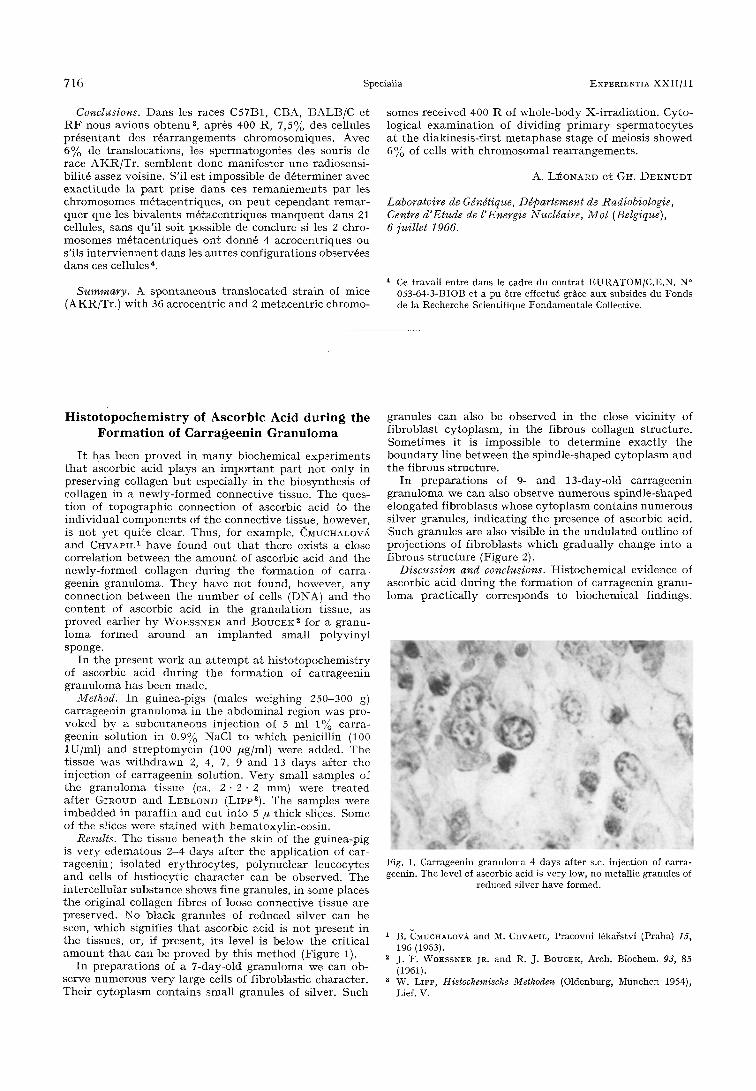
Results. The t issue b e n e a t h t h e skin of t he gu inea-p ig is v e r y e d e m a t o u s 2 -4 days a f t e r t he app l i ca t ion of car- r ageen in ; i so la ted e ry th rocy te s , po lynuc l ea r leucocytes a n d cells of h i s t iocy t ic c h a r a c t e r can be observed . The in te rce l lu la r s u b s t a n c e shows fine granules , in some places t h e or ig inal collagen f ibres of loose connec t ive t issue are preserved . No b l a c k granu les of reduced s i lver can be seen, wh ich signifies t h a t ascorbic acid is no t p re sen t in t h e t issues, or, if p resen t , i t s level is be low the cr i t ica l a m o u n t t h a t can be p r o v e d b y th i s m e t h o d (Figure 1).
I n p r e p a r a t i o n s of a 7-day-old g r a n u l o m a we can ob- serve n u m e r o u s v e r y large cells of f ib roblas t ic cha rac te r . T h e i r c y t o p l a s m con ta ins smal l g ranu les of silver. Such
granules c an also be obse rved in t h e close v ic in i ty of f i b rob la s t cy top lasm, in t h e f ibrous col lagen s t ruc tu re . Some t imes i t is imposs ib le to d e t e r m i n e exac t ly t h e b o u n d a r y l ine b e t w een the sp ind le - shaped c y t o p l a s m a n d t h e f ibrous s t ruc tu re .
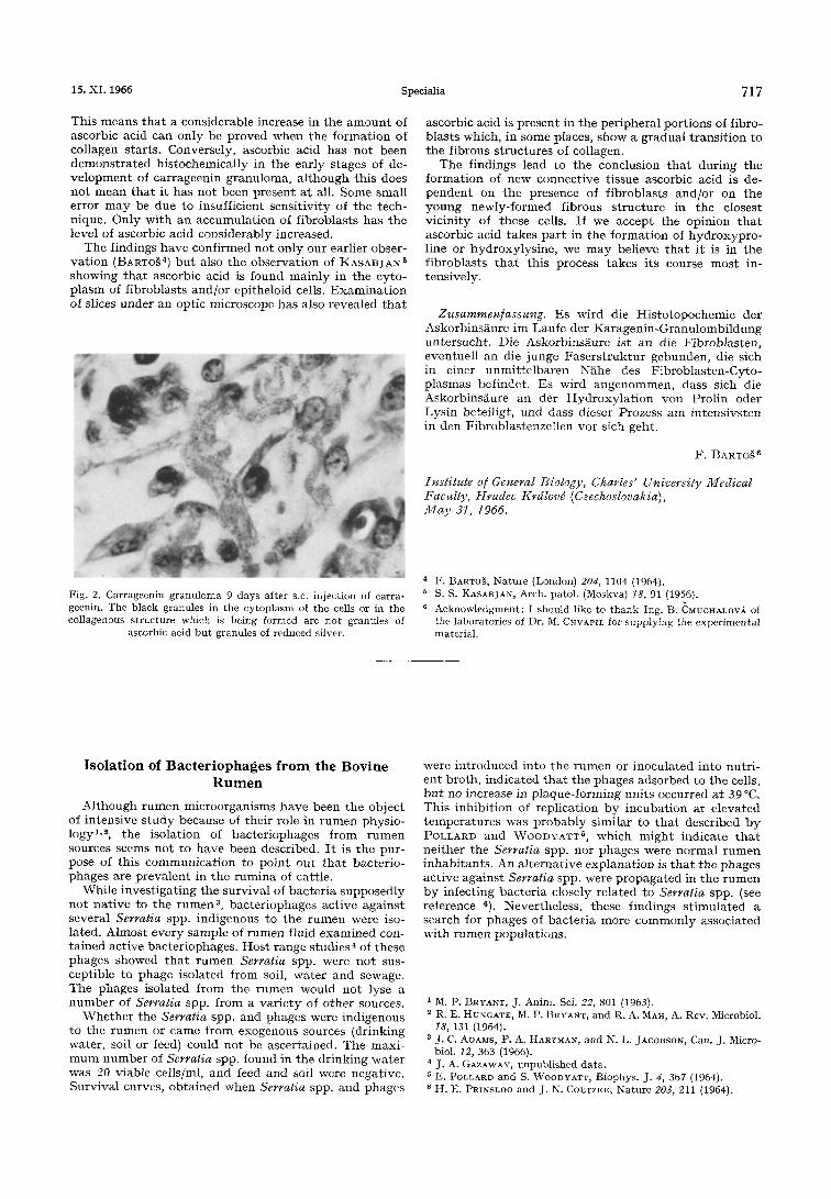
I n p r e p a r a t i o n s of 9- an d 13-day-old ca r r ageen in g r a n u l o m a we can also observe n u m e r o u s sp ind le - shaped e longa ted f ib rob las t s whose c y t o p l a s m con ta ins n u m e r o u s s i lver granules , i nd i ca t i ng t h e presence of ascorbic acid. Such granules are also vis ible in the u n d u l a t e d out l ine of p ro jec t ions of f ib rob las t s wh ich g r adua l l y change in to a f ibrous s t r u c t u r e (Figure 2).
Discussion and conclusions. His tochemica l ev idence of ascorbic acid du r ing t h e f o r m a t i o n of ca r rageen in g ranu- loma p rac t i ca l ly cor responds to b iochemica l f indings.
Fig. 1. Carrageenin granuloma 4 days after s.c. injection of carra- geenin. The level of ascorbic acid is very low, no metallic granules of
reduced silver have formed.
1 B. CMUCHALOV~ and M. CHVAPIL, Pracovni 16ka~stvi (Praha) 15, 196 (1963).
2 j . ]2. WOESSNER JR. and R. J. BOUCEK, Arch. Biochem. 93, 85 (1961).
a W. LIPP, Histochemische Methoden (Oldenburg, Mfinchen 1954), Lief. V.
15. XI. 1966 Specialia 717
This means t h a t a considerable increase in the a m o u n t of ascorbic acid can only be proved when the fo rma t ion of collagen s tar ts . Conversely, ascorbic acid has no t been d e m o n s t r a t e d h i s tochemica l ly in the ear ly s tages of de- v e l o p m e n t of car rageenin granuloma, a l though this does no t mean t h a t i t has no t been presen t a t all. Some small error m a y be due to insuff icient sens i t iv i ty of the tech- nique. Only wi th an accumula t ion of f ibroblas ts has the level of ascorbic acid cons iderably increased.
The f indings have conf i rmed not only our earlier obser- va t ion (BARTO~ 4) bu t also the observa t ion of KASABJAN 5 showing t h a t ascorbic acid is found main ly in tile cyto- p lasm of f ibroblas ts and /o r epi theloid cells. E x a m i n a t i o n of slices under an optic microscope has also revealed t h a t
ascorbic acid is p resen t in the per iphera l por t ions of f ibro- bIasts which, in some places, show a gradual t r ans i t ion to the f ibrous s t ruc tu res of collagen,
The f indings lead to t he conclusion t h a t dur ing the fo rmat ion of new connect ive t issue ascorbic acid is de- p e n d e n t on the presence of f ibroblas ts and /or on the young newly- fo rmed f ibrous s t ruc tu re in the closest v ic in i ty of these cells. If we accept the opinion t h a t ascorbic acid takes p a r t in the fo rma t ion of h y d r o x y p r o - line or hydroxylys ine , we m a y believe t h a t i t is in the f ibroblas ts t h a t this process takes i ts course mos t in- tensively.
Zusammen/assung. Es wird die Hi s to topochemie der AskorbinsXure im Laufe der Karagen in -Granu lombi ldung untersucht . Die Askorbins~ure ist an die F ibroblas ten , eventuel l an die junge F a s e r s t r u k t u r gebunden, die sich in einer u n m i t t e l b a r e n N~Lhe des F ib rob las ten-Cyto- p lasmas bef indet . Es wird angenommen , dass sich die Askorbins/ iure an der H y d r o x y l a t i o n yon Prol in oder Lys in beteil igt , und dass dieser Prozess am in tens ivs ten in den Fibrobtas tenze l len vor sich geht .
V. BARTO~ 6
Institute o/ General Biology, Charles' University Medical Faculty, Hradec Birdlovd (Czechoslovakia), May 31, 1966.
Fig. 2. Carrageenin granuloma 9 days after s.c. injection of carra- geenin. The black granules in the cytoplasm of the ceils or in the collagenous structure which is being formed are not granules of
ascorbic acid but granules of reduced silver.
4 F. BARTO~, Nature (London) 20d, 1104 (1964). 5 S. S. KASABJAN, Arch. patol. (Moskva) 78, 91 (1956). 6 Acknowledgment: I should like to thank Ing. B. CMUCHALOV~ of
the laboratories of Dr. M. CHVAPIL for supplying the experimental material.
Isolat ion of Bacter iophages f rom the Bovine R u m e n
Although rumen microorganisms have been the objec t of in tens ive s tudy because of the i r role in r u m e n physio- logy 1,2, the isolat ion of bac te r iophages f rom r u m e n sources seems no t to have been described. I t is the pur- pose of th is communica t ion to po in t ou t t h a t bacter io- phages are p reva len t in the rumina of cat t le .
Whi le inves t iga t ing the survival of bac te r ia supposed ly no t na t ive to t he r u m e n 3, bac te r iophages act ive aga ins t several Serratia spp. indigenous to the r u m e n were iso- lated. Almost every sample of rumen fluid examined con- t a ined act ive bacter iophages . Hos t range s tudies 4 of these phages showed t h a t r u m e n Serratia spp. were no t sus- cept ible to phage isolated f rom soil, wa te r and sewage. The phages isolated f rom the rumen would no t lyse a n u m b e r of Serratia spp. f rom a va r i e ty of o the r sources.
W h e t h e r the Serratia spp. and phages were indigenous to the rumen or came f rom exogenous sources (drinking water , soil or feed) could no t be ascer tained. The maxi - m u m n u m b e r of Serratia spp. found in the dr inking wa te r was 20 viable cells/ml, and feed and soil were negat ive. Survival curves, ob ta ined when Serratia spp. and phages
were in t roduced into the r u m e n or inocula ted into nut r i - en t broth , ind ica ted t h a t the phages adsorbed to the ceils, bu t no increase in p laque- fo rming uni ts occurred a t 39 ~ This inhib i t ion of repl icat ion by incubat ion at e leva ted t e m p e r a t u r e s was p ro b ab l y s imilar to t h a t descr ibed b y POLLARD and WOODYATT g, which migh t indica te t h a t ne i ther the Serratia spp. nor phages were normal r u m e n inhab i tan t s . An a l t e rna t ive exp lana t ion is t h a t the phages ac t ive agains t Serratia spp. were p ropaga t ed in the r u m e n by infect ing bac te r ia closely re la ted to Serratia spp. (see reference 6). Never theless , these f indings s t imula ted a search for phages of bac te r ia more co mmo n l y associa ted wi th rumen popula t ions .
1 M. P. BRYANT, J. Anita. Sci. 22, 801 (1963). 2 R. E. HUNGATE, M. P. BRYANT, and R. A. MAH, A. Rev. Microbiol.
18, 131 (1964). a j . C. ADAMS, P. A. HARTMAN, and N. L. JACOBSON, Can. J. Micro-
biol. 12, 363 (1966). 4 j, A. GAZAWAY, unpublished data. 5 E. POLLARD and S. WOODYATT, Biophys. J. 4, 367 (1964).
H. E. PRINSLOO and J. N. COETZEE, Nature 203, 211 (1964).