Embed Size (px)
Citation preview
In Vivo and In Vitro Neurotoxicity of the Human PrionProtein (PrP) Fragment P118 –135 Independently ofPrP Expression
Joelle Chabry,1 Christiane Ratsimanohatra,2* Isabelle Sponne,3* Pierre-Paul Elena,2 Jean-Pierre Vincent,1 andThierry Pillot3
1Institut de Pharmacologie Moleculaire et Cellulaire, Unite Mixte de Recherche 6097, Centre National de la Recherche Scientifique, 06560 Valbonne, France,2Iris Pharma, Les Nertieres, 06610 La Gaude, France, and 3Institut National de la Sante et de la Recherche Medicale, Universite de Nancy I, 54505Vandœuvre-les-Nancy, France
We recently demonstrated that the 118 –135 putative transmembrane domain of prion protein (PrP) exhibited membrane fusogenicproperties and induced apoptotic neuronal cell death of rat cortical neurons, independently of its aggregation state. The aim of thepresent study was to analyze the in vivo neurotoxicity of the prion fragment P118 –135 and to evaluate the potential role of the physio-logical isoform of PrP in the P118 –135-induced cell death. Here, we demonstrate that the nonfibrillar P118 –135 is cytotoxic to retinalneurons in vivo as monitored by intravitreal inoculation and recording of the electrical activity of retina and tissue examination.Moreover, knock-out PrP gene mice exhibit similar sensitivity to the nonfibrillar P118 –135-induced cell death and electrical perturba-tions, strongly suggesting that cell death occurs independently of PrP expression. Interestingly, a variant nonfusogenic P118 –135 peptide(termed P118 –135�) had no effects on in vivo neuronal viability, suggesting that the P118 –135-induced cell death is mediated by itsmembrane destabilizing properties. These data have further been confirmed in vitro. We show that the fusogenic peptide P118 –135induces death of cultured neurons from both wild-type and knock-out PrP gene mice via an apoptotic-mediated pathway, involving earlycaspase activation and DNA fragmentation. Altogether these results emphasize the neurotoxicity of the fusogenic nonfibrillar PrPtransmembrane domain and indicate that fibril formation and PrP expression are not obligatory requirements for neuronal cell death.The use of synthetic prion peptides could provide insights into the understanding of neuronal loss mechanisms that take place during thedevelopment of the various types of spongiform encephalopathies.
Key words: prion peptide; retina; apoptosis; in vivo; caspase activities; electroretinogram
IntroductionTransmissible spongiform encephalopathies (TSE) are fatal neu-rological disorders including Creutzfeldt-Jakob disease andGerstmann-Straussler-Scheinker syndrome (GSS) in humans,scrapie in sheep and goats, and bovine spongiform encephalop-athy in cattle. Common pathological characteristics of TSE are,among others, vacuolization of the neuropils, severe gliosis, anddegeneration of neurons (Fraser, 1993). The accumulation in thebrain of scrapie-infected animals of large aggregates of a patho-logical isoform [prion protein (PrP)sc, also called PrP-res] of thenormal cellular prion protein (PrPc or PrP-sen) is usually ob-served (Kretzschmar et al., 1986; Locht et al., 1986). AlthoughPrP-res has been proposed to be responsible for both transmis-
sion and pathogenicity of TSEs, the occurrence of natural andexperimental TSEs in the absence of PrP-res accumulation dem-onstrates that the aggregation step is not an obligatory require-ment for neurodegeneration. Moreover, PrP-res preparation in-jected intracerebrally into PrP gene knock-out mice (PrP 0/0) failsto cause disease and to provoke neural damages. These observa-tions raise the possibility that PrP-res is not directly toxic by itselfand support the idea that the development of TSEs requires thepresence of both prion protein isoforms, PrP-res and PrP-sen.
Other aspects of PrP-sen biosynthesis such as aberrant subcel-lular location and topology may greatly influence the pathogenic-ity. Cell-free translation system studies (Hay et al., 1987; Lopez etal., 1990) demonstrated that PrP could be found in several topo-logic forms, including a transmembrane spanning domain iso-form (termed CtmPrP). Mice carrying a mutated PrP gene thatfavors markedly the synthesis of CtmPrP show spontaneous neu-rodegeneration without detectable PrP-res accumulation (Hegdeet al., 1998, 1999). The extent of neurodegeneration correlateswith the amount of CtmPrP, suggesting that CtmPrP is directlyresponsible for neuronal cell death. Brains of GSS-affected pa-tients bearing the A117V PrP mutation contain higher levels ofCtmPrP as compared with other human TSE-affected brains, butthey fail to present PrP-res deposits. It is likely that the A117V PrPmutation may influence CtmPrP accumulation, resulting in the
Received Aug. 22, 2002; revised Oct. 18, 2002; accepted Oct. 22, 2002.This work was supported in part by French Government grants from the Action Concertee Incitive Jeunes Cher-
cheurs (2000), the Action Thematique Concertee-Prions (Institut National de la Sante et de la Recherche Medicale),and the Groupement d’Interet Scientifique: infections a prions (2001). We are grateful to Dr. Charles Weissmann forproviding the PrP 0/0 mice. We thank Jean-Daniel Barde for animal care, Roxane Pichot for technical assistance, andNicole Zsurger for help with the preparation of the figures. We thank Drs. Nathalie Daude and Jean-Louis Nahon forcritical reading of this manuscript.
*C.R. and I.S. contributed equally to this work.Correspondence should be addressed to Joelle Chabry, Institut de Pharmacologie Moleculaire et Cellulaire, Unite
Mixte de Recherche 6097, Centre National de la Recherche Scientifique, 660 Route des Lucioles, 06560 Valbonne,France. E-mail: [email protected] © 2003 Society for Neuroscience 0270-6474/03/230462-08$15.00/0
462 • The Journal of Neuroscience, January 15, 2003 • 23(2):462– 469
neuropathological changes observed in GSS-affected brain; how-ever, the molecular events triggered by the nonaggregated CtmPrPand leading to neurodegeneration remain unknown.
The CtmPrP isoform spans the endoplasmic reticulum mem-brane at residues 113–135 with its N-terminus domain facing thecytosolic compartment (Hegde et al., 1998). We and others haveused the putative transmembrane domain of CtmPrP, e.g., aminoacids 118 –135, to model and characterize apoptotic neuronaldeath associated with topological variants of PrP-sen (Haik et al.,2000; Pillot et al., 2000). In contrast to the PrP fragment 106 –126,which required both fibrillation and the presence of PrP-sen toexert its neurotoxicity (Brown et al., 1996), we demonstrated thatthe nonfibrillar peptide P118 –135 induced apoptosis of corticalneurons (Pillot et al., 2000), an effect mediated in part by itsmembrane perturbation properties (Pillot et al., 1997) (for re-view, see Brasseur et al., 1997).
In the present study, we address the question of the in vivoneurotoxicity of the nonfibrillar prion peptide P118 –135 on bothwild-type and PrP-devoid mice. We demonstrate that direct in-jection of soluble P118 –135 into mouse eyes induces cell death ofretinal neurons via an apoptotic pathway independent of PrPexpression. Moreover, primary cultures of neurons from brainsof both wild-type and PrP gene knock-out mice were treated withthe fusogenic P118 –135 peptide, and morphological and bio-chemical hallmarks of apoptotic cell death were investigated.
Materials and MethodsMaterials. The caspase substrates Asp-Glu-Val-Asp-p-nitroanilide(DEVD-pNA; caspase-3 substrate), Tyr-Val-Ala-Asp-p-nitroanilide(YVAD-pNA; caspase-1 substrate), IEPD-AMC (caspase-8 substrate),and LEHD-AMC (caspase-9 substrate) were purchased from Bachem.Serum-free medium Neurobasal, N2 supplement, and penicillin–strep-tomycin mixture were from Invitrogen (Gaithersburg, MD). All otherreagents were of highly purified grade from Sigma (St. Louis, MO).
Peptides. The human sequences of the prion protein fragments P118 –135 (AGAVVGGLGGYMLGSAMS, fusogenic) and P118 –135� (AGGV-VGGLGGYMLASAMS, nonfusogenic) were synthesized as describedpreviously (Pillot et al., 1996, 1997). These peptides differ at two posi-tions (underlined). Peptides were dissolved in PBS at a concentration of1 mM, distributed into 20 �l aliquots, and stored at �20°C until use.Under these conditions, the peptides remain soluble (Pillot et al., 2000).To obtain amyloid fibrils, the P118 –135 peptide should be incubated at a1 mM concentration for 72 hr at room temperature (Pillot et al., 1997,2000). The human sequence of the prion protein fragment (P106 –126,KTNMKHMAGAAAAGAVVGGLG) was purchased from Bachem.
Intravitreal injection. Adult male C57-black wild-type or PrP 0/0 mice[named Zurich I; Bueler et al. (1992)] (10 –12 weeks old) were anesthe-tized with an intraperitoneal injection of 60 mg/kg sodium pentobarbi-tal. Injections (1 �l) of peptides or vehicle (PBS) were done unilaterallywith a 33 gauge needle introduced into the posterior chamber on theupper pole of the eye directed toward the center of the vitreous. Theinjections were performed slowly to allow a better diffusion of the pep-tide and to avoid any ocular hypertension. For negative controls, micewere injected in the same conditions with 1 �l of vehicle (PBS). Nosignificant differences in the a- and b-wave values were observed betweenthe noninjected eyes and the eyes injected with 1 �l of PBS. At least threeanimals were used for each experimental condition.
Electroretinograms. Full-field electroretinogram (ERG) responses wereobtained with overnight dark-adapted mice prepared under dim redlight before recording. The pupils of anesthetized mice were dilated witha drop of 0.5% Mydriaticum. A silver chloride ring-recording electrodewas placed on the cornea, and the reference electrode, with a silver–silverchloride tip, was introduced into the mouth. Light stimulus (15 msec)was provided by a single flash placed 0.25 m in front of the animal. TheERGs were recorded using the EPIC-2000 (LKC Technologies) visualelectroretinogram test system and then stored and analyzed. Amplitude
of the a-wave was measured from the baseline to the bottom of thea-wave; b-wave amplitude was measured from the bottom of the a-waveto the peak of the b-wave. The averaged responses represent the mean oftwo white flashes delivered 2 min apart. Electroretinograms were re-corded before peptide treatments and then 1 and 7 d after injection. Atthe end of the ERG recording experiments, the histology and the in situterminal deoxynucleotidyl transferase-mediated biotinylated UTP nickend labeling (TUNEL) method were performed as described previously(Ettaiche et al., 2000). Briefly, mice were euthanized with an overdose ofsodium pentobarbital 1 and 7 d after intravitreal injections. The eyes wereenucleated and fixed in ice-cold 4% paraformaldehyde in PBS for 24 hrand then cryoprotected overnight in PBS containing 20% sucrose. Theeyes were then embedded in O.C.T. compound (Tissue-Tek, SAKURA,Tokyo, Japan), and frozen sections (10 �m) were cut on a cryostat (Leica,Nussloch, Germany). In situ cell death detection was performed on 1 dtreated eyes, following the manufacturer’s recommendations (Boeh-ringer Mannheim, Mannheim, Germany) and then revealed using a 3,3�-diaminobenzidine (DAB) substrate kit (Vector Laboratories, Burlin-game, CA). Morphological and histological observations of 7 d treatedretinas were done on stained sections with 1% cresyl violet and thenprocessed for detailed examination by light microscopy.
Cell culture. Cortical neurons from embryonic day (E) 13–14 C57-black wild-type or PrP 0/0 mice were prepared as described previously(Chabry et al., 1990) with minors modifications. Briefly, cells were dis-sociated mechanically with a Pasteur pipette in a chemically definedNeurobasal medium containing N2 supplement and penicillin–strepto-mycin. Dissociated cells were then plated at a density of 3 � 10 6 cells in 35mm tissue plastic dishes (or 5 � 10 4 cells in 96-well tissue plates) pre-coated with polylysine (10 �g/ml) and grown at 37°C in humidifiedatmosphere of 5% CO2, 95% air. After 3 d in vitro, cells were incubatedwith cytosine arabinoside (50 �M), an inhibitor of mitosis, to preventglial cell proliferation. For the different experiments described below,neurons were used after 4 –5 d of in vitro culture.
Cytotoxicity assay. Ninety-six-well tissue-plated neurons were treatedin the absence or presence of various concentrations of indicated pep-tides for different periods of time. The neurotoxicity of the peptides wasassessed quantitatively by the (3-(4,5-dimethylthiazol-2-yl)-5-(3-carboxymethoxyphenyl)-2-(4-sulfophenyl)-2H tetra-zolium (MTS)(Promega, Madison, WI) assay according to the manufacturer’s recom-mendations. The absorbance, measured at 492 nm from 96-well assayplates, is directly proportional to the number of living cells.
In situ labeling with TUNEL method. After 4 –5 d of culture, neuronsplated into 35 mm dishes were incubated in the absence or presence of 20�M P118 –135 at 37°C for 12 or 24 hr. At the end of the incubation time,cells were fixed in ice-cold 4% paraformaldehyde in PBS for 10 min andthen rinsed twice with PBS and incubated for 30 min at room tempera-ture in methanol containing 0.3% H2O2 to quench endogenous peroxi-dase activities. The in situ cell death detection was performed followingthe recommendations of the manufacturer (Boehringer Mannheim) andthen revealed using the DAB substrate kit (Vector). Cells were stainedwith eosin, coverslipped with glycerol, and processed for detailed exam-ination by light microscopy.
DNA fragmentation analysis. Neurons treated with 20 �M of P118 –135for 12 and 24 hr at 37°C were rinsed twice with PBS and then scraped offin 1 ml of lysis buffer (10 mM Tris, pH 7.4, 5 mM EDTA, 1% SDS). Proteinswere digested with 200 �g/ml proteinase K in lysis buffer for 2 hr at 55°C.Neuronal DNA was extracted with phenol-chloroform, and the aqueousphase was incubated with DNase-free RNase A (100 �g/ml) and thenprecipitated in a solution of 0.3 M sodium acetate in ethanol overnight at�20°C. Precipitated DNA samples were resuspended in distilled water,and the DNA concentration was determined by measuring the absor-bance at 260 nm. Samples of 10 �g of DNA were electrophoresed through1.2% agarose gel containing 1 �g/ml ethidium bromide. DNA bandswere visualized by UV light-transilluminator and photographed.
Measurement of caspase-like proteolytic activities. Caspase activitieswere measured by means of the cleavage of the substrates DEVD-pNA,YVAD-pNa, LEHD-AMC, and IEPD-AMC (Bachem). Briefly, after theindicated times of peptide treatment, cells were rinsed three times withice-cold PBS and incubated for 20 min on ice in a 25 mM HEPES buffer,
Chabry et al. • Cytotoxicity of the Prion Protein Fragment 118 –135 J. Neurosci., January 15, 2003 • 23(2):462– 469 • 463
pH 7.5, containing 1% (v/v) Triton X-100, 5mM EDTA, 1 mM EGTA, 5 mM MgCl2, 5 mM
dithiothreitol, 1 mM phenylmethylsulfonyl flu-oride, 10 �g/ml each of pepstatin and leupep-tine, and 5 �g/ml aprotinin. The lysate wascentrifuged for 15 min at 12,000 rpm and as-sayed for protein by the Bradford method (Bio-Rad, Hercules, CA). Fifty micrograms of pro-tein were incubated for 2 hr with 100 �M
caspase substrate initially dissolved in DMSO.The cleavage of caspase substrates was moni-tored by absorbance measurements at 405nm for DEVD-pNA and YVAD-pNa, and byfluorescence emission at 460 nm after excita-tion at 360 nm for LEHD-AMC and IEPD-AMC, using a Fluostar reader plate (BMG-Labtechnologies).
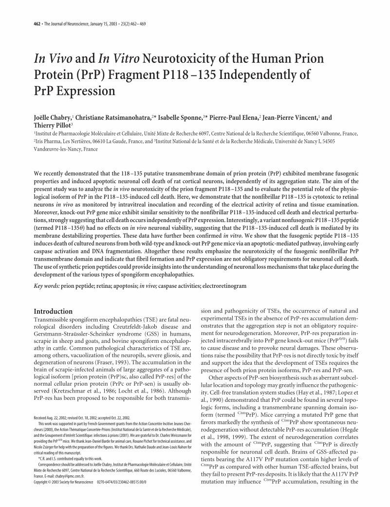
ResultsIn vivo cytotoxicity of thesoluble P118 –135The in vivo cytotoxicity of the prion pro-tein fragment P118 –135 was estimated bymeasuring the electrical activity ofpeptide- or vehicle-treated retinas of bothwild-type and PrP 0/0 mice. The electrical activity was monitoredby recording the ERGs 1 and 7 d after intravitreal inoculation.Figure 1 shows the effect of peptide injections on the ERG ofPrP 0/0 and wild-type mice as measured by the variation of theb-wave amplitudes. When compared with the vehicle-treatedgroup, the P118 –135-treated group exhibited significant deficitsin b-wave amplitudes, direct evidence of persistent and long-term damages to retinal function. The b-wave amplitude mark-edly decreased at day 1 and day 7 after injection in a dose-dependent manner. The maximal decrease of b-wave amplitudewas observed with the highest concentration of P118 –135 andreached up to 70% of the control value. Unlike the b-wave, thea-wave amplitude was not modified significantly even 7 d afterinjection of P118 –135 (data not shown). Interestingly, PrP 0/0
mice were sensitive to intravitreal injection of nonfibrillar P118 –135 (Fig. 1). Modifications of b-wave recorded in PrP 0/0 micefollow kinetics similar to those observed with wild-type mice,suggesting strongly that the in vivo cytotoxicity induced by P118 –135 does not require the presence of PrP-sen.
The peptide P118 –135� presents the same amino acid com-position as the P118 –135 peptide, but two amino acid residuesare permutated at positions 120 and 131 (see Materials and Meth-ods). It has been determined previously that this nonfusogenicfragment displays no neurotoxicity in vitro (Pillot et al., 2000).Accordingly, no significant decrease on the b-wave amplitudescould be observed after injection of the control peptide P118 –135� (Fig. 1).
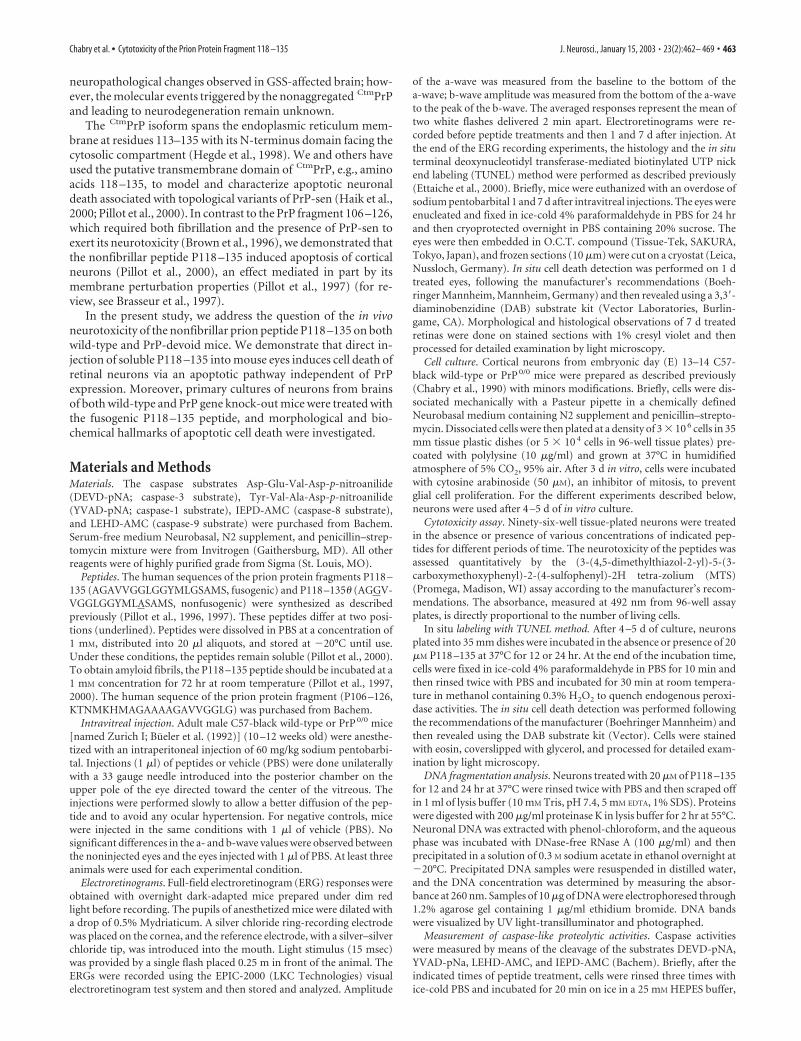
Histology studies performed on slides from control andpeptide-treated retinas 7 d after administration confirm the dra-matic damages caused by the nonfibrillar P118 –135 on retinalcells (Fig. 2A). All of the retinal changes observed occurred rela-tively close to the injection point, indicating a weak diffusion of thepeptide. Figure 2B shows representative photomicrographs of PBS-treated or P118–135-treated retina sections processed through theTUNEL labeling assay. One day after intravitreal injections of P118–135 (0.3 nmol), TUNEL-positive cells were observed in the inner andouter nuclear layers as well as in the ganglion cells layer (Fig. 2B). Nosignificant differences in TUNEL-labeling patterns were observedwhen injections were made into eyes of PrP0/0 and wild-type mice.
Intact retinas (data not shown) and retinas inoculated with PBSalone (Fig. 2B, control) showed only occasional TUNEL-positivecells.
In vitro cytotoxicity of P118 –135 on primarycultured neuronsWe established neuronal cultures from both PrP 0/0 and wild-typemice at E14. Under basal conditions, growth and survival of pri-mary neuronal cultures from PrP 0/0 mice did not differ signifi-cantly from PrP�/� neurons, up to 8 d in serum-free N2-supplemented medium (Fig. 3).
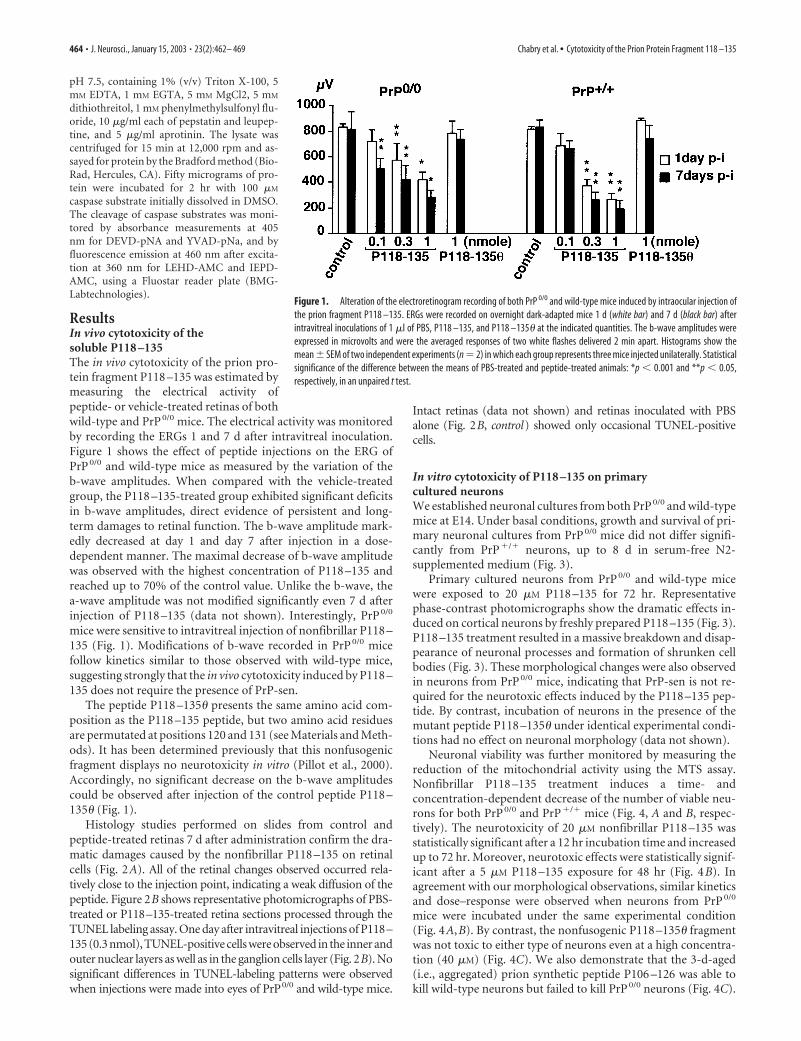
Primary cultured neurons from PrP 0/0 and wild-type micewere exposed to 20 �M P118 –135 for 72 hr. Representativephase-contrast photomicrographs show the dramatic effects in-duced on cortical neurons by freshly prepared P118 –135 (Fig. 3).P118 –135 treatment resulted in a massive breakdown and disap-pearance of neuronal processes and formation of shrunken cellbodies (Fig. 3). These morphological changes were also observedin neurons from PrP 0/0 mice, indicating that PrP-sen is not re-quired for the neurotoxic effects induced by the P118 –135 pep-tide. By contrast, incubation of neurons in the presence of themutant peptide P118 –135� under identical experimental condi-tions had no effect on neuronal morphology (data not shown).
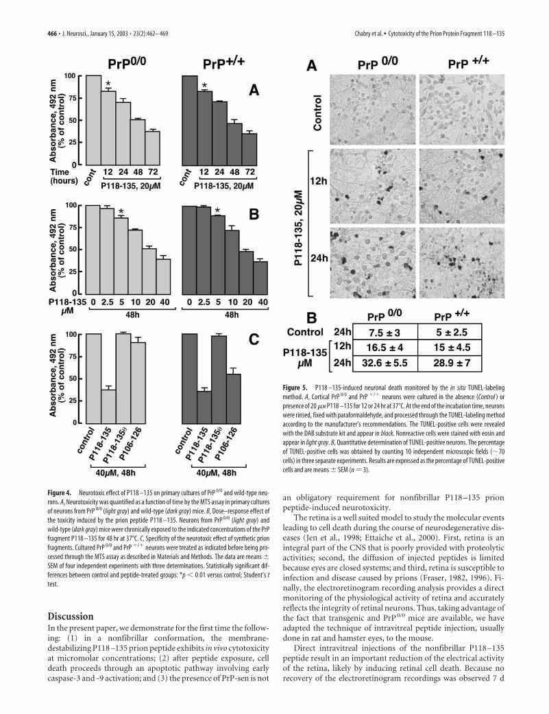
Neuronal viability was further monitored by measuring thereduction of the mitochondrial activity using the MTS assay.Nonfibrillar P118 –135 treatment induces a time- andconcentration-dependent decrease of the number of viable neu-rons for both PrP 0/0 and PrP�/� mice (Fig. 4, A and B, respec-tively). The neurotoxicity of 20 �M nonfibrillar P118 –135 wasstatistically significant after a 12 hr incubation time and increasedup to 72 hr. Moreover, neurotoxic effects were statistically signif-icant after a 5 �M P118 –135 exposure for 48 hr (Fig. 4B). Inagreement with our morphological observations, similar kineticsand dose–response were observed when neurons from PrP 0/0
mice were incubated under the same experimental condition(Fig. 4A,B). By contrast, the nonfusogenic P118 –135� fragmentwas not toxic to either type of neurons even at a high concentra-tion (40 �M) (Fig. 4C). We also demonstrate that the 3-d-aged(i.e., aggregated) prion synthetic peptide P106 –126 was able tokill wild-type neurons but failed to kill PrP 0/0 neurons (Fig. 4C).
Figure 1. Alteration of the electroretinogram recording of both PrP 0/0 and wild-type mice induced by intraocular injection ofthe prion fragment P118 –135. ERGs were recorded on overnight dark-adapted mice 1 d (white bar) and 7 d (black bar) afterintravitreal inoculations of 1 �l of PBS, P118 –135, and P118 –135� at the indicated quantities. The b-wave amplitudes wereexpressed in microvolts and were the averaged responses of two white flashes delivered 2 min apart. Histograms show themean � SEM of two independent experiments (n � 2) in which each group represents three mice injected unilaterally. Statisticalsignificance of the difference between the means of PBS-treated and peptide-treated animals: *p � 0.001 and **p � 0.05,respectively, in an unpaired t test.
464 • J. Neurosci., January 15, 2003 • 23(2):462– 469 Chabry et al. • Cytotoxicity of the Prion Protein Fragment 118 –135
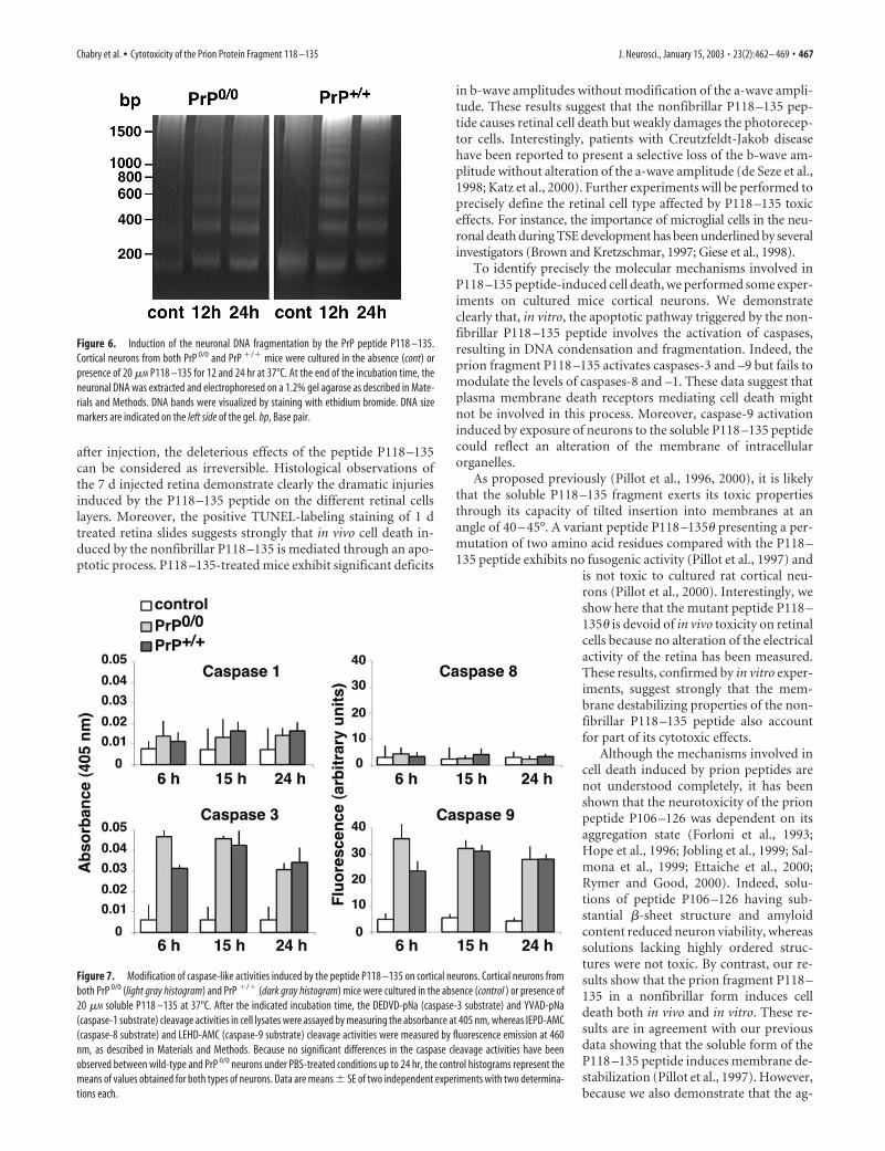
Nonfibrillar P118 –135 induces apoptosis to cortical neuronsTo further characterize the cell death mechanisms induced by thepeptide P118 –135, PBS- and peptide-treated cultures of neuronswere processed through the TUNEL-labeling assay (Fig. 5). After12 hr exposure in the presence of 20 �M nonfibrillar P118 –135,TUNEL-positive neurons were observed in both wild-type andPrP 0/0 neurons. The number of TUNEL-positive neurons in-creases after P118 –135 peptide exposure, suggesting that celldeath induced by the soluble P118 –135 proceeds through apo-ptosis. PrP 0/0 and wild-type cortical neurons exposed to 20 �M
soluble P118 –135 for 12 hr exhibited �16.5 and �15% TUNEL-positive cells, respectively. The number of apoptotic cells reached�32.6 and �28.9% after 24 hr incubation time for both PrP 0/0
and PrP�/� cultured neurons, respectively (Fig. 5B). Neuronstreated with PBS alone (Fig. 5, control) or with the nonfusogenicpeptide P118 –135� (data not shown) showed only occasionalTUNEL-positive cells.
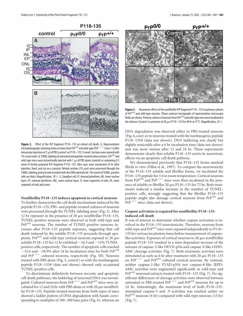
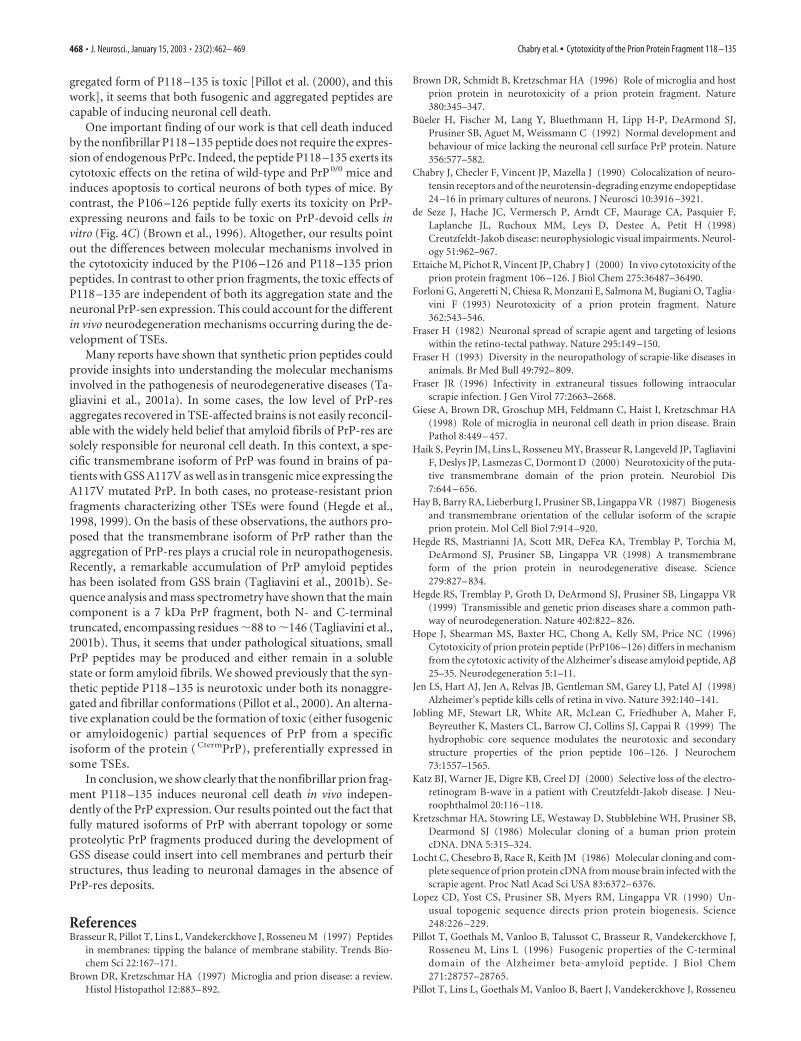
To discriminate definitively between necrotic and apoptoticcell death pathways, the laddering of neuronal DNA was investi-gated. Cultured neurons from PrP�/� and PrP 0/0 mice were in-cubated for 12 and 24 hr with PBS alone or with 20 �M nonfibril-lar P118 –135. Peptide-treated neurons from both types of miceshowed a ladder pattern of DNA degradation with bands corre-sponding to multiples of 180 –200 base pairs (Fig. 6), whereas no
DNA degradation was observed either in PBS-treated neurons(Fig. 6, cont) or in neurons treated with the nonfusogenic peptideP118 –135� (data not shown). DNA laddering was clearly butslightly noticeable after a 6 hr incubation time (data not shown)and was more intense after 12 and 24 hr. These experimentsdemonstrate clearly that soluble P118 –135 exerts its neurotoxiceffects via an apoptotic cell death pathway.
We demonstrated previously that P118 –135 forms amyloidfibrils in vitro (Pillot et al., 1997). To compare the neurotoxicityof the P118 –135 soluble and fibrillar forms, we incubated theP118 –135 peptide for 3 d at room temperature. Cortical neuronsfrom PrP 0/0 and PrP�/� mice were then incubated in the pres-ence of soluble or fibrillar 20 �M P118 –135 for 72 hr. Both treat-ments induced a similar increase in the number of TUNEL-positive cells, strongly suggesting that the fibrillar P118 –135peptide might also damage cortical neurons from PrP 0/0 andPrP�/� mice (data not shown).
Caspase activation is required for nonfibrillar P118 –135-induced cell deathIt was of interest to determine whether caspase activation is in-volved in the P118 –135 neurotoxicity. Therefore, neurons fromwild-type and PrP 0/0 mice were exposed independently to P118 –135 for various incubation times before measurement of caspase-like activities. Exposure of cortical neurons to 20 �M nonfibrillarpeptide P118 –135 resulted in a time-dependent increase of theamount of caspase-3-like DEVD-pNa and caspase-9-like LEHD-AMC cleavage activities (Fig. 7). Both enzymatic activities werestimulated as early as 6 hr after treatment with 20 �M P118 –135on PrP�/� and PrP 0/0 cultured cortical neurons. By contrast,neither caspase-1-like YVAD-pNA nor caspase-8-like IEPD-AMC activities were augmented significantly in wild-type andPrP 0/0 neuronal extracts treated with P118 –135 (Fig. 7). No sig-nificant differences of cleavage activities were observed betweenuntreated or PBS-treated PrP�/� and PrP 0/0 neurons for up to24 hr. Interestingly, the maximum level of both P118 –135-stimulated caspase-3 and -9 activities was reached earlier onPrP 0/0 neurons (6 hr) compared with wild-type neurons (15 hr)(Fig. 7).
Figure 2. Effect of the PrP fragment P118 –135 on retinal cell death. A, Representativemicrophotographs showing retina sections from PrP 0/0 and wild-type (PrP �/�) mice 7 d afterintraocular injections of 1 �l of PBS (control ) or P118 –135 (1 nmol). Sections were stained with1% cresyl violet. B, TUNEL labeling of untreated and peptide-treated retina sections. PrP 0/0 andwild-type mice were intravitreally injected with 1 �l of PBS alone (control) or containing 0.3nmol of freshly prepared PrP fragment P118 –135. Mice eyes were enucleated 24 hr afterinjection, fixed, and cut on a cryostat. Retinal sections (10 �m) were processed through theTUNEL-labeling protocol and revealed with the DAB substrate kit. The nuclei of TUNEL-positivecells are black. Magnification, 10�. G, Ganglion cell; IP, internal plexiform; INL, inner nuclearlayer; EP, external plexiform; ONL, outer nuclear layer; IS, inner segments of rods; OS, outersegments of rods and cones.
Figure 3. Neurotoxic effects of the nonfibrillar PrP fragment P118 –135 on primary culturesof PrP 0/0 and wild-type neurons. Phase-contrast micrographs of representative microscopicfields are shown. Primary cultures of neurons from PrP 0/0 and wild-type mice were incubated inthe absence (Control ) or presence of 20 �M P118 –135 for 48 hr at 37°C. Magnification, 25�.
Chabry et al. • Cytotoxicity of the Prion Protein Fragment 118 –135 J. Neurosci., January 15, 2003 • 23(2):462– 469 • 465
DiscussionIn the present paper, we demonstrate for the first time the follow-ing: (1) in a nonfibrillar conformation, the membrane-destabilizing P118 –135 prion peptide exhibits in vivo cytotoxicityat micromolar concentrations; (2) after peptide exposure, celldeath proceeds through an apoptotic pathway involving earlycaspase-3 and -9 activation; and (3) the presence of PrP-sen is not
an obligatory requirement for nonfibrillar P118 –135 prionpeptide-induced neurotoxicity.
The retina is a well suited model to study the molecular eventsleading to cell death during the course of neurodegenerative dis-eases (Jen et al., 1998; Ettaiche et al., 2000). First, retina is anintegral part of the CNS that is poorly provided with proteolyticactivities; second, the diffusion of injected peptides is limitedbecause eyes are closed systems; and third, retina is susceptible toinfection and disease caused by prions (Fraser, 1982, 1996). Fi-nally, the electroretinogram recording analysis provides a directmonitoring of the physiological activity of retina and accuratelyreflects the integrity of retinal neurons. Thus, taking advantage ofthe fact that transgenic and PrP 0/0 mice are available, we haveadapted the technique of intravitreal peptide injection, usuallydone in rat and hamster eyes, to the mouse.
Direct intravitreal injections of the nonfibrillar P118 –135peptide result in an important reduction of the electrical activityof the retina, likely by inducing retinal cell death. Because norecovery of the electroretinogram recordings was observed 7 d
Figure 4. Neurotoxic effect of P118 –135 on primary cultures of PrP 0/0 and wild-type neu-rons. A, Neurotoxicity was quantified as a function of time by the MTS assay in primary culturesof neurons from PrP 0/0 (light gray) and wild-type (dark gray) mice. B, Dose–response effect ofthe toxicity induced by the prion peptide P118 –135. Neurons from PrP 0/0 (light gray) andwild-type (dark gray) mice were chronically exposed to the indicated concentrations of the PrPfragment P118 –135 for 48 hr at 37°C. C, Specificity of the neurotoxic effect of synthetic prionfragments. Cultured PrP 0/0 and PrP �/� neurons were treated as indicated before being pro-cessed through the MTS assay as described in Materials and Methods. The data are means �SEM of four independent experiments with three determinations. Statistically significant dif-ferences between control and peptide-treated groups: *p � 0.01 versus control; Student’s ttest.
Figure 5. P118 –135-induced neuronal death monitored by the in situ TUNEL-labelingmethod. A, Cortical PrP 0/0 and PrP �/� neurons were cultured in the absence (Control ) orpresence of 20 �M P118 –135 for 12 or 24 hr at 37°C. At the end of the incubation time, neuronswere rinsed, fixed with paraformaldehyde, and processed through the TUNEL-labeling methodaccording to the manufacturer’s recommendations. The TUNEL-positive cells were revealedwith the DAB substrate kit and appear in black. Nonreactive cells were stained with eosin andappear in light gray. B, Quantitative determination of TUNEL-positive neurons. The percentageof TUNEL-positive cells was obtained by counting 10 independent microscopic fields (�70cells) in three separate experiments. Results are expressed as the percentage of TUNEL-positivecells and are means � SEM (n � 3).
466 • J. Neurosci., January 15, 2003 • 23(2):462– 469 Chabry et al. • Cytotoxicity of the Prion Protein Fragment 118 –135
after injection, the deleterious effects of the peptide P118 –135can be considered as irreversible. Histological observations ofthe 7 d injected retina demonstrate clearly the dramatic injuriesinduced by the P118 –135 peptide on the different retinal cellslayers. Moreover, the positive TUNEL-labeling staining of 1 dtreated retina slides suggests strongly that in vivo cell death in-duced by the nonfibrillar P118 –135 is mediated through an apo-ptotic process. P118 –135-treated mice exhibit significant deficits
in b-wave amplitudes without modification of the a-wave ampli-tude. These results suggest that the nonfibrillar P118 –135 pep-tide causes retinal cell death but weakly damages the photorecep-tor cells. Interestingly, patients with Creutzfeldt-Jakob diseasehave been reported to present a selective loss of the b-wave am-plitude without alteration of the a-wave amplitude (de Seze et al.,1998; Katz et al., 2000). Further experiments will be performed toprecisely define the retinal cell type affected by P118 –135 toxiceffects. For instance, the importance of microglial cells in the neu-ronal death during TSE development has been underlined by severalinvestigators (Brown and Kretzschmar, 1997; Giese et al., 1998).
To identify precisely the molecular mechanisms involved inP118 –135 peptide-induced cell death, we performed some exper-iments on cultured mice cortical neurons. We demonstrateclearly that, in vitro, the apoptotic pathway triggered by the non-fibrillar P118 –135 peptide involves the activation of caspases,resulting in DNA condensation and fragmentation. Indeed, theprion fragment P118 –135 activates caspases-3 and –9 but fails tomodulate the levels of caspases-8 and –1. These data suggest thatplasma membrane death receptors mediating cell death mightnot be involved in this process. Moreover, caspase-9 activationinduced by exposure of neurons to the soluble P118 –135 peptidecould reflect an alteration of the membrane of intracellularorganelles.
As proposed previously (Pillot et al., 1996, 2000), it is likelythat the soluble P118 –135 fragment exerts its toxic propertiesthrough its capacity of tilted insertion into membranes at anangle of 40 – 45°. A variant peptide P118 –135� presenting a per-mutation of two amino acid residues compared with the P118 –135 peptide exhibits no fusogenic activity (Pillot et al., 1997) and
is not toxic to cultured rat cortical neu-rons (Pillot et al., 2000). Interestingly, weshow here that the mutant peptide P118 –135� is devoid of in vivo toxicity on retinalcells because no alteration of the electricalactivity of the retina has been measured.These results, confirmed by in vitro exper-iments, suggest strongly that the mem-brane destabilizing properties of the non-fibrillar P118 –135 peptide also accountfor part of its cytotoxic effects.
Although the mechanisms involved incell death induced by prion peptides arenot understood completely, it has beenshown that the neurotoxicity of the prionpeptide P106 –126 was dependent on itsaggregation state (Forloni et al., 1993;Hope et al., 1996; Jobling et al., 1999; Sal-mona et al., 1999; Ettaiche et al., 2000;Rymer and Good, 2000). Indeed, solu-tions of peptide P106 –126 having sub-stantial �-sheet structure and amyloidcontent reduced neuron viability, whereassolutions lacking highly ordered struc-tures were not toxic. By contrast, our re-sults show that the prion fragment P118 –135 in a nonfibrillar form induces celldeath both in vivo and in vitro. These re-sults are in agreement with our previousdata showing that the soluble form of theP118 –135 peptide induces membrane de-stabilization (Pillot et al., 1997). However,because we also demonstrate that the ag-
Figure 6. Induction of the neuronal DNA fragmentation by the PrP peptide P118 –135.Cortical neurons from both PrP 0/0 and PrP �/� mice were cultured in the absence (cont) orpresence of 20 �M P118 –135 for 12 and 24 hr at 37°C. At the end of the incubation time, theneuronal DNA was extracted and electrophoresed on a 1.2% gel agarose as described in Mate-rials and Methods. DNA bands were visualized by staining with ethidium bromide. DNA sizemarkers are indicated on the left side of the gel. bp, Base pair.
Figure 7. Modification of caspase-like activities induced by the peptide P118 –135 on cortical neurons. Cortical neurons fromboth PrP 0/0 (light gray histogram) and PrP �/� (dark gray histogram) mice were cultured in the absence (control ) or presence of20 �M soluble P118 –135 at 37°C. After the indicated incubation time, the DEDVD-pNa (caspase-3 substrate) and YVAD-pNa(caspase-1 substrate) cleavage activities in cell lysates were assayed by measuring the absorbance at 405 nm, whereas IEPD-AMC(caspase-8 substrate) and LEHD-AMC (caspase-9 substrate) cleavage activities were measured by fluorescence emission at 460nm, as described in Materials and Methods. Because no significant differences in the caspase cleavage activities have beenobserved between wild-type and PrP 0/0 neurons under PBS-treated conditions up to 24 hr, the control histograms represent themeans of values obtained for both types of neurons. Data are means � SE of two independent experiments with two determina-tions each.
Chabry et al. • Cytotoxicity of the Prion Protein Fragment 118 –135 J. Neurosci., January 15, 2003 • 23(2):462– 469 • 467
gregated form of P118 –135 is toxic [Pillot et al. (2000), and thiswork], it seems that both fusogenic and aggregated peptides arecapable of inducing neuronal cell death.
One important finding of our work is that cell death inducedby the nonfibrillar P118 –135 peptide does not require the expres-sion of endogenous PrPc. Indeed, the peptide P118 –135 exerts itscytotoxic effects on the retina of wild-type and PrP 0/0 mice andinduces apoptosis to cortical neurons of both types of mice. Bycontrast, the P106 –126 peptide fully exerts its toxicity on PrP-expressing neurons and fails to be toxic on PrP-devoid cells invitro (Fig. 4C) (Brown et al., 1996). Altogether, our results pointout the differences between molecular mechanisms involved inthe cytotoxicity induced by the P106 –126 and P118 –135 prionpeptides. In contrast to other prion fragments, the toxic effects ofP118 –135 are independent of both its aggregation state and theneuronal PrP-sen expression. This could account for the differentin vivo neurodegeneration mechanisms occurring during the de-velopment of TSEs.
Many reports have shown that synthetic prion peptides couldprovide insights into understanding the molecular mechanismsinvolved in the pathogenesis of neurodegenerative diseases (Ta-gliavini et al., 2001a). In some cases, the low level of PrP-resaggregates recovered in TSE-affected brains is not easily reconcil-able with the widely held belief that amyloid fibrils of PrP-res aresolely responsible for neuronal cell death. In this context, a spe-cific transmembrane isoform of PrP was found in brains of pa-tients with GSS A117V as well as in transgenic mice expressing theA117V mutated PrP. In both cases, no protease-resistant prionfragments characterizing other TSEs were found (Hegde et al.,1998, 1999). On the basis of these observations, the authors pro-posed that the transmembrane isoform of PrP rather than theaggregation of PrP-res plays a crucial role in neuropathogenesis.Recently, a remarkable accumulation of PrP amyloid peptideshas been isolated from GSS brain (Tagliavini et al., 2001b). Se-quence analysis and mass spectrometry have shown that the maincomponent is a 7 kDa PrP fragment, both N- and C-terminaltruncated, encompassing residues �88 to �146 (Tagliavini et al.,2001b). Thus, it seems that under pathological situations, smallPrP peptides may be produced and either remain in a solublestate or form amyloid fibrils. We showed previously that the syn-thetic peptide P118 –135 is neurotoxic under both its nonaggre-gated and fibrillar conformations (Pillot et al., 2000). An alterna-tive explanation could be the formation of toxic (either fusogenicor amyloidogenic) partial sequences of PrP from a specificisoform of the protein ( CtermPrP), preferentially expressed insome TSEs.
In conclusion, we show clearly that the nonfibrillar prion frag-ment P118 –135 induces neuronal cell death in vivo indepen-dently of the PrP expression. Our results pointed out the fact thatfully matured isoforms of PrP with aberrant topology or someproteolytic PrP fragments produced during the development ofGSS disease could insert into cell membranes and perturb theirstructures, thus leading to neuronal damages in the absence ofPrP-res deposits.
ReferencesBrasseur R, Pillot T, Lins L, Vandekerckhove J, Rosseneu M (1997) Peptides
in membranes: tipping the balance of membrane stability. Trends Bio-chem Sci 22:167–171.
Brown DR, Kretzschmar HA (1997) Microglia and prion disease: a review.Histol Histopathol 12:883– 892.
Brown DR, Schmidt B, Kretzschmar HA (1996) Role of microglia and hostprion protein in neurotoxicity of a prion protein fragment. Nature380:345–347.
Bueler H, Fischer M, Lang Y, Bluethmann H, Lipp H-P, DeArmond SJ,Prusiner SB, Aguet M, Weissmann C (1992) Normal development andbehaviour of mice lacking the neuronal cell surface PrP protein. Nature356:577–582.
Chabry J, Checler F, Vincent JP, Mazella J (1990) Colocalization of neuro-tensin receptors and of the neurotensin-degrading enzyme endopeptidase24 –16 in primary cultures of neurons. J Neurosci 10:3916 –3921.
de Seze J, Hache JC, Vermersch P, Arndt CF, Maurage CA, Pasquier F,Laplanche JL, Ruchoux MM, Leys D, Destee A, Petit H (1998)Creutzfeldt-Jakob disease: neurophysiologic visual impairments. Neurol-ogy 51:962–967.
Ettaiche M, Pichot R, Vincent JP, Chabry J (2000) In vivo cytotoxicity of theprion protein fragment 106 –126. J Biol Chem 275:36487–36490.
Forloni G, Angeretti N, Chiesa R, Monzani E, Salmona M, Bugiani O, Taglia-vini F (1993) Neurotoxicity of a prion protein fragment. Nature362:543–546.
Fraser H (1982) Neuronal spread of scrapie agent and targeting of lesionswithin the retino-tectal pathway. Nature 295:149 –150.
Fraser H (1993) Diversity in the neuropathology of scrapie-like diseases inanimals. Br Med Bull 49:792– 809.
Fraser JR (1996) Infectivity in extraneural tissues following intraocularscrapie infection. J Gen Virol 77:2663–2668.
Giese A, Brown DR, Groschup MH, Feldmann C, Haist I, Kretzschmar HA(1998) Role of microglia in neuronal cell death in prion disease. BrainPathol 8:449 – 457.
Haik S, Peyrin JM, Lins L, Rosseneu MY, Brasseur R, Langeveld JP, TagliaviniF, Deslys JP, Lasmezas C, Dormont D (2000) Neurotoxicity of the puta-tive transmembrane domain of the prion protein. Neurobiol Dis7:644 – 656.
Hay B, Barry RA, Lieberburg I, Prusiner SB, Lingappa VR (1987) Biogenesisand transmembrane orientation of the cellular isoform of the scrapieprion protein. Mol Cell Biol 7:914 –920.
Hegde RS, Mastrianni JA, Scott MR, DeFea KA, Tremblay P, Torchia M,DeArmond SJ, Prusiner SB, Lingappa VR (1998) A transmembraneform of the prion protein in neurodegenerative disease. Science279:827– 834.
Hegde RS, Tremblay P, Groth D, DeArmond SJ, Prusiner SB, Lingappa VR(1999) Transmissible and genetic prion diseases share a common path-way of neurodegeneration. Nature 402:822– 826.
Hope J, Shearman MS, Baxter HC, Chong A, Kelly SM, Price NC (1996)Cytotoxicity of prion protein peptide (PrP106 –126) differs in mechanismfrom the cytotoxic activity of the Alzheimer’s disease amyloid peptide, A�25–35. Neurodegeneration 5:1–11.
Jen LS, Hart AJ, Jen A, Relvas JB, Gentleman SM, Garey LJ, Patel AJ (1998)Alzheimer’s peptide kills cells of retina in vivo. Nature 392:140 –141.
Jobling MF, Stewart LR, White AR, McLean C, Friedhuber A, Maher F,Beyreuther K, Masters CL, Barrow CJ, Collins SJ, Cappai R (1999) Thehydrophobic core sequence modulates the neurotoxic and secondarystructure properties of the prion peptide 106 –126. J Neurochem73:1557–1565.
Katz BJ, Warner JE, Digre KB, Creel DJ (2000) Selective loss of the electro-retinogram B-wave in a patient with Creutzfeldt-Jakob disease. J Neu-roophthalmol 20:116 –118.
Kretzschmar HA, Stowring LE, Westaway D, Stubblebine WH, Prusiner SB,Dearmond SJ (1986) Molecular cloning of a human prion proteincDNA. DNA 5:315–324.
Locht C, Chesebro B, Race R, Keith JM (1986) Molecular cloning and com-plete sequence of prion protein cDNA from mouse brain infected with thescrapie agent. Proc Natl Acad Sci USA 83:6372– 6376.
Lopez CD, Yost CS, Prusiner SB, Myers RM, Lingappa VR (1990) Un-usual topogenic sequence directs prion protein biogenesis. Science248:226 –229.
Pillot T, Goethals M, Vanloo B, Talussot C, Brasseur R, Vandekerckhove J,Rosseneu M, Lins L (1996) Fusogenic properties of the C-terminaldomain of the Alzheimer beta-amyloid peptide. J Biol Chem271:28757–28765.
Pillot T, Lins L, Goethals M, Vanloo B, Baert J, Vandekerckhove J, Rosseneu
468 • J. Neurosci., January 15, 2003 • 23(2):462– 469 Chabry et al. • Cytotoxicity of the Prion Protein Fragment 118 –135

M, Brasseur R (1997) The 118 –135 peptide of the human prion proteinforms amyloid fibrils and induces liposome fusion. J Mol Biol274:381–393.
Pillot T, Drouet B, Pincon-Raymond M, Vandekerckhove J, Rosseneu M,Chambaz J (2000) A nonfibrillar form of the fusogenic prion proteinfragment [118 –135] induces apoptotic cell death in rat cortical neurons.J Neurochem 75:2298 –2308.
Rymer DL, Good TA (2000) The role of prion peptide structure and aggre-gation in toxicity and membrane binding. J Neurochem 75:2536 –2545.
Salmona M, Malesani P, De Gioia L, Gorla S, Bruschi M, Molinari A, DellaVedova F, Pedrotti B, Marrari MA, Awan T, Bugiani O, Forloni G, Taglia-
vini F (1999) Molecular determinants of the physicochemical propertiesof a critical prion protein region comprising residues 106 –126. BiochemJ 342:207–214.
Tagliavini F, Forloni G, D’Ursi P, Bugiani O, Salmona M (2001a) Studies onpeptide fragments of prion proteins. Adv Protein Chem 57:171–201.
Tagliavini F, Lievens PM, Tranchant C, Warter JM, Mohr M, Giaccone G,Perini F, Rossi G, Salmona M, Piccardo P, Ghetti B, Beavis RC, Bugiani O,Frangione B, Prelli F (2001b) A 7-kDa prion protein (PrP) fragment, anintegral component of the PrP region required for infectivity, is the majoramyloid protein in Gerstmann-Straussler-Scheinker disease A117V. J BiolChem 276:6009 – 6015.
Chabry et al. • Cytotoxicity of the Prion Protein Fragment 118 –135 J. Neurosci., January 15, 2003 • 23(2):462– 469 • 469