Embed Size (px)
Citation preview
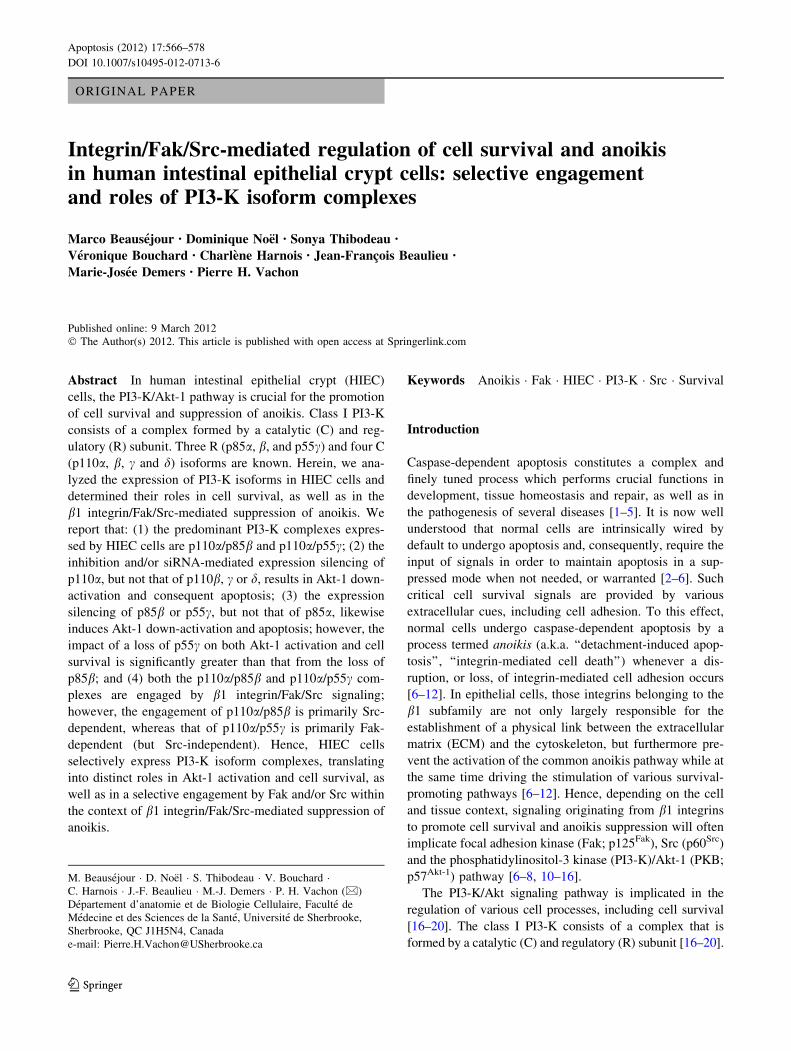
ORIGINAL PAPER
Integrin/Fak/Src-mediated regulation of cell survival and anoikisin human intestinal epithelial crypt cells: selective engagementand roles of PI3-K isoform complexes
Marco Beausejour • Dominique Noel • Sonya Thibodeau •
Veronique Bouchard • Charlene Harnois • Jean-Francois Beaulieu •
Marie-Josee Demers • Pierre H. Vachon
Published online: 9 March 2012
� The Author(s) 2012. This article is published with open access at Springerlink.com
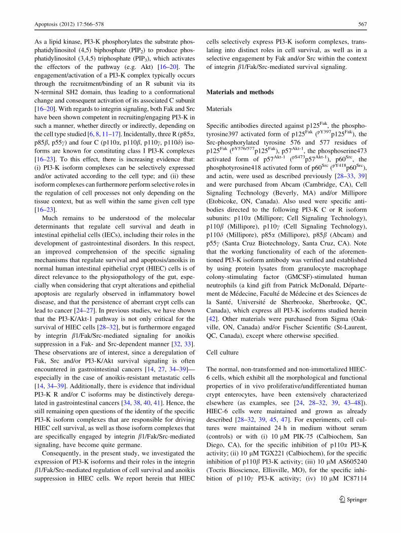
Abstract In human intestinal epithelial crypt (HIEC)
cells, the PI3-K/Akt-1 pathway is crucial for the promotion
of cell survival and suppression of anoikis. Class I PI3-K
consists of a complex formed by a catalytic (C) and reg-
ulatory (R) subunit. Three R (p85a, b, and p55c) and four C
(p110a, b, c and d) isoforms are known. Herein, we ana-
lyzed the expression of PI3-K isoforms in HIEC cells and
determined their roles in cell survival, as well as in the
b1 integrin/Fak/Src-mediated suppression of anoikis. We
report that: (1) the predominant PI3-K complexes expres-
sed by HIEC cells are p110a/p85b and p110a/p55c; (2) the
inhibition and/or siRNA-mediated expression silencing of
p110a, but not that of p110b, c or d, results in Akt-1 down-
activation and consequent apoptosis; (3) the expression
silencing of p85b or p55c, but not that of p85a, likewise
induces Akt-1 down-activation and apoptosis; however, the
impact of a loss of p55c on both Akt-1 activation and cell
survival is significantly greater than that from the loss of
p85b; and (4) both the p110a/p85b and p110a/p55c com-
plexes are engaged by b1 integrin/Fak/Src signaling;
however, the engagement of p110a/p85b is primarily Src-
dependent, whereas that of p110a/p55c is primarily Fak-
dependent (but Src-independent). Hence, HIEC cells
selectively express PI3-K isoform complexes, translating
into distinct roles in Akt-1 activation and cell survival, as
well as in a selective engagement by Fak and/or Src within
the context of b1 integrin/Fak/Src-mediated suppression of
anoikis.
Keywords Anoikis � Fak � HIEC � PI3-K � Src � Survival
Introduction
Caspase-dependent apoptosis constitutes a complex and
finely tuned process which performs crucial functions in
development, tissue homeostasis and repair, as well as in
the pathogenesis of several diseases [1–5]. It is now well
understood that normal cells are intrinsically wired by
default to undergo apoptosis and, consequently, require the
input of signals in order to maintain apoptosis in a sup-
pressed mode when not needed, or warranted [2–6]. Such
critical cell survival signals are provided by various
extracellular cues, including cell adhesion. To this effect,
normal cells undergo caspase-dependent apoptosis by a
process termed anoikis (a.k.a. ‘‘detachment-induced apop-
tosis’’, ‘‘integrin-mediated cell death’’) whenever a dis-
ruption, or loss, of integrin-mediated cell adhesion occurs
[6–12]. In epithelial cells, those integrins belonging to the
b1 subfamily are not only largely responsible for the
establishment of a physical link between the extracellular
matrix (ECM) and the cytoskeleton, but furthermore pre-
vent the activation of the common anoikis pathway while at
the same time driving the stimulation of various survival-
promoting pathways [6–12]. Hence, depending on the cell
and tissue context, signaling originating from b1 integrins
to promote cell survival and anoikis suppression will often
implicate focal adhesion kinase (Fak; p125Fak), Src (p60Src)
and the phosphatidylinositol-3 kinase (PI3-K)/Akt-1 (PKB;
p57Akt-1) pathway [6–8, 10–16].
The PI3-K/Akt signaling pathway is implicated in the
regulation of various cell processes, including cell survival
[16–20]. The class I PI3-K consists of a complex that is
formed by a catalytic (C) and regulatory (R) subunit [16–20].
M. Beausejour � D. Noel � S. Thibodeau � V. Bouchard �C. Harnois � J.-F. Beaulieu � M.-J. Demers � P. H. Vachon (&)
Departement d’anatomie et de Biologie Cellulaire, Faculte de
Medecine et des Sciences de la Sante, Universite de Sherbrooke,
Sherbrooke, QC J1H5N4, Canada
e-mail: [email protected]
123
Apoptosis (2012) 17:566–578
DOI 10.1007/s10495-012-0713-6
As a lipid kinase, PI3-K phosphorylates the substrate phos-
phatidylinositol (4,5) biphosphate (PIP2) to produce phos-
phatidylinositol (3,4,5) triphosphate (PIP3), which activates
the effectors of the pathway (e.g. Akt) [16–20]. The
engagement/activation of a PI3-K complex typically occurs
through the recruitment/binding of an R subunit via its
N-terminal SH2 domain, thus leading to a conformational
change and consequent activation of its associated C subunit
[16–20]. With regards to integrin signaling, both Fak and Src
have been shown competent in recruiting/engaging PI3-K in
such a manner, whether directly or indirectly, depending on
the cell type studied [6, 8, 11–17]. Incidentally, three R (p85a,
p85b, p55c) and four C (p110a, p110b, p110c, p110d) iso-
forms are known for constituting class I PI3-K complexes
[16–23]. To this effect, there is increasing evidence that:
(i) PI3-K isoform complexes can be selectively expressed
and/or activated according to the cell type; and (ii) these
isoform complexes can furthermore perform selective roles in
the regulation of cell processes not only depending on the
tissue context, but as well within the same given cell type
[16–23].
Much remains to be understood of the molecular
determinants that regulate cell survival and death in
intestinal epithelial cells (IECs), including their roles in the
development of gastrointestinal disorders. In this respect,
an improved comprehension of the specific signaling
mechanisms that regulate survival and apoptosis/anoikis in
normal human intestinal epithelial crypt (HIEC) cells is of
direct relevance to the physiopathology of the gut, espe-
cially when considering that crypt alterations and epithelial
apoptosis are regularly observed in inflammatory bowel
disease, and that the persistence of aberrant crypt cells can
lead to cancer [24–27]. In previous studies, we have shown
that the PI3-K/Akt-1 pathway is not only critical for the
survival of HIEC cells [28–32], but is furthermore engaged
by integrin b1/Fak/Src-mediated signaling for anoikis
suppression in a Fak- and Src-dependent manner [32, 33].
These observations are of interest, since a deregulation of
Fak, Src and/or PI3-K/Akt survival signaling is often
encountered in gastrointestinal cancers [14, 27, 34–39]—
especially in the case of anoikis-resistant metastatic cells
[14, 34–39]. Additionally, there is evidence that individual
PI3-K R and/or C isoforms may be distinctively deregu-
lated in gastrointestinal cancers [34, 38, 40, 41]. Hence, the
still remaining open questions of the identity of the specific
PI3-K isoform complexes that are responsible for driving
HIEC cell survival, as well as those isoform complexes that
are specifically engaged by integrin b1/Fak/Src-mediated
signaling, have become quite germane.
Consequently, in the present study, we investigated the
expression of PI3-K isoforms and their roles in the integrin
b1/Fak/Src-mediated regulation of cell survival and anoikis
suppression in HIEC cells. We report herein that HIEC
cells selectively express PI3-K isoform complexes, trans-
lating into distinct roles in cell survival, as well as in a
selective engagement by Fak and/or Src within the context
of integrin b1/Fak/Src-mediated survival signaling.
Materials and methods
Materials
Specific antibodies directed against p125Fak, the phospho-
tyrosine397 activated form of p125Fak (pY397p125Fak), the
Src-phosphorylated tyrosine 576 and 577 residues of
p125Fak (pY576/577p125Fak), p57Akt-1, the phosphoserine473
activated form of p57Akt-1 (pS473p57Akt-1), p60Src, the
phosphotyrosine418 activated form of p60Src (pY418p60Src),
and actin, were used as described previously [28–33, 39]
and were purchased from Abcam (Cambridge, CA), Cell
Signaling Technology (Beverly, MA) and/or Millipore
(Etobicoke, ON, Canada). Also used were specific anti-
bodies directed to the following PI3-K C or R isoform
subunits: p110a (Millipore; Cell Signaling Technology),
p110b (Millipore), p110c (Cell Signaling Technology),
p110d (Millipore), p85a (Millipore), p85b (Abcam) and
p55c (Santa Cruz Biotechnology, Santa Cruz, CA). Note
that the working functionality of each of the aforemen-
tioned PI3-K isoform antibody was verified and established
by using protein lysates from granulocyte macrophage
colony-stimulating factor (GMCSF)-stimulated human
neutrophils (a kind gift from Patrick McDonald, Departe-
ment de Medecine, Faculte de Medecine et des Sciences de
la Sante, Universite de Sherbrooke, Sherbrooke, QC,
Canada), which express all PI3-K isoforms studied herein
[42]. Other materials were purchased from Sigma (Oak-
ville, ON, Canada) and/or Fischer Scientific (St-Laurent,
QC, Canada), except where otherwise specified.
Cell culture
The normal, non-transformed and non-immortalized HIEC-
6 cells, which exhibit all the morphological and functional
properties of in vivo proliferative/undifferentiated human
crypt enterocytes, have been extensively characterized
elsewhere (as examples, see [24, 28–32, 39, 43–48]).
HIEC-6 cells were maintained and grown as already
described [28–32, 39, 45, 47]. For experiments, cell cul-
tures were maintained 24 h in medium without serum
(controls) or with (i) 10 lM PIK-75 (Calbiochem, San
Diego, CA), for the specific inhibition of p110a PI3-K
activity; (ii) 10 lM TGX221 (Calbiochem), for the specific
inhibition of p110b PI3-K activity; (iii) 10 lM AS605240
(Tocris Bioscience, Ellisville, MO), for the specific inhi-
bition of p110c PI3-K activity; (iv) 10 lM IC87114
Apoptosis (2012) 17:566–578 567
123
(Calbiochem), for the specific inhibition of p110d PI3-K
activity; (v) 30 lM Ly294002 (Calbiochem), for the ‘‘pan’’
inhibition of PI3-K activity enacted by p110a-d; (vi) 1 lM
PF573228 (Tocris Bioscience), for the specific inhibition of
Fak; (vii) 20 lM PP2 (Calbiochem), for the inhibition of
Src; (viii) 100 lg/ml of the monoclonal antibody P4C10 (a
kind gift of Erkki Ruoslahti, The Sanford-Burnham Med-
ical Research Institute, LaJolla, CA), which inhibits the
binding activity of the b1 integrin subunit [28–33]; or (ix)
100 lg/ml non-immune mouse IgGs (Sigma), as control
for the P4C10 blocking antibody. The working concentra-
tions of the inhibitors used were determined previously
with dose–response assays (not shown). It is noteworthy
that control cultures included exposure to the same solvent
as that used for inhibitors and showed no significant dif-
ferences with cultures maintained in serum-free medium
only (not shown). For full anoikis, cells were kept in sus-
pension 24 h (serum-free) in poly-2-hydroxyethyl meth-
acrylate (polyHEMA)-coated dishes, as already described
[28–30, 32, 33, 39].
Caspase-activated DNAse (CAD)-mediated DNA
laddering assays
DNA was isolated and the visualization of CAD-mediated
internucleosomal DNA fragmentation (DNA laddering) on
2% agarose gels (20 lg DNA/lane) was performed as
described elsewhere [28–33]. Note that the method used for
DNA extraction employs Triton rather than SDS, thus
leaving behind most intact genomic DNA [28–33, 49].
In situ terminal deoxynucleotidyl transferase (TDT)-
mediated dUTP nick-end labeling (ISEL) assays
Coverslip-grown HIEC cells were processed and ISEL was
carried out as previously described [28–33, 39]. Evaluation
of ISEL-positive cells counterstained with 40,6-diamidino-
2-phenylindole (DAPI) was performed as described else-
where [28–33, 39]. Typically, apoptotic indices were
compared to those of control cultures, 9100 (expressed as
‘‘% of control’’).
Fluorometric caspase-3 (CASP-3) activity assays
The CASP-3 fluorometric assay used herein is based on the
hydrolysis of acetyl Asp-Glu-Val-Asp 7-amido-4-methyl-
coumarin (Ac-DEVD-AMC) by CASP-3, resulting in the
release of the fluorescent 7-amino-4-methylcoumarin
(AMC) catalysis product. The excitation and emission
wavelengths of AMC are 380 nm and 430–460 nm,
respectively. Cells from treated and non-treated/control
cultures were solubilized in modified cold IP buffer (see
below), from which phenylmethylsulfonyl fluoride was
omitted. From each assayed sample, 30 lg proteins were
added to 500 ll of freshly prepared CASP-3 reaction buffer
(100 mM HEPES (pH 7.5), 20% glycerol and 5 mM
dithiothreitol), followed by adding 2 ll of a 5 mM stock
solution of Ac-DEVD-AMC (Calbiochem), and a sub-
sequent 2 h incubation at 37�C. A blank, constituting of
modified IP buffer, reaction buffer and Ac-DEVD-AMC,
was likewise incubated in parallel to the reaction mixtures.
After incubation, another 500 ll of reaction buffer was
added and reactions (blank included) were read in a Hitachi
S-2500 Spectrofluorometer (graciously made available to
us by Martin Bisaillon, Departement de Biochimie, Faculte
de Medecine et des Sciences de la Sante, Universite de
Sherbrooke, Sherbrooke, QC, Canada), at an excitation
wavelength of 380 nm. For each assay, the exact AMC
emission apex within the 430–460 nm range was first
determined with the blank, which in turn dictated at which
emission wavelength the reactions were read. The relative
CASP-3 activity was determined thereafter by applying the
formula DF/F0 = (FE - F0)/F0, where F0 = levels of
emitted fluorescence by the blank, FE = levels of emitted
fluorescence by the assayed reaction, and DF/F0 = relative
CASP-3 activity of the assayed reaction. In turn, the rela-
tive CASP-3 activity from treated cultures was compared
to that of non-treated/controls, 9100 (expressed as ‘‘% of
control’’).
Reverse transcriptase-polymerase chain reaction
(RT-PCR)
Total RNA extraction and subsequent RT-PCR were car-
ried out as described previously [29, 30]. Specific primers
for the amplification of p110a, p110b, p110c, p110d, p85a,
p85b, p55c and actin were purchased from Invitrogen Life
Techologies (Grand Island, NY). Controls for reactions
were: (a) DNA without adding primers; and (b) primers
without adding DNA (not shown) [29, 30]. Relative
expression levels of PI3-K isoform mRNAs were deter-
mined by comparison with actin mRNA as a reference.
Band intensities of amplified fragments were scanned and
semi-quantified using an Alpha Imager 1200 Documenta-
tion and Analysis system (Alpha Innotech, San Leandro,
CA), in order to establish the ratios ‘‘PI3-K isoform/
Actin’’.
Western blotting (WB)
Cell cultures were lysed in sample buffer (2.3% SDS, 10%
glycerol, and 0.001% bromphenol blue in 62.5 mM Tris–
HCl (pH 6.8) containing 5% b-mercaptoethanol) and pro-
cessed as described previously [28–33, 39]. Proteins were
resolved by SDS-PAGE (50 lg proteins/lane), electro-
transferred and probed as already described [28–33, 39].
568 Apoptosis (2012) 17:566–578
123
Immunoreactive bands were semi-quantified with Scion
Image (Scion, Frederick, MD), as already described [28–
33, 39], and the relative expression levels of PI3-K iso-
forms were determined by comparison with actin as a
reference, in order to establish the ratios ‘‘PI3-K isoform/
Actin’’.
Immunoprecipitation (IP)/co-IP analyses and relative
kinase activation assays
Cell cultures were lysed in cold IP buffer [50 mM Tris–HCl
(pH 7.2), 150 mM NaCl, 1 mM dithiothreitol, 0.5 mM
EDTA, 1% Nonidet P-40, 0.5% sodium deoxycholate,
0.1% SDS, 100 lM Na3VO4, 1 mM phenylmethylsulfonyl
fluoride, 0.5 lg/ml leupeptin, 0.5 lg/ml aprotinin, 0.7 lg/ml
pestadin, 40 mM b-glycerophosphate, and 10 mM Na2P4O7]
and processed for IP as described previously [28–33, 39].
Immunoprecipitates were solubilized in sample buffer,
resolved by SDS-PAGE and probed by WB (see above).
Relative kinase activation analyses were performed as
already described [28–33, 39]. Typically, immunoreactive
bands were semi-quantified with Scion Image (Scion), as
already described [28–33, 39], and the relative activated
levels of kinases were established with the ratios phos-
phorylated kinase/total kinase, which in turn were compared
to control cultures, 9100 (expressed as ‘‘% of control’’).
Small interference RNA (siRNA)-mediated expression
silencing assays
siRNAs specifically directed against the mRNAs of p110a(sip110a), p110b (sip110b), p85a (sip85a), p85b (sip85b)
or p55c (sip55c) were purchased from OriGene (Rockville,
MD). A non-silencing control siRNA (siCNS) was pur-
chased from Qiagen (Mississauga, ON, Canada). HIECs
were transfected with either of each siRNA at a final
concentration of 10 nM, according to the protocol descri-
bed previously [45]. Three siRNAs for each isoform ana-
lyzed were tested. Only those siRNAs that resulted in a
reduction of relative protein expression levels of at least
75% (as assessed by WB; see Fig. 4a as example) were
used herein. 48 h following transfection, cells were pro-
cessed for analyses. In some experiments, combinations of
two siRNAs directed against PI3-K isoform subunits were
used, still at a final concentration of 10 nM each. Under
such circumstances, the control siCNS was used at a final
concentration of 20 nM instead.
Data processing
Results and values shown represent mean ± SEM for at
least three (n C 3) separate experiments and/or cultures.
Statistically significant differences were determined by the
Student t test, with SigmaSTAT (Systat Software, San Jose,
CA). Data were compiled, analyzed and processed with
Excel (Microsoft, Redmond, WA). Except otherwise spec-
ified, images from blots, gels and scans were processed with
Vistascan (Umax Technologies, Fremont, CA), Photoshop
(Adobe, San Jose, CA) and PowerPoint (Microsoft).
Results
HIEC cells selectively express PI3-K subunit isoforms
and isoform complexes
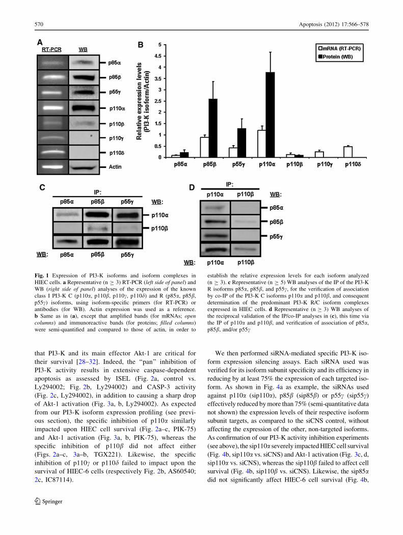
We first established the expression profile of class I PI3-K
R and C isoforms in HIEC-6 cells. Semi-quantitative RT-
PCR analyses indicate that these cells predominantly
express the mRNAs for p85b, p55c, p110a and p110d,
whereas those for p85a, p110b and p110c are weakly
expressed (Fig. 1a, left side of panel; 1b, open columns).
Verification of protein expression by WB confirmed that
p85b, p55c and p110a are indeed predominantly expressed,
and that p85a and p110b are effectively weakly expressed
(Fig. 1a, right side of panel; 1b, filled columns). However,
no protein products for either p110c or p110d were
detected in HIEC-6 cells (Fig. 1a, right side of panel; 1b,
filled columns), as confirmed by the strong detection of
these two isoforms in GMCSF-stimulated human neutro-
phils using the same specific antibodies (not shown).
We then verified which PI3-K R/C isoform complexes
are found in HIEC cells by performing IP analyses of R
subunits and verification of association of C subunits via
co-IP. As expected from our expression studies (see
above), the IP of either p85b or p55c revealed a strong
association with p110a, but little to no association with
p110b (Fig. 1c). Also as expected from our expression
studies, what little of p85a that was IP yielded weakly
detectable p110a and no detectable p110b (Fig. 1c). We
confirmed these IP/co-IP observations by performing
reciprocal analyses whereby C subunits were IP and the
co-IP association of R subunits was verified (Fig. 1d).
Therefore, our expression profiling and IP/co-IP analy-
ses altogether indicate that HIEC cells selectively express
PI3-K R and C subunit isoforms, which in turn translates
into a selective expression of PI3-K R/C isoform com-
plexes. Specifically, p110a/p85b and p110a/p55c are the
largely predominant PI3-K isoform complexes found in
these cells.
Selective roles of PI3-K subunit isoforms and isoform
complexes in HIEC cell survival
We functionally analyzed the roles of PI3-K isoforms in
the maintenance of survival in HIEC-6 cells, considering
Apoptosis (2012) 17:566–578 569
123
that PI3-K and its main effector Akt-1 are critical for
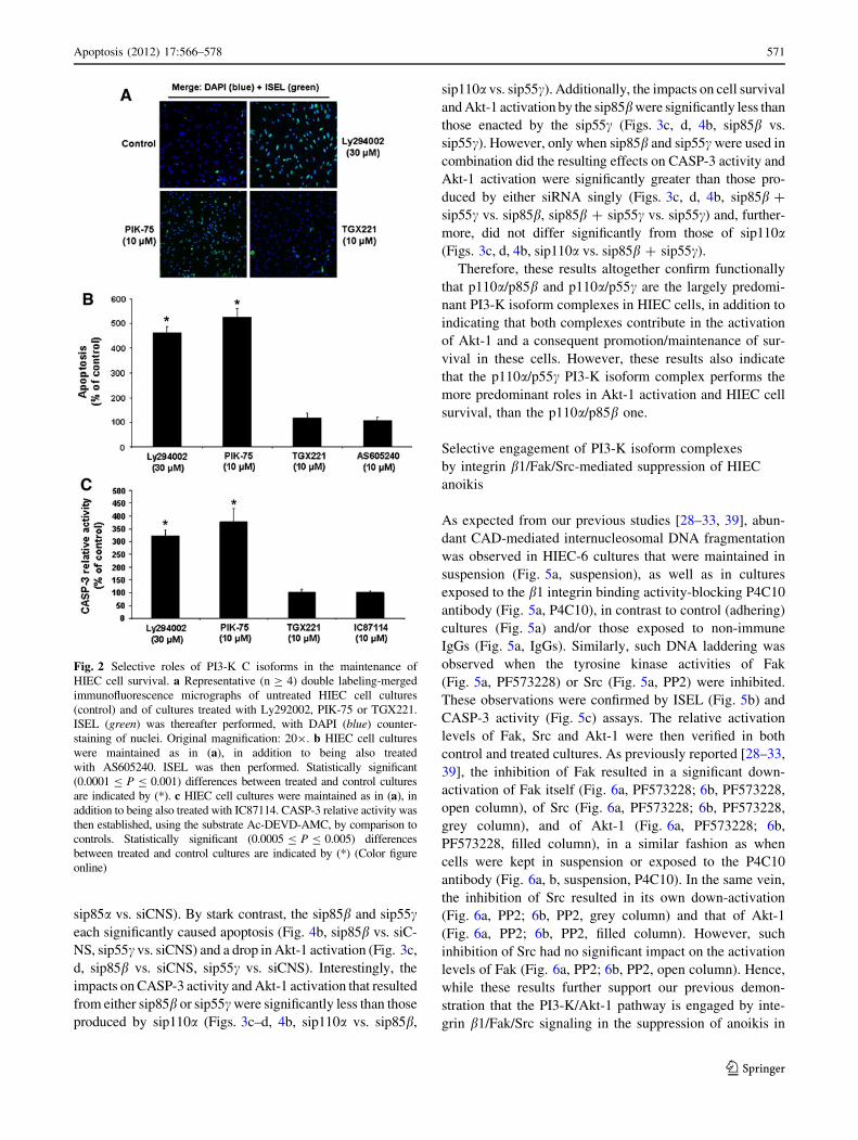
their survival [28–32]. Indeed, the ‘‘pan’’ inhibition of
PI3-K activity results in extensive caspase-dependent
apoptosis as assessed by ISEL (Fig. 2a, control vs.
Ly294002; Fig. 2b, Ly294002) and CASP-3 activity
(Fig. 2c, Ly294002), in addition to causing a sharp drop
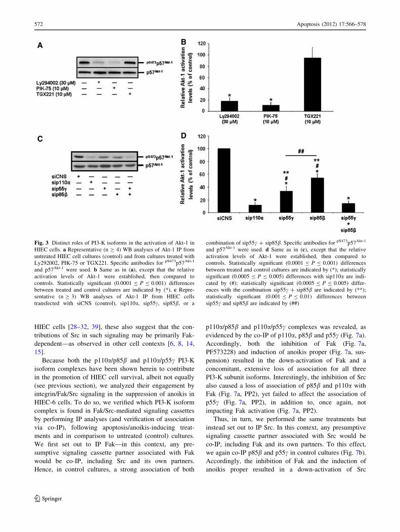
of Akt-1 activation (Fig. 3a, b, Ly294002). As expected
from our PI3-K isoform expression profiling (see previ-
ous section), the specific inhibition of p110a similarly
impacted upon HIEC cell survival (Fig. 2a–c, PIK-75)
and Akt-1 activation (Fig. 3a, b, PIK-75), whereas the
specific inhibition of p110b did not affect either
(Figs. 2a–c, 3a–b, TGX221). Likewise, the specific
inhibition of p110c or p110d failed to impact upon the
survival of HIEC-6 cells (respectively Fig. 2b, AS60540;
2c, IC87114).
We then performed siRNA-mediated specific PI3-K iso-
form expression silencing assays. Each siRNA used was
verified for its isoform subunit specificity and its efficiency in
reducing by at least 75% the expression of each targeted iso-
form. As shown in Fig. 4a as example, the siRNAs used
against p110a (sip110a), p85b (sip85b) or p55c (sip55c)
effectively reduced by more than 75% (semi-quantitative data
not shown) the expression levels of their respective isoform
subunit targets, as compared to the siCNS control, without
affecting the expression of the other, non-targeted isoforms.
As confirmation of our PI3-K activity inhibition experiments
(see above), the sip110a severely impacted HIEC cell survival
(Fig. 4b, sip110a vs. siCNS) and Akt-1 activation (Fig. 3c, d,
sip110a vs. siCNS), whereas the sip110b failed to affect cell
survival (Fig. 4b, sip110b vs. siCNS). Likewise, the sip85adid not significantly affect HIEC-6 cell survival (Fig. 4b,
Fig. 1 Expression of PI3-K isoforms and isoform complexes in
HIEC cells. a Representative (n C 3) RT-PCR (left side of panel) and
WB (right side of panel) analyses of the expression of the known
class I PI3-K C (p110a, p110b, p110c, p110d) and R (p85a, p85b,
p55c) isoforms, using isoform-specific primers (for RT-PCR) or
antibodies (for WB). Actin expression was used as a reference.
b Same as in (a), except that amplified bands (for mRNAs; opencolumns) and immunoreactive bands (for proteins; filled columns)
were semi-quantified and compared to those of actin, in order to
establish the relative expression levels for each isoform analyzed
(n C 3). c Representative (n C 5) WB analyses of the IP of the PI3-K
R isoforms p85a, p85b, and p55c, for the verification of association
by co-IP of the PI3-K C isoforms p110a and p110b, and consequent
determination of the predominant PI3-K R/C isoform complexes
expressed in HIEC cells. d Representative (n C 3) WB analyses of
the reciprocal validation of the IP/co-IP analyses in (c), this time via
the IP of p110a and p110b, and verification of association of p85a,
p85b, and/or p55c
570 Apoptosis (2012) 17:566–578
123
sip85a vs. siCNS). By stark contrast, the sip85b and sip55ceach significantly caused apoptosis (Fig. 4b, sip85b vs. siC-
NS, sip55c vs. siCNS) and a drop in Akt-1 activation (Fig. 3c,
d, sip85b vs. siCNS, sip55c vs. siCNS). Interestingly, the
impacts on CASP-3 activity and Akt-1 activation that resulted
from either sip85b or sip55c were significantly less than those
produced by sip110a (Figs. 3c–d, 4b, sip110a vs. sip85b,
sip110a vs. sip55c). Additionally, the impacts on cell survival
and Akt-1 activation by the sip85b were significantly less than
those enacted by the sip55c (Figs. 3c, d, 4b, sip85b vs.
sip55c). However, only when sip85b and sip55c were used in
combination did the resulting effects on CASP-3 activity and
Akt-1 activation were significantly greater than those pro-
duced by either siRNA singly (Figs. 3c, d, 4b, sip85b ?
sip55c vs. sip85b, sip85b ? sip55c vs. sip55c) and, further-
more, did not differ significantly from those of sip110a(Figs. 3c, d, 4b, sip110a vs. sip85b ? sip55c).
Therefore, these results altogether confirm functionally
that p110a/p85b and p110a/p55c are the largely predomi-
nant PI3-K isoform complexes in HIEC cells, in addition to
indicating that both complexes contribute in the activation
of Akt-1 and a consequent promotion/maintenance of sur-
vival in these cells. However, these results also indicate
that the p110a/p55c PI3-K isoform complex performs the
more predominant roles in Akt-1 activation and HIEC cell
survival, than the p110a/p85b one.
Selective engagement of PI3-K isoform complexes
by integrin b1/Fak/Src-mediated suppression of HIEC
anoikis
As expected from our previous studies [28–33, 39], abun-
dant CAD-mediated internucleosomal DNA fragmentation
was observed in HIEC-6 cultures that were maintained in
suspension (Fig. 5a, suspension), as well as in cultures
exposed to the b1 integrin binding activity-blocking P4C10
antibody (Fig. 5a, P4C10), in contrast to control (adhering)
cultures (Fig. 5a) and/or those exposed to non-immune
IgGs (Fig. 5a, IgGs). Similarly, such DNA laddering was
observed when the tyrosine kinase activities of Fak
(Fig. 5a, PF573228) or Src (Fig. 5a, PP2) were inhibited.
These observations were confirmed by ISEL (Fig. 5b) and
CASP-3 activity (Fig. 5c) assays. The relative activation
levels of Fak, Src and Akt-1 were then verified in both
control and treated cultures. As previously reported [28–33,
39], the inhibition of Fak resulted in a significant down-
activation of Fak itself (Fig. 6a, PF573228; 6b, PF573228,
open column), of Src (Fig. 6a, PF573228; 6b, PF573228,
grey column), and of Akt-1 (Fig. 6a, PF573228; 6b,
PF573228, filled column), in a similar fashion as when
cells were kept in suspension or exposed to the P4C10
antibody (Fig. 6a, b, suspension, P4C10). In the same vein,
the inhibition of Src resulted in its own down-activation
(Fig. 6a, PP2; 6b, PP2, grey column) and that of Akt-1
(Fig. 6a, PP2; 6b, PP2, filled column). However, such
inhibition of Src had no significant impact on the activation
levels of Fak (Fig. 6a, PP2; 6b, PP2, open column). Hence,
while these results further support our previous demon-
stration that the PI3-K/Akt-1 pathway is engaged by inte-
grin b1/Fak/Src signaling in the suppression of anoikis in
Fig. 2 Selective roles of PI3-K C isoforms in the maintenance of
HIEC cell survival. a Representative (n C 4) double labeling-merged
immunofluorescence micrographs of untreated HIEC cell cultures
(control) and of cultures treated with Ly292002, PIK-75 or TGX221.
ISEL (green) was thereafter performed, with DAPI (blue) counter-
staining of nuclei. Original magnification: 209. b HIEC cell cultures
were maintained as in (a), in addition to being also treated
with AS605240. ISEL was then performed. Statistically significant
(0.0001 B P B 0.001) differences between treated and control cultures
are indicated by (*). c HIEC cell cultures were maintained as in (a), in
addition to being also treated with IC87114. CASP-3 relative activity was
then established, using the substrate Ac-DEVD-AMC, by comparison to
controls. Statistically significant (0.0005 B P B 0.005) differences
between treated and control cultures are indicated by (*) (Color figure
online)
Apoptosis (2012) 17:566–578 571
123
HIEC cells [28–32, 39], these also suggest that the con-
tributions of Src in such signaling may be primarily Fak-
dependent—as observed in other cell contexts [6, 8, 14,
15].
Because both the p110a/p85b and p110a/p55c PI3-K
isoform complexes have been shown herein to contribute
in the promotion of HIEC cell survival, albeit not equally
(see previous section), we analyzed their engagement by
integrin/Fak/Src signaling in the suppression of anoikis in
HIEC-6 cells. To do so, we verified which PI3-K isoform
complex is found in Fak/Src-mediated signaling cassettes
by performing IP analyses (and verification of association
via co-IP), following apoptosis/anoikis-inducing treat-
ments and in comparison to untreated (control) cultures.
We first set out to IP Fak—in this context, any pre-
sumptive signaling cassette partner associated with Fak
would be co-IP, including Src and its own partners.
Hence, in control cultures, a strong association of both
p110a/p85b and p110a/p55c complexes was revealed, as
evidenced by the co-IP of p110a, p85b and p55c (Fig. 7a).
Accordingly, both the inhibition of Fak (Fig. 7a,
PF573228) and induction of anoikis proper (Fig. 7a, sus-
pension) resulted in the down-activation of Fak and a
concomitant, extensive loss of association for all three
PI3-K subunit isoforms. Interestingly, the inhibition of Src
also caused a loss of association of p85b and p110a with
Fak (Fig. 7a, PP2), yet failed to affect the association of
p55c (Fig. 7a, PP2), in addition to, once again, not
impacting Fak activation (Fig. 7a, PP2).
Thus, in turn, we performed the same treatments but
instead set out to IP Src. In this context, any presumptive
signaling cassette partner associated with Src would be
co-IP, including Fak and its own partners. To this effect,
we again co-IP p85b and p55c in control cultures (Fig. 7b).
Accordingly, the inhibition of Fak and the induction of
anoikis proper resulted in a down-activation of Src
Fig. 3 Distinct roles of PI3-K isoforms in the activation of Akt-1 in
HIEC cells. a Representative (n C 4) WB analyses of Akt-1 IP from
untreated HIEC cell cultures (control) and from cultures treated with
Ly292002, PIK-75 or TGX221. Specific antibodies for pS473p57Akt-1
and p57Akt-1 were used. b Same as in (a), except that the relative
activation levels of Akt-1 were established, then compared to
controls. Statistically significant (0.0001 B P B 0.001) differences
between treated and control cultures are indicated by (*). c Repre-
sentative (n C 3) WB analyses of Akt-1 IP from HIEC cells
transfected with siCNS (control), sip110a, sip55c, sip85b, or a
combination of sip55c ? sip85b. Specific antibodies for pS473p57Akt-1
and p57Akt-1 were used. d Same as in (c), except that the relative
activation levels of Akt-1 were established, then compared to
controls. Statistically significant (0.0001 B P B 0.001) differences
between treated and control cultures are indicated by (*); statistically
significant (0.0005 B P B 0.005) differences with sip110a are indi-
cated by (#); statistically significant (0.0005 B P B 0.005) differ-
ences with the combination sip55c ? sip85b are indicated by (**);
statistically significant (0.001 B P B 0.01) differences between
sip55c and sip85b are indicated by (##)
572 Apoptosis (2012) 17:566–578
123
(Fig. 7b, PF573228, suspension), in a loss of Fak-Src
interactions (as assessed by the Src-mediated phosphory-
lation of the Y576/577 residues of Fak [6, 14, 15, 32, 33,
35–37, 39]; Fig. 7b, PF573228, suspension), and a loss of
association of p85b and p55c (Fig. 7b, PF573228, sus-
pension). Conversely, the inhibition of Src caused its own
down-activation (Fig. 7b, PP2), a loss of interactions with
Fak (Fig. 7b, PP2) and, consequently, a loss of association
of not only p85b, but of p55c as well (Fig. 7b, PP2).
Therefore, these results altogether indicate that both the
p110a/p85b and p110a/p55c PI3-K isoform complexes are
engaged by integrin b1/Fak/Src signaling in the suppres-
sion of anoikis in HIEC cells. However, these results fur-
ther reveal that such engagement of the two complexes in
Fak/Src signaling cassettes is selective in nature. Indeed,
the engagement of p110a/p85b is primarily Src-dependent
(the engagement of which is itself primarily Fak-depen-
dent), whereas the engagement of p110a/p55c is primarily
Fak-dependent (but Src-independent).
Discussion
In the present study, we investigated the expression of PI3-
K isoforms and their roles in the integrin b1/Fak/Src-
mediated regulation of HIEC cell survival and suppression
of anoikis. Herein, we demonstrate that p110a/p85b and
p110a/p55c are the largely predominant PI3-K isoform
complexes in HIEC cells, whereas the individual isoforms
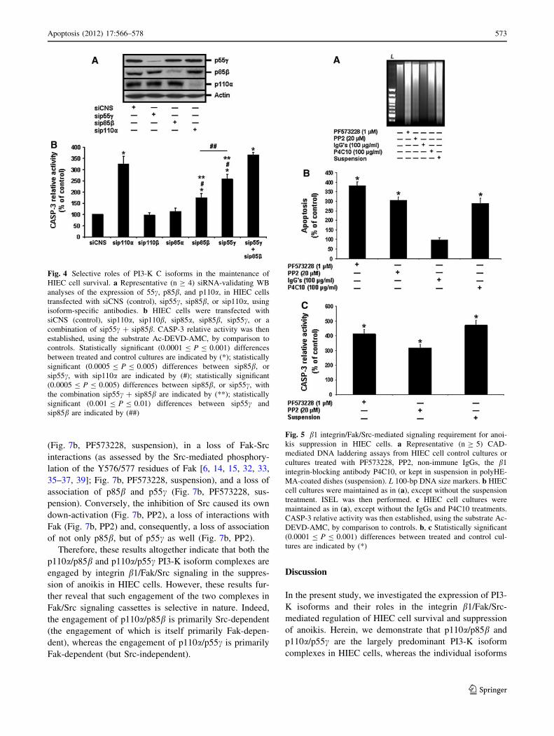
Fig. 4 Selective roles of PI3-K C isoforms in the maintenance of
HIEC cell survival. a Representative (n C 4) siRNA-validating WB
analyses of the expression of 55c, p85b, and p110a, in HIEC cells
transfected with siCNS (control), sip55c, sip85b, or sip110a, using
isoform-specific antibodies. b HIEC cells were transfected with
siCNS (control), sip110a, sip110b, sip85a, sip85b, sip55c, or a
combination of sip55c ? sip85b. CASP-3 relative activity was then
established, using the substrate Ac-DEVD-AMC, by comparison to
controls. Statistically significant (0.0001 B P B 0.001) differences
between treated and control cultures are indicated by (*); statistically
significant (0.0005 B P B 0.005) differences between sip85b, or
sip55c, with sip110a are indicated by (#); statistically significant
(0.0005 B P B 0.005) differences between sip85b, or sip55c, with
the combination sip55c ? sip85b are indicated by (**); statistically
significant (0.001 B P B 0.01) differences between sip55c and
sip85b are indicated by (##)
Fig. 5 b1 integrin/Fak/Src-mediated signaling requirement for anoi-
kis suppression in HIEC cells. a Representative (n C 5) CAD-
mediated DNA laddering assays from HIEC cell control cultures or
cultures treated with PF573228, PP2, non-immune IgGs, the b1
integrin-blocking antibody P4C10, or kept in suspension in polyHE-
MA-coated dishes (suspension). L 100-bp DNA size markers. b HIEC
cell cultures were maintained as in (a), except without the suspension
treatment. ISEL was then performed. c HIEC cell cultures were
maintained as in (a), except without the IgGs and P4C10 treatments.
CASP-3 relative activity was then established, using the substrate Ac-
DEVD-AMC, by comparison to controls. b, c Statistically significant
(0.0001 B P B 0.001) differences between treated and control cul-
tures are indicated by (*)
Apoptosis (2012) 17:566–578 573
123
p85a and p110b are expressed weakly, and p110c and
p110d are not expressed. Concordantly, only the p110a/
p85b and p110a/p55c complexes perform the critical
functions of Akt-1 activation and subsequent maintenance
of HIEC cell survival. However, the contributions of
p110a/p55c in Akt-1 activation and cell survival are sig-
nificantly greater than those of p110a/p85b. We also pro-
vide further evidence that the maintenance of HIEC cell
survival and suppression of anoikis by b1 integrins is
dependent on associated Fak signaling cassettes, in which
Src is recruited. To this effect, we show that the p110a/
p85b and p110a/p55c PI3-K isoform complexes are
selectively engaged by such integrin/Fak/Src signaling,
whereby the engagement of p110a/p85b is primarily Src-
dependent and that of p110a/p55c is primarily Fak-
dependent (but Src-independent). Hence, as summarized in
Fig. 8, HIEC cells selectively express PI3-K R and C
subunit isoforms, which translates into a selective
expression of PI3-K R/C isoform complexes, which in turn
results into isoform-distinct roles in the activation of Akt-1
and the promotion of HIEC cell survival and, additionally,
in their selective engagement by b1 integrin/Fak/Src sig-
naling in the suppression of anoikis.
It is now well established that PI3-K R and C isoforms
can be distinctively expressed according to the cell type
[16–23, 50]. In this respect, it is also accepted that one
regulatory mechanism of the roles of PI3-K isoform com-
plexes occurs at the gene expression level, in order to
determine which isoform complexes are formed [16–23,
50]. This is well illustrated herein with regards to HIEC
cells, as their selective expression profile of PI3-K R and C
isoforms directly impacts on the constitution of the pre-
dominant class I PI3-K isoform complexes expressed by
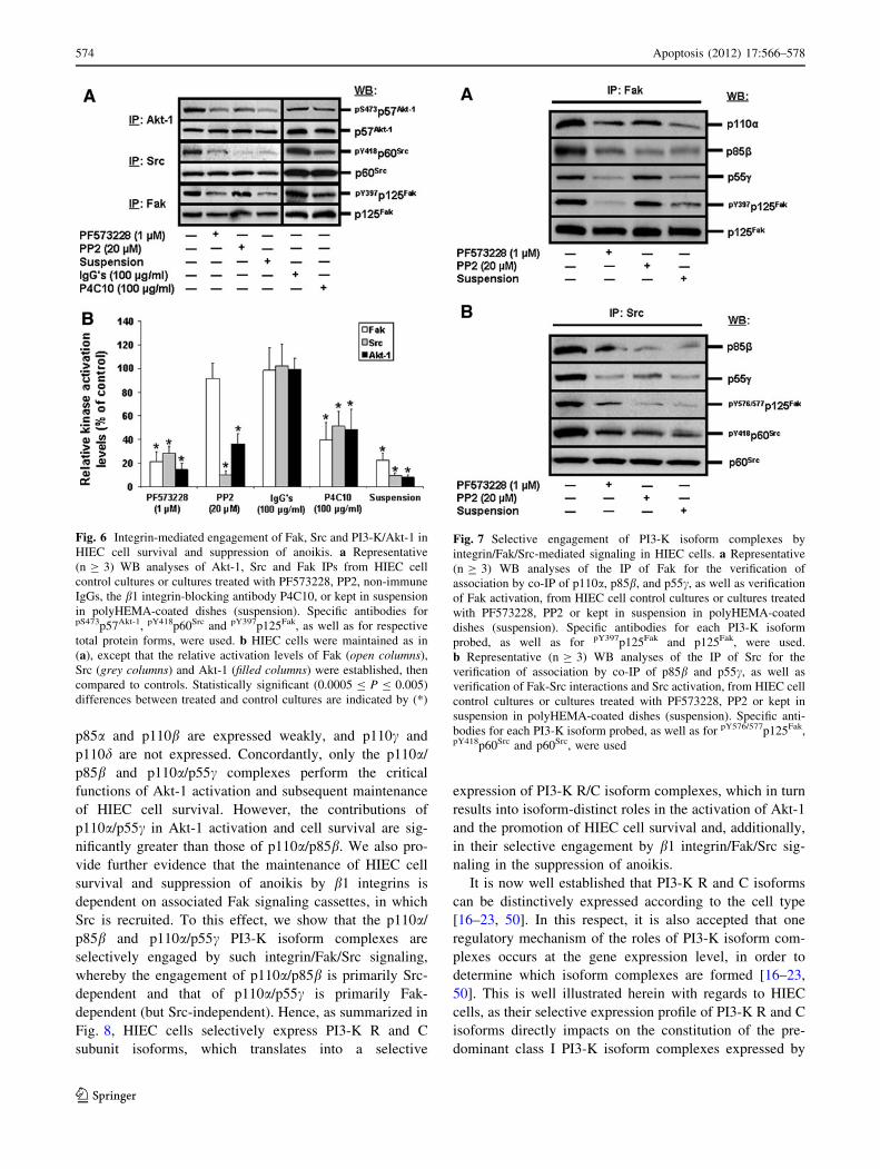
Fig. 6 Integrin-mediated engagement of Fak, Src and PI3-K/Akt-1 in
HIEC cell survival and suppression of anoikis. a Representative
(n C 3) WB analyses of Akt-1, Src and Fak IPs from HIEC cell
control cultures or cultures treated with PF573228, PP2, non-immune
IgGs, the b1 integrin-blocking antibody P4C10, or kept in suspension
in polyHEMA-coated dishes (suspension). Specific antibodies forpS473p57Akt-1, pY418p60Src and pY397p125Fak, as well as for respective
total protein forms, were used. b HIEC cells were maintained as in
(a), except that the relative activation levels of Fak (open columns),
Src (grey columns) and Akt-1 (filled columns) were established, then
compared to controls. Statistically significant (0.0005 B P B 0.005)
differences between treated and control cultures are indicated by (*)
Fig. 7 Selective engagement of PI3-K isoform complexes by
integrin/Fak/Src-mediated signaling in HIEC cells. a Representative
(n C 3) WB analyses of the IP of Fak for the verification of
association by co-IP of p110a, p85b, and p55c, as well as verification
of Fak activation, from HIEC cell control cultures or cultures treated
with PF573228, PP2 or kept in suspension in polyHEMA-coated
dishes (suspension). Specific antibodies for each PI3-K isoform
probed, as well as for pY397p125Fak and p125Fak, were used.
b Representative (n C 3) WB analyses of the IP of Src for the
verification of association by co-IP of p85b and p55c, as well as
verification of Fak-Src interactions and Src activation, from HIEC cell
control cultures or cultures treated with PF573228, PP2 or kept in
suspension in polyHEMA-coated dishes (suspension). Specific anti-
bodies for each PI3-K isoform probed, as well as for pY576/577p125Fak,pY418p60Src and p60Src, were used
574 Apoptosis (2012) 17:566–578
123
them (Fig. 8). Interestingly, our findings suggest that such
a selective expression profile of PI3-K isoforms in HIEC
cells is established not only via transcriptional regulation
(e.g. weak mRNA levels, and consequently weak protein
levels, for p85a and p110b), but furthermore via post-
transcriptional and/or translational regulation (e.g. weak or
strong mRNA levels for p110c and p110d respectively, yet
absence of protein expression for both). Hence, these
observations in HIEC cells emphasize the already
acknowledged complex nature of the regulatory mecha-
nisms that are responsible for the gene regulation of PI3-K
R and C isoforms [16–23, 50], in addition to providing one
more note added in proof to warrant further studies on the
transcriptional and post-transcriptional/translational regu-
lation of their expression. Such studies would be quite
relevant to colorectal cancer (CRC), considering that
although p110a is the predominant PI3-K C subunit in
HIEC cells (this study), and that mutations of p110a
conferring elevated/constitutive activity are found in one
third of CRC tumors [34, 38, 40, 41], the expression of
both p110a and p110b is nonetheless frequently elevated in
CRC [34, 38, 40, 41, 51]. Similarly, p85a (but not,
apparently, p85b) is likewise frequently elevated in CRC
tumors [34, 38, 40, 41, 51]. More strikingly, a previous
study reported that the predominant PI3-K isoform com-
plexes in CRC cells are p110a/p85a and p110b/p85a [52],
instead of p110a/p85b and p110a/p55c as shown herein in
HIEC cells. While the status of p55c in CRC tumors and/or
cells remains unknown, such findings in CRC altogether
stand in stark contrast to our own in normal, non-trans-
formed and non-immortalized HIEC cells—therefore
underlying the need to investigate fully the expression,
regulation and roles of PI3-K isoforms under the normal
physiological context, in order to achieve a better com-
prehension of the aberrant expression and/or deregulation
of these isoforms in cancer and cancer cell lines [16, 18–
22, 38, 50, 51]. The same axiom would likewise apply with
regards to other gastrointestinal disorders that display sig-
nificant deregulation of IEC survival, such as inflammatory
bowel diseases or necrotic enterocolitis [24, 25].
Although our knowledge of the regulation and roles of
specific PI3-K isoform complexes remains poor [19–23,
50], there is nonetheless increasing evidence that such
complexes can perform distinct functions in the regulation
of various cell processes not only depending on the tissue
context, but as well within the same given cell type [16–23,
50]. To this effect, the two predominant PI3-K complexes
in HIEC cells (p110a/p85b and p110a/p55c) not only
contribute distinctively in the activation of Akt-1 and the
maintenance of HIEC cell survival, but are furthermore
engaged selectively by b1 integrin/Fak/Src signaling in the
suppression of anoikis (Fig. 8). It is noteworthy that such
functional identification of distinct PI3-K isoform com-
plexes engaged by integrin/Fak/Src signaling, as well as the
selective engagement by Fak and Src of said distinct iso-
form complexes, has never been observed or reported
previously. Likewise, the identification of a p55c-contain-
ing PI3-K complex engaged by b1 integrin/Fak-mediated
signaling is novel. It is currently accepted that the R sub-
units are largely responsible for the specificity of engage-
ment, as well as the distinctiveness of the roles enacted, of
class I PI3-K isoform complexes [19–23, 50, 51]. As
example, reports have shown that p85a, p85b and p55cexhibit differential binding capacity to activated growth
factor tyrosine kinase receptors (RTKs), such as those for
insulin, epidermal growth factor (EGF) and platelet-
derived growth factor (PDGF) [19–23, 50, 51, 53]. This is
likely due first and foremost to individual structural and/or
functional domain differences among R subunits, although
this remains to be investigated more systematically [19–23,
50, 51]. For instance, while both p85b and p55c bear two
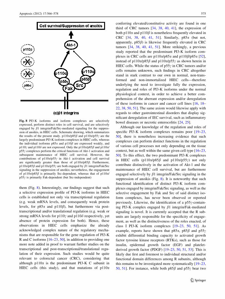
Fig. 8 PI3-K isoforms and isoform complexes are selectively
expressed, perform distinct roles in cell survival, and are selectively
engaged by b1 integrin/Fak/Src-mediated signaling for the suppres-
sion of anoikis, in HIEC cells. Schematic drawing, which summarizes
the results of the present study. p110a/p85b and p110a/p55c are the
largely predominant PI3-K isoform complexes in HIEC cells, whereas
the individual isoforms p85a and p110b are expressed weakly, and
p110c and p110d are not expressed. Only the p110a/p85b and p110a/
p55c complexes perform the critical functions of Akt-1 activation and
subsequent maintenance of HIEC cell survival. However, the
contributions of p110a/p55c in Akt-1 activation and cell survival
are significantly greater than those of p110a/p85b. Furthermore,
p110a/p85b and p110a/p55c are both engaged by b1 integrin/Fak/Src
signaling in the suppression of anoikis; nevertheless, the engagement
of p110a/p85b is primarily Src-dependent, whereas that of p110a/
p55c is primarily Fak-dependent (but Src-independent)
Apoptosis (2012) 17:566–578 575
123
SH2 domains (one C-terminal—iSH2—for regulation of
the C subunit, and one N-terminal—nSH2—through which
recruitment/binding of the R subunit in a PI3-K complex
occurs), p85b contains in its N-terminus one additional
proline-rich motif, a BCR homology domain and an SH3
domain [19–22]. Therefore, such structural distinctions
between p85b and p55c may be largely responsible for
their selective engagement observed herein by Src and Fak,
respectively (Fig. 8). Furthermore, basic structural and
functional differences between Fak and Src are also likely
to contribute into such selective engagement of PI3-K
isoform complexes. Depending on the cell context, Fak and
Src have been shown able to directly recruit PI3-K [6–8,
13–15, 26, 36, 37, 54]. Alternately, Fak or Src can recruit
PI3-K via various signaling cassette partners, such as the
adaptors Shc or IRS-1 [6–8, 13–15, 26, 36, 37, 54]. Con-
sidering that Fak, or Src, or both, are often deregulated in
cancer (including CRC) [6, 14, 36, 37, 39], further studies
will be required in order to unravel the determinants that
are responsible for the selective engagement of p110a/
p85b and p110a/p55c by Src and Fak, respectively, as
reported herein. Additionally, roles in cell processes (other
than survival and anoikis suppression) that p110a/p85b and
p110a/p55c may enact in HIEC cells remain to be
investigated.
The relationship between selective PI3-K isoform
expression and consequent distinct engagement/roles in
cell survival shown in the present study is reminiscent of
our previous findings in HIEC cells with regards to two
other kinase isoform families—namely p38 and Akt.
Indeed, HIEC cells express p38a, b and c (but not p38d),
whereby the activation of p38b is antagonized by the PI3-
K/Akt-1 pathway as it drives apoptosis/anoikis when acti-
vated, while the other two p38 isoforms play no role in
either HIEC cell survival or death [29, 31, 32]. Similarly,
HIEC cells express Akt-1 and -2 (but not Akt-3), whereby
Akt-1 is b1 integrin/Fak/PI3-K-dependent for its activation
and is required for cell survival, while Akt-2 activation is
b1 integrin/Fak-dependent (but PI3-K-independent) and
yet plays no role in HIEC cell survival or death [30–32]. To
this effect, the siRNAs used herein and directed against
either p110a, p85b or p55c failed to affect Akt-2 activation
(phosphorylation on the S474 residue) in any significant
manner (data not shown). Hence, these previous observa-
tions concerning p38 and Akt isoforms, coupled to the
current ones with regards to PI3-K isoforms and isoform
complexes, further underlie the undeniable fact that the
regulation of cell survival and anoikis constitutes a highly
complex issue that implicates distinct mechanisms
according to the cell type—at the very least [3–6, 9, 10].
However, the question now arises as to why p110a/p55cperforms the greater contributions, than p110a/p85b, to
Akt-1 activation and HIEC cell survival, as well as why
neither PI3-K isoform complexes influence Akt-2 activa-
tion. On the one hand, the precise determinants of the
activation of each known Akt isoform specifically remain
poorly understood [6, 16–18, 55, 56]. For instance, ‘‘Akt’’
activation can require its binding of PIP3 and the serine-
threonine kinase activity of another PI3-K effector, PDK1,
or can be altogether PI3-K-independent [16–18, 30, 55–
57]. Although our findings herein confirm the requirement
of PI3-K activity (specifically, that of p110a) for the acti-
vation of Akt-1 (but not Akt-2) in HIEC cells, we can now
set aside another putative determinant of Akt-1 activation,
namely the requirement for ILK (another PI3-K effector).
Indeed, the siRNA-mediated expression silencing of ILK
does not affect HIEC cell survival [45], as opposed to the
suppression of Akt-1’s own activity through the forced
expression of a dominant negative, kinase-dead Akt-1
mutant [30–32]. On the other hand, we have previously
shown that the engagement of the PI3-K/Akt-1 pathway,
including by b1 integrin/Fak/Src-signaling in the suppres-
sion of anoikis, is not only critical for HIEC cell survival,
but furthermore translates into complex regulatory mech-
anisms of the expression and/or activity of cell survival
determinants, such as individual anti- and pro-apoptotic
Bcl-2 homologs [6, 28, 31, 32]. Therefore, additional
studies will be required to elucidate the bases of Akt-1
activation by p110a/p85b and p110a/p55c in HIEC cells, in
addition to functionally identifying their roles in the reg-
ulation of Bcl-2 homologs, thus leading to a better under-
standing of their distinct contributions in HIEC cell
survival.
Conclusion
The present study allows for a clearer picture of the
molecular determinants that are involved in the regulation
of HIEC cell survival. Specifically, the findings herein
provide evidence for the selective expression of PI3-K
isoform complexes and a consequent distinct engagement
of said expressed complexes by b1 integrin/Fak/Src-sig-
naling, in turn translating into distinct contributions of
these PI3-K complexes in the activation of Akt-1, the
promotion of cell survival and the suppression of anoikis
(Fig. 8). In addition to these novel findings, further studies
should provide a greater understanding of the inherent
complexities in the roles of PI3-K in the control of cell
survival and apoptosis/anoikis not only within the normal
physiological context of the epithelium of the gut, but as
well within the physiopathological context of gastrointes-
tinal disorders—such as CRC.
Acknowledgments The authors thank Drs. P. McDonald (Depart-
ement de Medecine, Faculte de Medecine et des Sciences de la Sante,
576 Apoptosis (2012) 17:566–578
123
Universite de Sherbrooke, Sherbrooke, Quebec, Canada), E. Ruoslahti
(The Sanford-Burnham Medical Research Institute, LaJolla, Califor-
nia, USA), and M. Bisaillon (Departement de Biochimie, Faculte de
Medecine et des Sciences de la Sante, Universite de Sherbrooke,
Sherbrooke, Quebec, Canada) for their generous gifts of tools,
reagents and/or apparatuses. This work was supported in part by a
grant from the Canadian Institutes of Health Research (CIHR) and a
grant from the Faculte de Medicine et des Sciences de la Sante de
l’Universite de Sherbrooke/Centre de Recherche Clinique Etienne-
Lebel (both to P.H.V.). M.B. was supported by the Centre de
Recherche en Biologie des Epitheliums (CRBe). P.H.V. is also a
Researcher of the Canadian Foundation for Innovation (CFI).
Conflict of interest The authors declare that they have no conflict
of interest.
Open Access This article is distributed under the terms of the
Creative Commons Attribution License which permits any use, dis-
tribution, and reproduction in any medium, provided the original
author(s) and the source are credited.
References
1. Edinger AL, Thompson DB (2004) Death by design: apoptosis,
necrosis and autophagy. Curr Opin Cell Biol 16:663–669
2. Penaloza C, Orlanski S, Ye Y et al (2008) Cell death in mam-
malian development. Curr Pharm Des 14:184–196
3. Hellwig CT, Passante E, Rehm M (2011) The molecular
machinery regulating apoptosis signal transduction and its
implication in human physiology and pathophysiologies. Curr
Mol Med 11:31–47
4. Kelly GL, Strasser A (2011) The essential role of evasion from
cell death in cancer. Adv Cancer Res 111:39–96
5. Ulukaya E, Acilan C, Yilmaz Y (2011) Apoptosis: why and how
does it occur in biology? Cell Biochem Funct 29:468–480
6. Vachon PH (2011) Integrin signaling, cell survival, and anoikis:
distinctions, differences, and differentiation. J Signal Transduct
2011:738137
7. Grossmann J (2002) Molecular mechanisms of ‘‘detachment-
induced apoptosis-anoikis’’. Apoptosis 7:247–260
8. Martin SS, Vuori K (2004) Regulation of Bcl-2 proteins during
anoikis and amorphosis. Biochim Biophys Acta 1692:145–157
9. Gilmore AP (2005) Anoikis. Cell Death Differ 12:1473–1477
10. Gilmore AP, Owens TW, Foster FM et al (2009) How adhesion
signals reach mitochondrial conclusion-ECM regulation of
apoptosis. Curr Opin Cell Biol 21:654–661
11. Streuli SH (2009) Integrins and cell-fate determination. J Cell Sci
122:171–177
12. Horbinski C, Mojesky C, Kyprianou N (2010) Live free or die:
tales of homeless (cells) in cancer. Am J Pathol 177:1044–1052
13. Stupack DG, Cheresh DA (2002) Get a ligand, get a life: inte-
grins, signaling and cell survival. J Cell Sci 115:3729–3738
14. Mitra SK, Schlaepfer DD (2006) Integrin-regulated FAK-Src
signaling in normal and cancer cells. Curr Opin Cell Biol 18:
516–523
15. Harburger DS, Calderwood DA (2009) Integrin signalling at a
glance. J Cell Sci 122:159–163
16. Osaki M, Oshimura M, Ito H (2004) PI3-K/Akt pathway: its
functions and alterations in human cancer. Apoptosis 9:667–676
17. Duronio V (2008) The life of a cell: apoptosis regulation by the
PI3-K/PKB pathway. Biochem J 415:333–344
18. Franke TF (2008) PI3-K/Akt: getting it right matters. Oncogene
27:6473–6488
19. Zhao L, Vogt PK (2008) Class I PI3K in oncogenic cellular
transformation. Oncogene 27:5486–5496
20. Engelman JA (2009) Targeting PI3K signalling in cancer:
opportunities, challenges and limitations. Nat Rev Cancer 9:
550–562
21. Vanhaesebroeck B, Ali K, Bilancio A et al (2005) Signalling by
PI3-K isoforms: insights from gene-targeted mice. Trends Bio-
chem Sci 30:194–204
22. Vanhaesebroeck B, Guillermet-Guibert J, Graupera M et al
(2010) The emerging mechanisms of isoform-specific PI3K sig-
nalling. Nat Rev Mol Cell Biol 11:329–341
23. Błajecka K, Borgstrom A, Arcaro A (2011) Phosphatidylinositol
3-kinase isoforms as novel drug targets. Curr Drug Targets
12:1056–1058
24. Menard D, Beaulieu J-F, Boudreau F et al (2005) Gastrointestinal
tract. In: Unsicker K, Kriegelstein K (eds) Cell signaling and
growth factors in development II. Wiley-VCH, Verlag,
pp 755–790
25. Edelblum KL, Yan F, Yamaoka T et al (2006) Regulation of
apoptosis during homeostasis and disease in the intestinal epi-
thelium. Inflamm Bowel Dis 12:413–424
26. Vachon PH (2006) Cell survival: differences and differentiation.
Med Sci (Paris) 22:423–429
27. Saif MW, Chu E (2010) Biology of colorectal cancer. Cancer J
16:196–201
28. Gauthier R, Harnois C, Drolet J-F et al (2001) Human intestinal
epithelial cell survival: differentiation state-specific control
mechanisms. Am J Physiol Cell Physiol 280:C1540–C1554
29. Vachon PH, Harnois C, Grenier A et al (2002) Differentiation
state-selective roles of p38 isoforms in human intestinal epithelial
cell anoikis. Gastroenterology 123:1980–1991
30. Dufour G, Demers M-J, Gagne D et al (2004) Human intestinal
epithelial cell survival and anoikis. Differentiation state-distinct
regulation and roles of protein kinase B/Akt isoforms. J Biol
Chem 279:44113–44122
31. Harnois C, Demers M-J, Bouchard V et al (2004) Human intes-
tinal epithelial crypt cell survival and death: complex modula-
tions of Bcl-2 homologs by Fak, PI3-K/Akt-1, MEK/Erk, and p38
signaling pathways. J Cell Physiol 198:209–222
32. Bouchard V, Harnois C, Demers M-J et al (2008) b1 integrin/Fak/
Src signaling in intestinal epithelial crypt cell survival: integra-
tion of complex regulatory mechanisms. Apoptosis 13:531–542
33. Bouchard V, Demers M-J, Thibodeau S et al (2007) Fak/Src
signaling in human intestinal epithelial cell survival and anoikis:
differentiation state-specific uncoupling with the PI3-K/Akt-1
and MEK/Erk pathways. J Cell Physiol 212:717–728
34. Michl P, Downward J (2005) Mechanisms of disease: PI3K/AKT
signaling in gastrointestinal cancers. Z Gastroenterol 43:1133–1139
35. Le Tourneau C, Faivre S, Raymond E (2007) The role of inte-
grins in colorectal cancer. Oncology 21:21–24
36. Chen J (2008) Is Src the key to understanding metastasis and
developing new treatments for colon cancer? Nat Clin Pract
Gastroenterol Hepatol 5:306–307
37. Golubovskaya VM, Kweh FA, Cance WG (2009) Focal adhesion
kinase and cancer. Histol Histopathol 24:503–510
38. Zhang J, Roberts TM, Shivdasani RA (2011) Targeting PI3K
signaling as a therapeutic approach for colorectal cancer. Gas-
troenterology 141:50–61
39. Demers M-J, Thibodeau S, Noel D et al (2009) Intestinal epi-
thelial cancer cell anoikis resistance: EGFR-mediated sustained
activation of Src overrides Fak-dependent signaling to MEK/Erk
and/or PI3-K/Akt-1. J Cell Biochem 107:639–654
40. Rychahou PG, Jackson LN, Silva SR et al (2006) Targeted
molecular therapy of the PI3K pathway: therapeutic significance
of PI3K subunit targeting in colorectal carcinoma. Ann Surg
243:833–844
Apoptosis (2012) 17:566–578 577
123
41. Ihle NT, Powis G, Kopetz S (2011) PI-3-Kinase inhibitors in
colorectal cancer. Curr Cancer Drug Targets 11:190–198
42. Hawkins PT, Stephens LR, Suire S et al (2010) PI3K signaling in
neutrophils. Curr Top Microbiol Immunol 346:183–202
43. Perreault N, Beaulieu J-F (1996) Use of the dissociating enzyme
thermolysin to generate viable human normal intestinal epithelial
cell cultures. Exp Cell Res 224:354–364
44. Pageot L-P, Perreault N, Basora N et al (2000) Human cell
models to study small intestinal functions. Recapitulation of the
crypt-villus axis. Microsc Res Tech 49:394–406
45. Gagne D, Groulx J-F, Benoit YD et al (2010) Integrin-linked
kinase regulates migration and proliferation of human intestinal
cells under a fibronectin-dependent mechanism. J Cell Physiol
222:387–400
46. Levy E, Delvin E, Menard D, Beaulieu JF (2009) Functional
development of human fetal gastrointestinal tract. Methods Mol
Biol 550:205–224
47. Benoit YD, Larrivee J-F, Groulx J-F et al (2010) Integrin
alpha8beta1 confers anoikis susceptibility to human intestinal
epithelial crypt cells. Biochem Biophys Res Commun 399:
434–439
48. Beaulieu J-F, Menard D (2012) Isolation, characterization, and
culture of normal human intestinal crypt and villus cells. Methods
Mol Biol 806:157–173
49. Frisch SM (1999) Methods for studying anoikis. In: Howlett AR
(ed) Methods in molecular biology, vol 129: integrin protocols.
Humana Press, Totowa, pp 251–256
50. Jia S, Roberts TM, Zhao JJ (2009) Should individual PI3 kinase
isoforms be targeted in cancer? Curr Opin Cell Biol 21:199–208
51. Samuels Y, Ericson K (2006) Oncogenic PI3-K and its role in
cancer. Curr Opin Oncol 18:77–82
52. Benistant C, Chapuis H, Roche S (2000) A specific function for
phosphatidylinositol 3-kinase alpha (p85alpha-p110alpha) in cell
survival and for phosphatidylinositol 3-kinase beta (p85alpha-
p110beta) in de novo DNA synthesis of human colon carcinoma
cells. Oncogene 19:5083–5090
53. Inukai K, Funaki M, Anai M et al (2001) Five isoforms of the
phosphatidylinositol 3-kinase regulatory subunit exhibit different
associations with receptor tyrosine kinases and their tyrosine
phosphorylations. FEBS Lett 490:32–38
54. Cabodi S, Di Stefano P, Leal Mdel P et al (2010) Integrins and
signal transduction. Adv Exp Med Biol 674:43–54
55. Du K, Tsichlis PN (2005) Regulation of the Akt kinase by
interacting proteins. Oncogene 24:7401–7409
56. Bozulic L, Hemmings BA (2009) PIKKing on PKB: regulation of
PKB activity by phosphorylation. Curr Opin Cell Biol 21:
256–261
57. Hers I, Vincent EE, Tavare JM (2011) Akt signalling in health
and disease. Cell Signal 23:1515–1527
578 Apoptosis (2012) 17:566–578
123