Embed Size (px)
Citation preview
جاهعت فرحاث عباس سطيف
UNIVERSITE FERHAT ABBES SETIF
كليت علوم الطبيعت والحياة
FACULTE DES SCEINCES DE LA NATURE ET DE LA VIE
قسن الكيوياء الحيويت
Département de Biochimie
Mémoire
Présenté par
BENCHEIKH Dalila
Pour l’obtention du titre de
MAGISTER en BIOCHIMIE
Option : Biochimie et Physiologie Expérimentale
THEME
Polyphenols and antioxidant properties of extracts
from Mentha pulegium L. and Matricaria camomilla L.
Soutenu le……………devant le jury :
Président : Pr. BAGHIANI Abdrrahmane Professeur U .F.A Sétif
Rapporteur : Pr. KHENNOUF Seddik Professeur U.F.A Sétif
Examinateur: Pr .MERBAH Meriem Professeur U.F.A Sétif
Dr. DAHAMNA Saliha Professeur U.F.A Sétif
2011/2012
ACKNOWLEDGMENTS
In the first place I would like to express my gratitude to my supervisor Dr.Seddik Khennouf
for his supervision, advice, and guidance in this thesis as well as giving me extraordinary
experiences through out the work. Above all and the most needed, he provided me
unflinching encouragement and support in various ways. His truly scientist intuition has made
him as a constant oasis of ideas and passions in science, which exceptionally inspire and
enrich my growth as a student, a researcher and a scientist want to be. I am indebted to him
more than he knows.
I gratefully thank Pr. BAGHIANI Abdrrahmane and Pr. MERBAH Meriem and
Dr. DAHAMNA Saliha for their constructive comments on this thesis. I am thankful that in
the midst of all their activity, they accepted to be members of the reading committee.
I gratefully acknowledge Miss Saliha Djidel for her advice, supervision, and crucial
contribution, which made her a backbone of this work. Her involvement with her originality
has triggered and nourished my intellectual maturity that I will benefit from, for a long time to
come. Saliha, I am grateful in every possible way and hope to keep up our collaboration in the
future.
It is a pleasure to pay tribute also to Saliha Dahamna for her help to bring the plant.
Many thanks go in particular to Professor Pr. Laouer Hocine for the identification of plant
species.
I would also like to thank my family for the support they provided me through my entire life
and in particular, I must acknowledge my husband and my brother for their encouragement
and for their help with their particular skill in handling precisely delicate assistants.
Many thanks go to all those who contributed for the successful of this thesis.
DEDICATION
This thesis is dedicated to my father, who taught me that the best kind of knowledge to have
is that which is learned for its own sake. It is also dedicated to my mother, who taught me that
even the largest task can be accomplished if it is done one step at a time.
This thesis is dedicated to my husband, Toufik, and my lovely daughter, Nada. I give my
deepest expression of love and appreciation for the encouragement that you gave and the
sacrifices you made during this graduate program. Thank you for the support and company
during late nights of typing.
To my sisters Myriem, Assia and Soumia. My brothers Mohammed and Abdnour. To all
those who know Dalila.
TABLE OF CONTENTS
SUMMARY……………………………………………………………………...I
I...……………………………………………………………………………ميخض
RESUME………………………………………………………………………..II
ABREVIATION………………………………………………………………..III
LIST OF FIGURES………………………………….…………………………IV
LIST OF TABLES………………………………………………………………V
INTRODUCTION……………………………………………………………….1
Chapter 1 - REVIEW OF THE LITERATURE
I. Oxidant stress…………………………………………………………….........2
I.1. Oxidative stress………………………………………………………….......2
I.1.1. Definition of stress……………………………………………………......2
I.2. Reactive oxygen species…………………………………………………….3
I.2.1. Definition………………………………………………………………….3
I.3. Source of free radicals……………………………………………………….3
I.1.3.The effect of free radicals………………………………………………....5
I.2. Anti-oxidant defense system………………………………………………...6
I.3. Polyphenolic compounds……………………………………………………6
I.3.1. Phenolic acids……………………………………………………………...7
I.3.1.1. Definition………………………………………………………………..7
I.3.1.2. Classification…………………………………………………………….7
I.3.2. Flavonoids…………………………………………………………………9
I.3.2.1. Definition………………………………………………………………..9
I.3.2.2. Classification…………………………………………………………...10
I.3.3. Tannins…………………………………………………………………...18
I.3.3.1. Definition………………………………………………………………18
I.3.3.2. Classification…………………………………………………………...18
1. Hydrolysable tannins………………………………………………………...18
2. Condensed tannin……………………………………………………………18
3. Complex tannin……………………………………………………………...19
II. Oxidative stress……………………………………………………………...20
II.1. Definition………………………………………………………………….20
III. Herbal therapy……………………………………………………………...21
III.1. Mentha pulegium L……………………………………………………….21
III.1.1. Monograph of Mentha pulegium L…………………………..................21
III.1.2. Systematic position……………………………………………………..22
III.1.3. Description……………………………………………………………..22
III.1.4. Botanical description……………………………………………...........23
III.1.5. Origin and distribution…………………………………………............23
III.1.6. Traditional medicinal uses………………………………………...........23
III.1.7. Medicinal uses………………………………………………….............24
III.1.8. Chemical constituents…………………………………………..............24
III.2. Camomille romaine………………………………………………………25
III.2.1. Monograph of Matricaria chamomilla L………………………………25
III.2.2. Systematic position…………………………………………….............25
III.2.3. Description…………………………………………………………….26
III.2.4. Botanical description……………………………………………..........26
III.2.5. Origin and distribution…………………………………………........26
III.2.6. The traditional use of Matricaria camomilla L………………….......27
III.2.7. Medicinal properties………………………………………………....28
III.2.8. Chemical constituents…………………….……………………….....28
Chapter 2: MATERIALS AND METHODS
I. MATERIAL…………………………………………………….....................29
I.1. Plant material………………………………………………………………29
I.2. Chemicals……………………………………………………………......29
II. METHODS………………………………………………………………….30
II.1. Choice of solvent………………………………………………………….30
II.1.1. Polyphenols extraction procedures……………………………...............30
II.2.2. Dosage of the metabolites in plants extracts…………………………….33
II.2.2.1 Determination of total polyphenols…………………………................33
II.2.2.2 Determination of flavonoids…………………………………………...34
II.2.2.3 Determination of tannins………………………………………………35
II.2.3. The antioxidant activity……………………………………………........37
II.2.3.1 DPPH radical scavenging activity……………………………………..37
II.2.3.1. Test of β-carotene- linoleic acid………………………………............38
Chapter 3: Results and discussion
III.1. Preparation of extracts from the plants…………………………………...40
III.1.1. Extracts from Mentha pulegium L ……………………………………..40
III.1.2. Extracts from Matricaria chamomilla L……………………………….42
III.2. Determination of total polyphenols, flavonoids and tannins in plants
extracts…………………………………………………………………….........43
III.2.1.Determination of total polyphenols, flavonoids and tannins in Mentha
pulegium L extracts …………………………………………………………....43
III.2.2. Determination of total polyphenols, flavonoids and tannins in Matricaria
chamomilla L extracts…………….....................................................................45
III.3. Antioxidant activity…………………………………………………........48
III.3.1. Test of DPPH…………………………………………………...............48
III.3.1.1. Basis of the Method……………………………………………..........48
III.3.1.2. DPPH scavenging of extracts of Mentha pulegium L.: ………………65
III.3.1.3.DPPH scavenging of extracts of Matricaria chamomilla L…...…….53
III.3.2. ß- carotene/ linoleic acid……………………………………….............56
III.3.2.1. Antioxidant activity of Mentha pulegium L extracts…………………56
III.3.2.2. Antioxidant activity of Matricaria chamomilla L extracts…………..59
CONCLUSION…………………………………………………………….......61
References……………………………………………………………………...62
SUMMARY
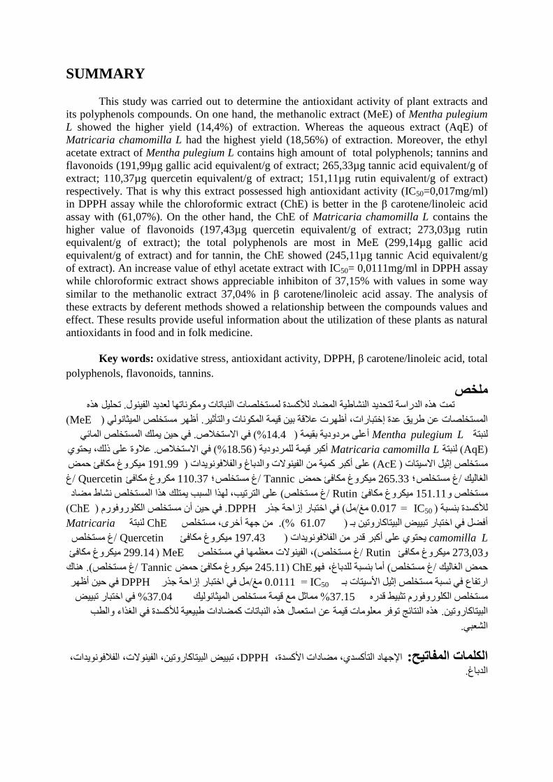
This study was carried out to determine the antioxidant activity of plant extracts and
its polyphenols compounds. On one hand, the methanolic extract (MeE) of Mentha pulegium
L showed the higher yield (14,4%) of extraction. Whereas the aqueous extract (AqE) of
Matricaria chamomilla L had the highest yield (18,56%) of extraction. Moreover, the ethyl
acetate extract of Mentha pulegium L contains high amount of total polyphenols; tannins and
flavonoids (191,99µg gallic acid equivalent/g of extract; 265,33µg tannic acid equivalent/g of
extract; 110,37µg quercetin equivalent/g of extract; 151,11µg rutin equivalent/g of extract)
respectively. That is why this extract possessed high antioxidant activity (IC50=0,017mg/ml)
in DPPH assay while the chloroformic extract (ChE) is better in the β carotene/linoleic acid
assay with (61,07%). On the other hand, the ChE of Matricaria chamomilla L contains the
higher value of flavonoids (197,43µg quercetin equivalent/g of extract; 273,03µg rutin
equivalent/g of extract); the total polyphenols are most in MeE (299,14µg gallic acid
equivalent/g of extract) and for tannin, the ChE showed (245,11µg tannic Acid equivalent/g
of extract). An increase value of ethyl acetate extract with IC50= 0,0111mg/ml in DPPH assay
while chloroformic extract shows appreciable inhibiton of 37,15% with values in some way
similar to the methanolic extract 37,04% in β carotene/linoleic acid assay. The analysis of
these extracts by deferent methods showed a relationship between the compounds values and
effect. These results provide useful information about the utilization of these plants as natural
antioxidants in food and in folk medicine.
Key words: oxidative stress, antioxidant activity, DPPH, β carotene/linoleic acid, total
polyphenols, flavonoids, tannins.
هلخصححيُو هزي . حمج هزي اىذساست ىخحذَذ اىىشاعُت اىمضاد ىألمسذة ىمسخخيصاث اىىباحاث ومنىواحها ىؼذَذ اىفُىىه
( MeE)أظهش مسخخيض اىمُثاوىىٍ . اىمسخخيصاث ػه عشَق ػذة إخخباساث، أظهشث ػالقت بُه قُمت اىمنىواث واىخأثُش
فٍ حُه َميل اىمسخخيض اىمائٍ . فٍ االسخخالص (%14.4) أػيً مشدودَت بقُمت Mentha pulegium Lىىبخت
(AqE) ىىبختMatricaria camomilla L ػالوة ػيً رىل، َحخىٌ . فٍ االسخخالص (%18.56) أمبش قُمت ىيمشدودَت
مُنشوؽ منافئ حمض 191.99)ػيً أمبش ممُت مه اىفُىىالث واىذباؽ واىفالفىوىَذاث (AcE)مسخخيض إثُو االسُخاث
ؽ / Quercetin منشوؽ منافئ110.37 ؛ؽ مسخخيض /Tannic مُنشوؽ منافئ حمض 265.33 ؛ؽ مسخخيض/اىغاىُل
ػيً اىخشحُب، ىهزا اىسبب َمخيل هزا اىمسخخيض وشاط مضاد (ؽ مسخخيض /Rutin مُنشوؽ منافئ 151.11مسخخيض و
( ChE)فٍ حُه أن مسخخيض اىنيىسوفىسً . DPPHفٍ اخخباس إصاحت جزس (مو/ مؾIC50 = 0.017 )ىألمسذة بىسبت
Matricaria ىىبخت ChEمه جهت أخشي، مسخخيض .(% 61.07)أفضو فٍ اخخباس حبُُض اىبُخاماسوحُه بـ
camomilla L مُنشوؽ منافئ 197.43) َحخىٌ ػيً أمبش قذس مه اىفالفىوىَذاث Quercetin / ؽ مسخخيض
مُنشوؽ منافئ MeE( 299.14، اىفُىىالث مؼظمها فٍ مسخخيض (ؽ مسخخيض /Rutin مُنشوؽ منافئ 273,03و
هىاك . (ؽ مسخخيض /Tannic مُنشوؽ منافئ حمض 245.11 )ChEفهىأما بىسبت ىيذباؽ، (ؽ مسخخيض/حمض اىغاىُل
فٍ حُه أظهش DPPHمو فٍ اخخباس إصاحت جزس / مؾIC50 = 0.0111اسحفاع فٍ وسبت مسخخيض إثُو األسُخاث بـ
فٍ اخخباس حبُُض %37.04 مماثو مغ قُمت مسخخيض اىمُثاوىىُل %37.15مسخخيض اىنيىسوفىسً حثبُظ قذسي
هزي اىىخائج حىفش مؼيىماث قُمت ػه اسخؼماه هزي اىىباحاث ممضاداث عبُؼُت ىألمسذة فٍ اىغزاء واىغب . اىبُخاماسوحُه
.اىشؼبٍ
، حبُُض اىبُخاماسوحُه، اىفُىىالث، اىفالفىوىَذاث، DPPHاإلجهاد اىخأمسذٌ، مضاداث األمسذة، :الكلواث الوفاتيح
.اىذباؽ
RESUME
Cette étude a été réalisée pour déterminer l'activité antioxydante des extraits des
plantes et leurs composés phénoliques. L‟analyse de ces extraits par différent tests a révélé une
relation entre les valeurs des composées et l‟effet. D‟une part, l'extrait méthanolique (EBr) de la
plante Mentha pulegium L est le plus élevés de rendement (14,4%) dans l‟extraction. Tandis
que l'extrait aqueux (EAq) de Matricaria chamomilla L a le rendement le plus élevé
(18,56%). Par ailleurs, l'extrait d'acétate d'éthyle a haute teneur en polyphénoles totaux,
tanins, et des flavonoïdes (191,99 microg equivalent d‟acide gallique/g d‟extrait ; 265, 33
microg equivalent d‟acide tannique/g d‟extrait ; 110,37 microg equivalent quercetine/g
d‟extrait ; 151,11 microg equivalent rutine/ g d‟extrait) respectivement. C‟est pour cela cet
extrait a possèdé une meilleure activité antioxydante (IC50=0017 mg/ ml) dans le test de
DPPH alors que l'extrait chloroformique (ECh) révéle une milleure activité dans le test de β
carotène/ l'acide linoléique (61,07%). D'autre part, l‟ECh de Matricaria chamomilla L a
possèdé la valeur la plus élevée en flavonoïdes (197,43 microg equivalent quercetine /g
d‟extrait ; 273,03 microg equivalent rutine / g d‟extrait), les polyphénoles sont les plus dans
EBr (299,14 microg/ g d'extrait) et pour le tanin, ECh a montré (245,11 microg equivalent
d‟acide tannique/ g d‟extrait). Une augmentation de la valeur d'extrait d‟acétate d'éthyle avec
0,0111 mg/ml dans le test DPPH alors que l‟extraire chloroformique a montré une inhibiton
appréciable de 37,15% avec des valeurs d'une certaine manière similaire à l'extrait
méthanolique 37,04% dans les résultats de test β-carotène/ acide linoléique. Ça fournit des
informations utiles sur l'utilisation de ces deux plantes comme antioxydants naturaux dans les
aliments et la phytothérapie.
Mots clés: stress oxydatif, l'activité antioxydante, DPPH, β-carotène/ acide linoléique,
polyphénoles totaux, flavonoïdes, tanins.
ABREVIATIONS
AcE: Ethyl acetate extract.
AlCl3: Aluminium trichloride.
AqE: Aqueous extract.
BHT: Butylated hydroxyToluene.
ChE: Chloroformic extract.
DPPH: 2, 2-diphenyl-1-picryl-hydrazyl.
FAPy: Formamidopyrimidine.
FAPyG: Formamidopyrimidine derivative of guanine.
H2O2: Hydrogen peroxide.
HO2 ·: Perhydroxyl radical.
HOCl: Hypochlorous acid.
HxE: Hexan extract.
I%: Inhibition percentage.
IC50: The concentration of the substrate which causes the loss of 50% of the activity of the
DPPH.
MeE: Methanolic extract.
MeOH: Methanol.
MPO: myeloperoxidase.
NO: The Nitric oxide.
O 2·–: Superoxide anion radical.
ROS: Reactive oxygen species.
SOD: Superoxide dismutase.
LIST OF FIGURES
Figure a1. The structure of phenolic acids(Harborne, 1986).
Figure a2. Basic skeleton stucture of flavonoids.
Figure a3. Chemical structure of flavonoids.
Figure a4. Structure of flavone.
Figure a5. Flavan structure.
Figure a6. Derivatives of flavan.
Figure a7. Structure of flavanonol.
Figure a8. Basic structures of isoflavonoids.
Figure a9. Flavylium skeleton of anthocyanidins.
Figure a10. The basic structures of chalcone, aurone.
Figure a11. Structure of flavanone.
Figure 1. The balance between the systems oxidant and antioxidant.
Figure 2. Photograph of Mentha pulegium L.
Figure 3. Photograph of Matricaria chamomilla L.
Figure 4. The structures of the major components of Matricaria chamomilla L.
Figure 5. Schematic diagram represents the process of extraction.
Figure 6. Standard curve of Gallic acid for the determination of total polyphenols.
Figure 7. Standard curve of Quercetin and Rutin for the determination of total flavonoids.
Figure 8. Standard curve of tannic acid for the determination of tannins.
Figure 9. The DPPH scavenging of extracts of Mentha pulegium L.
Figure 10. IC50 values of extracts of Mentha pulegium L. determined by DPPH assay.
Figure 11. The DPPH scavenging of extracts of Matricaria chamomilla L.
Figure 12. IC50 values of extracts of Matricaria chamomilla L. determined by DPPH assay.
Figure 13. Antioxidant activity of Mentha pulegium L. extracts by β-carotene/ linoleic acid
assay.
Figure 14. Inhibition percentage of divers extracts of Mentha pulegium L. in β-carotene
/linoleic acid assay.
Figure 15. Antioxidant activity of Matricaria chamomilla L. extracts in β-carotene/ linoleic
acid assay.
Figure 16. Inhibition percentage of divers extracts of Matricaria chamomilla L. in β-
carotene/ linoleic acid assay.
LIST OF TABLES
Table 1: Structures of the prominent naturally phenolic acids.
Table 2: Yeild of various extracts of Mentha pulegium L.
Table 3: Yeild of various extracts of Matricaria chamomilla L.
Table 4: Total polyphenols, flavonoids and tannins in Mentha pulegium L. extracts.
Table 5: Total polyphenols, flavonoids and tannins in Matricaria chamomilla L. extracts.
Table 5: Total polyphenols, flavonoids and tannins in Matricaria chamomilla L. extracts.
Table 6: DPPH scavenging of extracts of Mentha puleguim L.
Table 7: DPPH scavenging of extracts of Matricaria chamomilla L.

INTRODUCTION
INTRODUCTION
Reactive oxygen species (ROS) and their likely involvement in some human
physiopathologies have attracted growing interest from the health sector over the last few
decades. Recently, the interest increased in occurring naturally antioxidant which can be used
to protect the human beings against the damage from the oxidative stress (Scalbert et al.,
2005). Many plants contain natural antioxidants which act as metabolic response to the
endogenous production of free radicals and other oxidizing species (Grassmann et al., 2002).
These antioxidants mainly come from plants in the form of phenolic compounds (flavonoids,
phenolic acids, alcohols, stilbenes, tocopherols, tocotrienols), ascorbic acid and carotenoids.
For example, the aromatic herbs and spices were used for a long time in the Mediterranean
kitchen, not only to improve or modify the taste of food, but also to avoid its deterioration
(Proenca et al., 2003).
Plants have been the basis of traditional medicines throughout the world for thousands of
years and continue to provide new remedies to humankind; a great deal of effort has therefore
focused on using available experimental techniques to identify natural antioxidants from
plants. Several authors have reviewed the beneficial uses of these plant species (Speroni and
Scartezzini, 2000; Matkowski., 2008).
In this work, we studied the antioxidant and free radicals scavenging effects of plant extracts
from Mentha pulegium L. and Matricaria chamomilla L., which are largely used in the
Algerian folk medicine such as encouraging menstruation, in the treatement of painful related
to convulsions like the diarrhoeas. The contents of total polyphenols, flavonoids and tannins
in these extracts were also determined.
Chapter 1: REVIEW OF THE
LITERATURE
I. Oxidative stress:
I.1. Oxidative stress:
During the production of reactive species of oxygen (ROS) in the human beings, by
endogenous or external sources, for example tobacco smoke, certain pollutants, organic
solvents or pesticides, involves an oxydative stress (Gulcin et al., 2003).
I.1.1. Definition of stress:
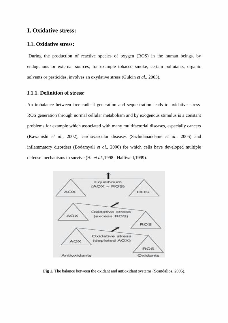
An imbalance between free radical generation and sequestration leads to oxidative stress.
ROS generation through normal cellular metabolism and by exogenous stimulus is a constant
problems for example which associated with many multifactorial diseases, especially cancers
(Kawanishi et al., 2002), cardiovascular diseases (Sachidanandame et al., 2005) and
inflammatory disorders (Bodamyali et al., 2000) for which cells have developed multiple
defense mechanisms to survive (Ha et al.,1998 ; Halliwell,1999).
Fig 1. The balance between the oxidant and antioxidant systems (Scandalios, 2005).
I.2. Reactive oxygen species:
I.2.1. Definition:
A free radical is any species capable of independent existence containing one or more
unpaired electrons (Halliwell and Gutteridge, 1984). The unpaired electron alters the chemical
reactivity of the molecule/atom, making it more reactive than the corresponding non-radical
form. The oxygen free radicals include superoxide anion radical (O 2·–), singlet oxygen (1O2),
hydroxyl radical (·OH), the nitric oxide (NO), and perhydroxyl radical (HO2 ·) are termed
collectively the „reactive oxygen species‟ (ROS). The usual route of O2 metabolism is through
its complete reduction to H2O by accepting four electrons. However, with a single electron
reduction several free radicals and hydrogen peroxide (H2O2) are formed (Grisham, 1992;
Nappi and Vass, 1998).
I.3. Source of free radicals:
In vivo, ROS are generated by oxidant enzymes, phagocytic cells, ionizing radiation, etc.
Superoxide anion is believed to be the first radical formed, mainly by the electron transport
chain when O2 picks up a single electron. Radicals such as ·OH, HO2· and H2O2 are formed
from O2·–
(Grisham, 1992 ; Nappi and Vass, 1998). O2·– undergoes a dismutation reaction
catalysed by the enzyme superoxide dismutase (SOD) to form H2O2, which by itself is not
reactive enough to cause damage to macromolecules. It is, however, a very important oxidant
since it can cross biological membranes and form the highly reactive ·OH by interaction with
transition metal ions such as Fe2+
or Cu+. H2O2 is reduced by three general mechanisms. First,
it is a substrate for two enzymes, catalase and glutathione peroxidase, that catalyse its
conversion to H2O and O2 (Maddipati and Marnett, 1987), a detoxification mechanism.
Secondly, H2O2 is converted by myeloperoxidase (MPO) in neutrophils to hypochlorous acid
(HOCl), a strong oxidant that acts as a bactericidal agent in phagocytic cells. Reaction of
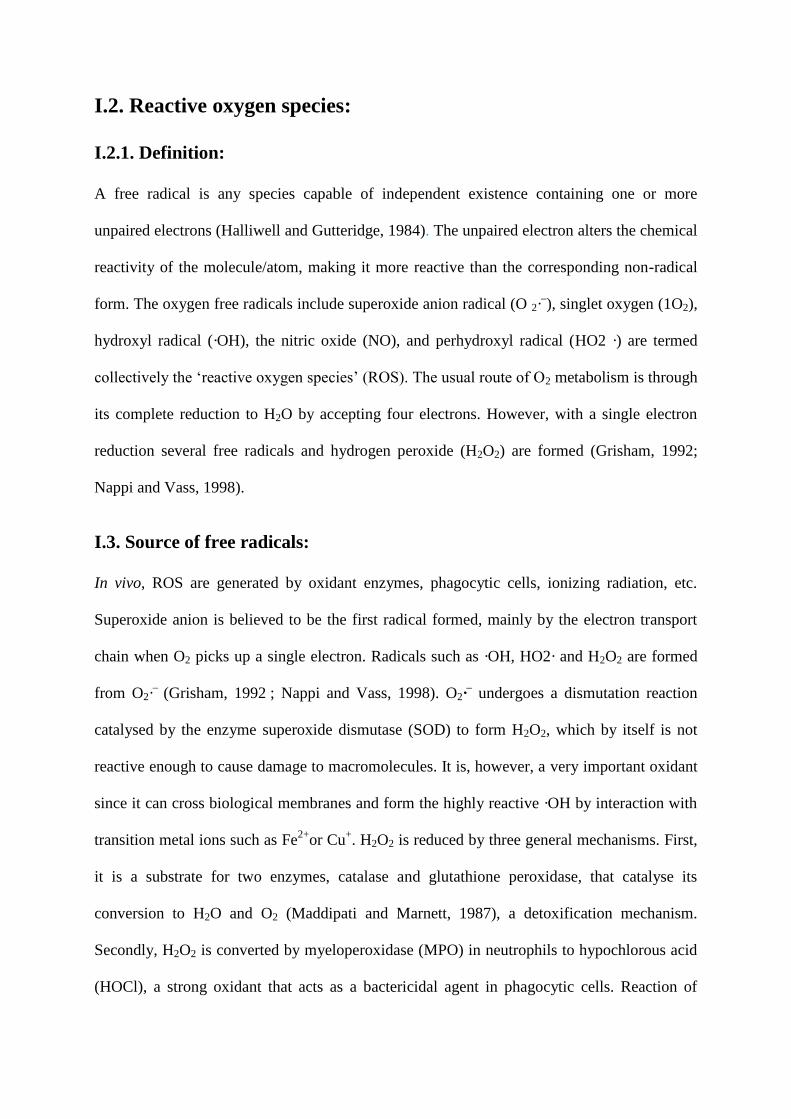
HOCl with H2O2 yields1O2. Thirdly, H2O2 is converted in a spontaneous reaction catalysed by
transition metal ions to the highly reactive ·OH.
HOCl 1O2 Cl
-
O2 O2.- H2O2 .OH H2O
Among the ROS, ·OH is the most potent damaging radical which can react with all biological
macromolecules (lipids, proteins, nucleic acids and carbohydrates). It is extremely reactive
and can lead to formation of DNA-protein cross-links, single- and double-strand breaks, base
damage, lipid peroxidation and protein fragmentation (Lloyd et al., 1997; Stohs and Bagchi,
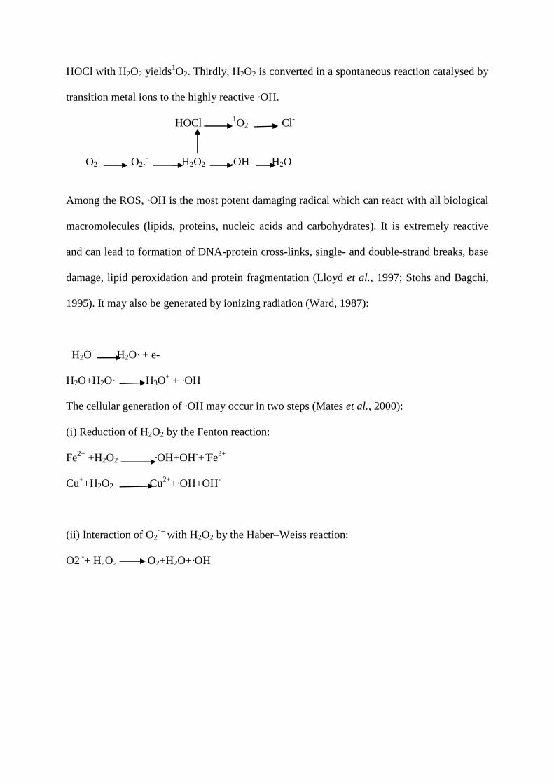
1995). It may also be generated by ionizing radiation (Ward, 1987):
H2O H2O· + e-
H2O+H2O· H3O+ + ·OH
The cellular generation of ·OH may occur in two steps (Mates et al., 2000):
(i) Reduction of H2O2 by the Fenton reaction:
Fe2+
+H2O2 ·OH+OH-+
-Fe
3+
Cu++H2O2 Cu
2++·OH+OH
-
(ii) Interaction of O2· –
with H2O2 by the Haber–Weiss reaction:
O2·-+ H2O2 O2+H2O+·OH
I.4. The effect of free radicals:
I.4.1. Oxidative damage to lipids:
Among the more susceptible targets of ·OH are polyunsaturated fatty acids. Abstraction of a
hydrogen atom from a molecule of polyunsaturated fatty acid initiates the process of lipid
peroxidation (Arouma, 1993). The peroxidation reactions differ among these fatty acids
depending on the number and position of the double bonds on the acyl chain and the reader is
referred to Frankel (1985). A hydrogen atom is abstracted from a second molecule, leading to
a new free radical. Aldehydes of lipid peroxidation can react with sulphydryl (cysteine) or
basic amino acids (histidine, lysine) affecting their biological characteristics (Arouma, 1993).
The peroxidation of lipids involves three distinct steps: initiation, propagation and termination
(Bradley and Minn, 1992).
I.4.2. Oxidative damage to proteins:
Oxidative attack on proteins results in site-specific amino acid modifications, fragmentation
of the peptide chain, aggregation of cross-linked reaction products, altered electrical charge
and increased susceptibility to proteolysis. The amino acids in a peptide differ in their
susceptibility to attack, and the various forms of activated oxygen differ in their potential
reactivity. Primary, secondary, and tertiary protein structures alter the relative susceptibility of
certain amino acids. In spite of this complexity, generalisations can be made. Sulphur
containing amino acids, and thiol groups specifically, are very susceptible sites. Activated
oxygen can abstract an H atom from cysteine residues to form a thiyl radical that will cross-
link to a second thiyl radical to form disulphide bridges. Alternatively, oxygen can add to a
methionine residue to form methionine sulphoxide derivatives. Reduction of both of these
may be accomplished in microbial systems by thioredoxin and thioredoxin reductase (Farr
and Kogama, 1991).
Other forms of free radical attack on proteins are not reversible. For example, the oxidation of
iron-sulphur centres by superoxide destroys enzymatic function (Gardner and Fridovich,
1991). Many amino acids undergo specific irreversible modifications when a protein is
oxidised. For example, tryptophan is readily cross-linked to form bityrosine products (Davies,
1987). Histidine, lysine, proline, arginine, and serine form carbonyl groups on oxidation
(Stadtman, 1986). The oxidative degradation of protein is enhanced in the presence of metal
cofactors that are capable of redox cycling, such as Fe. In these cases, the metal binds to a
divalent cation binding site on the protein. The metal then reacts with hydrogen peroxide in a
Fenton reaction to form a hydroxyl radical that rapidly oxidises an amino acid residue at or
near the cation binding site of the protein (Stadtman, 1986).
I.4.3. Oxidative damage to DNA:
Similarly, modification of individual nucleotide bases, single strand breaks and cross-linking
are the typical effects of ROS on nucleic acids (Arouma, 1993). The damage to DNA by ·OH
includes single-strand breaks, base modifications and conformational changes. Nitrogenous
bases react preferentially with ·OH rather than sugar moiety by 4–6-fold. Thymine and
guanine are most susceptible to modifications followed by cytosine and adenine. Thymine
glycol is the major oxidation product, its presence in urine serves as an indicator of
endogenous DNA damage. Cytosine glycols are also formed which can undergo deamination
to form uracil derivatives that base pair preferentially with adenine, instead of guanine.
Reduction of guanine leads to ring opening forming formamidopyrimidine (FAPy) derivative
of guanine (FAPyG). Oxidation leads to the formation of 8-oxo-deoxyguanine (8-oxodG), a
major product. Its measurement in urine is used as a biomarker of endogenous oxidative DNA
damage (Linn, 1998).
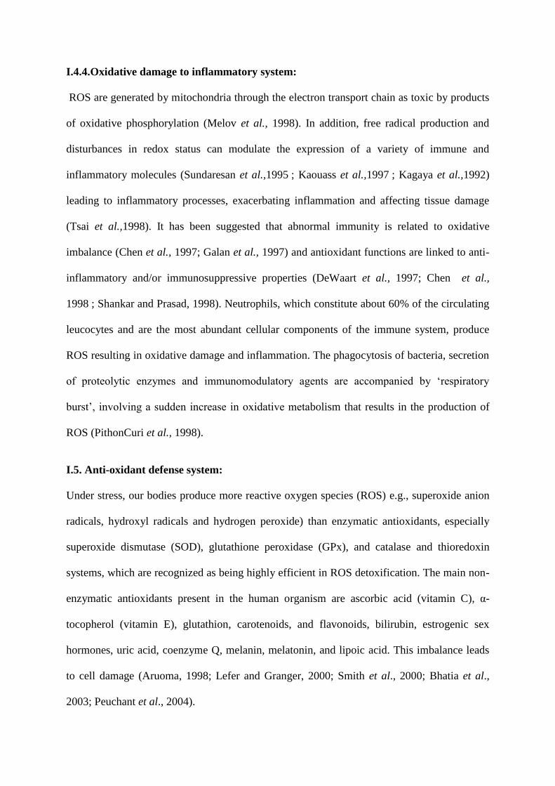
I.4.4.Oxidative damage to inflammatory system:
ROS are generated by mitochondria through the electron transport chain as toxic by products
of oxidative phosphorylation (Melov et al., 1998). In addition, free radical production and
disturbances in redox status can modulate the expression of a variety of immune and
inflammatory molecules (Sundaresan et al.,1995 ; Kaouass et al.,1997 ; Kagaya et al.,1992)
leading to inflammatory processes, exacerbating inflammation and affecting tissue damage
(Tsai et al.,1998). It has been suggested that abnormal immunity is related to oxidative
imbalance (Chen et al., 1997; Galan et al., 1997) and antioxidant functions are linked to anti-
inflammatory and/or immunosuppressive properties (DeWaart et al., 1997; Chen et al.,
1998 ; Shankar and Prasad, 1998). Neutrophils, which constitute about 60% of the circulating
leucocytes and are the most abundant cellular components of the immune system, produce
ROS resulting in oxidative damage and inflammation. The phagocytosis of bacteria, secretion
of proteolytic enzymes and immunomodulatory agents are accompanied by „respiratory
burst‟, involving a sudden increase in oxidative metabolism that results in the production of
ROS (PithonCuri et al., 1998).
I.5. Anti-oxidant defense system:
Under stress, our bodies produce more reactive oxygen species (ROS) e.g., superoxide anion
radicals, hydroxyl radicals and hydrogen peroxide) than enzymatic antioxidants, especially
superoxide dismutase (SOD), glutathione peroxidase (GPx), and catalase and thioredoxin
systems, which are recognized as being highly efficient in ROS detoxification. The main non-
enzymatic antioxidants present in the human organism are ascorbic acid (vitamin C), α-
tocopherol (vitamin E), glutathion, carotenoids, and flavonoids, bilirubin, estrogenic sex
hormones, uric acid, coenzyme Q, melanin, melatonin, and lipoic acid. This imbalance leads
to cell damage (Aruoma, 1998; Lefer and Granger, 2000; Smith et al., 2000; Bhatia et al.,
2003; Peuchant et al., 2004).
I.6. Polyphenolic compounds:
Many medicinal plants contain various bioactive compounds, such as polyphenolic
compounds, which are secondary metabolites. From a chemical point of view, polyphenols
can react with one-electron oxidants, which prevents free radical formation in biological
systems (Huang et al., 1992). This class includes phenolic acids, flavonoids and tannins
(Bruneton, 1993).
I.6.1. Phenolic acids:
I.6.1.1. Definition:
Phenolic acids are aromatic secondary plant metabolites widely distributed throughout the
plant kingdom (Hemmann, 1989). The term “phenolic acids”, in general, designates phenols
that possess one carboxylic acid functionality. However, when talking about plant
metabolites, it refers to a distinct group of organic acids (Shahidi and Wanasundara, 1992;
Robbins, 2003).
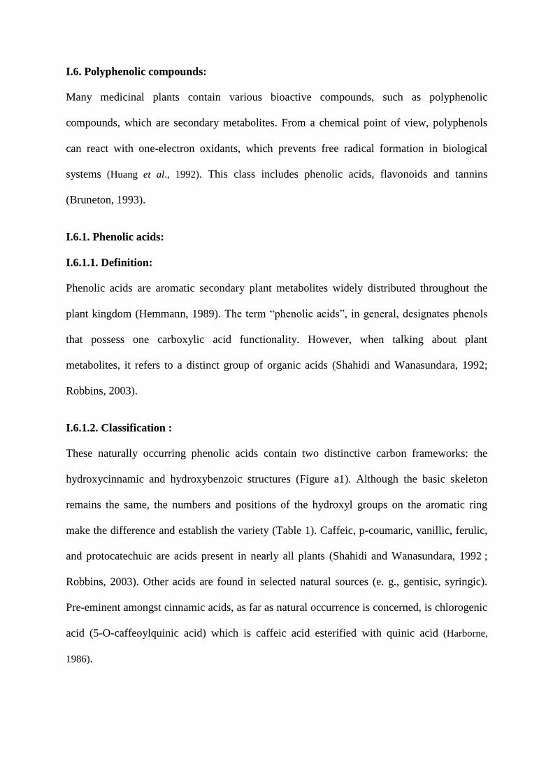
I.6.1.2. Classification :
These naturally occurring phenolic acids contain two distinctive carbon frameworks: the
hydroxycinnamic and hydroxybenzoic structures (Figure a1). Although the basic skeleton
remains the same, the numbers and positions of the hydroxyl groups on the aromatic ring
make the difference and establish the variety (Table 1). Caffeic, p-coumaric, vanillic, ferulic,
and protocatechuic are acids present in nearly all plants (Shahidi and Wanasundara, 1992 ;
Robbins, 2003). Other acids are found in selected natural sources (e. g., gentisic, syringic).
Pre-eminent amongst cinnamic acids, as far as natural occurrence is concerned, is chlorogenic
acid (5-O-caffeoylquinic acid) which is caffeic acid esterified with quinic acid (Harborne,
1986).
Hydroxybenzoic Acids Hydroxycinnamic Acids
Fig a1. The structure of phenolic acids(Harborne, 1986).
Table1. Structures of the prominent naturally phenolic acids (Harborne, 1986).
.
Name R1 R2 R3 R4
Bensoic acid H H H H
p-Hydroxybenzoic acid H H OH H
Vanillic acid H OCH3 OH H
Gallic acid H OH OH OH
Protocatechuic acid H OH OH H
Syringic acid H OCH3 OH OCH3
Gentisic acid OH H H OH
Veratric acid H OCH3 OCH3 H
Salicylic acid OH H H H
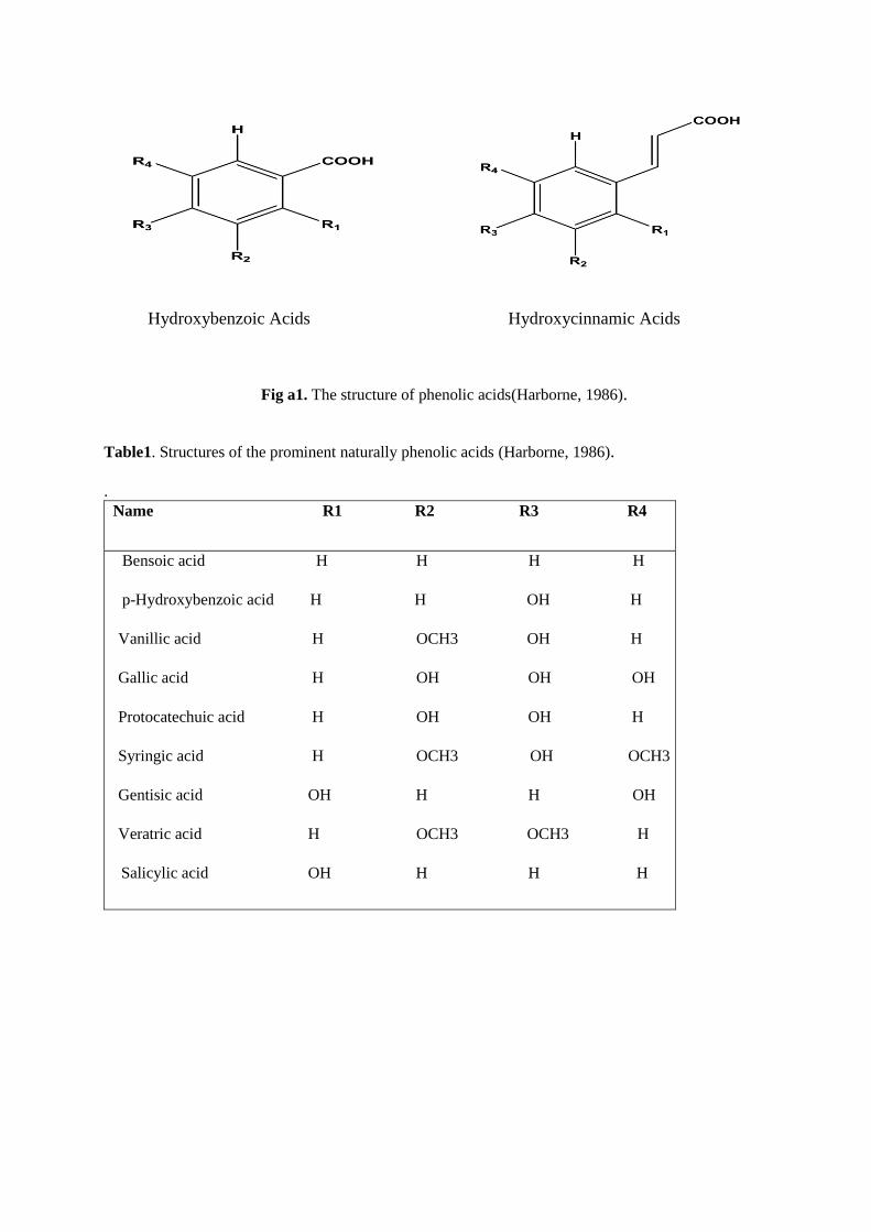
Name R1 R2 R3 R4
H Cinnamic acid H H H H
o-Coumaric acid OH H H H
m-Coumaric acid H OH H H
p-Coumaric acid H H OH H
Ferulic acid H OCH3 OH H
Sinapic acid H OCH3 OH OCH3
Caffeic acid H OH OH H
I.6.2. Flavonoids:
I.6.2.1. Definition:
Flavonoids are planar molecules ubiquitous in plants, formed from the aromatic amino acids
phenylalanine, tyrosine, and malonate (Harborne, 1986). Flavonoids as flower pigments
consist of two aromatic rings (A and B) and a heterocycle (C) with oxygen. Based on the
configuration and state of oxidation of the central C3 unit in the molecule, flavonoids are
divided into eight groups. The first to suggest this flavonoid structure was Robinson (1936).
This hypothesis was further confirmed by the formation and biosynthesis of quercetin in
tartary buckwheat (Underhill et al., 1957; Watkin et al., 1960).
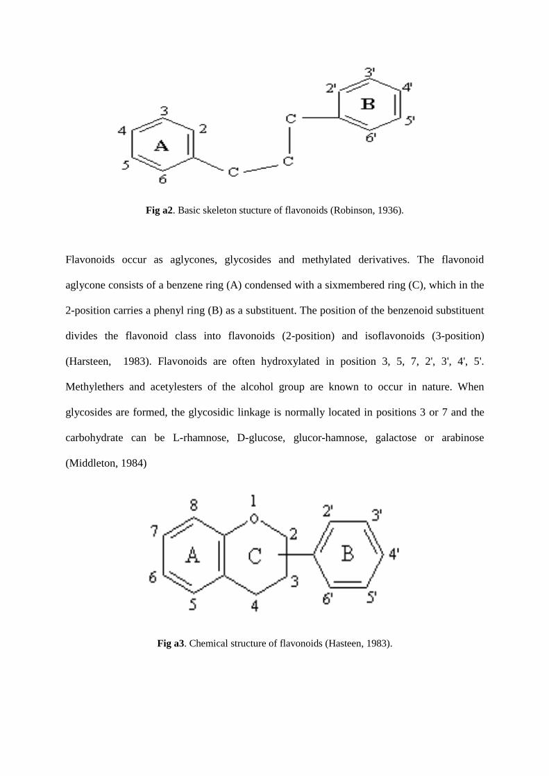
Fig a2. Basic skeleton stucture of flavonoids (Robinson, 1936).
Flavonoids occur as aglycones, glycosides and methylated derivatives. The flavonoid
aglycone consists of a benzene ring (A) condensed with a sixmembered ring (C), which in the
2-position carries a phenyl ring (B) as a substituent. The position of the benzenoid substituent
divides the flavonoid class into flavonoids (2-position) and isoflavonoids (3-position)
(Harsteen, 1983). Flavonoids are often hydroxylated in position 3, 5, 7, 2', 3', 4', 5'.
Methylethers and acetylesters of the alcohol group are known to occur in nature. When
glycosides are formed, the glycosidic linkage is normally located in positions 3 or 7 and the
carbohydrate can be L-rhamnose, D-glucose, glucor-hamnose, galactose or arabinose
(Middleton, 1984)
Fig a3. Chemical structure of flavonoids (Hasteen, 1983).
I.6.2.2. Classification:
Over 5000 naturally occurring flavonoids have been characterized from various plants. They
have been classified according to their chemical structure (Ververidis et al., 2007). According
to the oxidation condition of the pyran ring placed at the center of flavonoids, flavonoids can
be further subdivided into five major subclass as follows: flavonols, flavanols, flavones,
isoflavones, anthocyanidins (Moon et al., 2006).
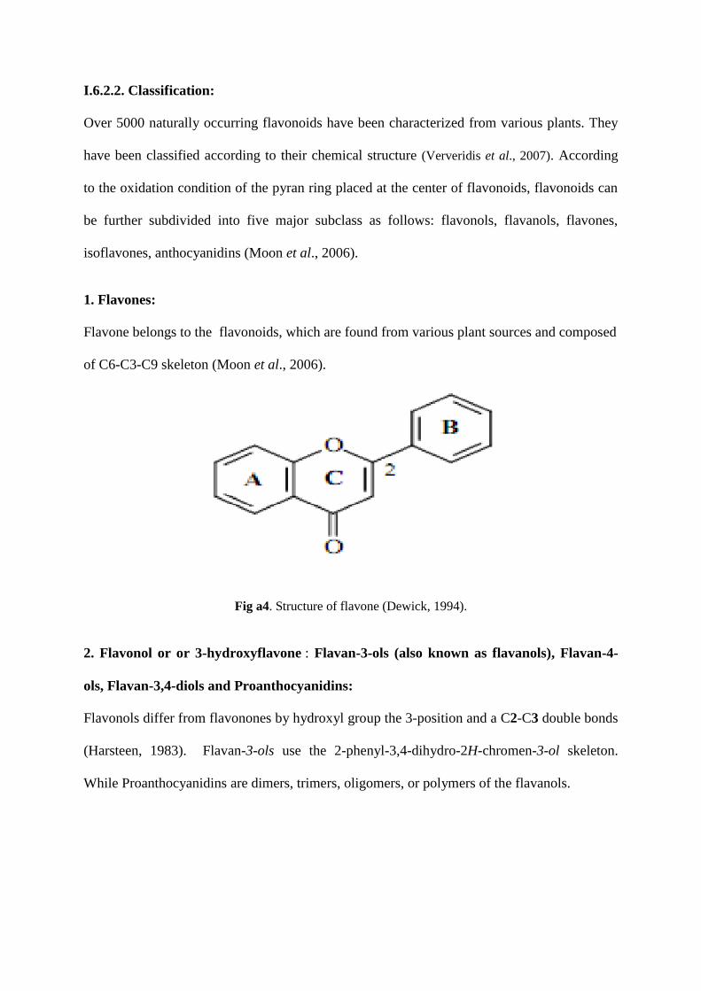
1. Flavones:
Flavone belongs to the flavonoids, which are found from various plant sources and composed
of C6-C3-C9 skeleton (Moon et al., 2006).
Fig a4. Structure of flavone (Dewick, 1994).
2. Flavonol or or 3-hydroxyflavone : Flavan-3-ols (also known as flavanols), Flavan-4-
ols, Flavan-3,4-diols and Proanthocyanidins:
Flavonols differ from flavonones by hydroxyl group the 3-position and a C2-C3 double bonds
(Harsteen, 1983). Flavan-3-ols use the 2-phenyl-3,4-dihydro-2H-chromen-3-ol skeleton.
While Proanthocyanidins are dimers, trimers, oligomers, or polymers of the flavanols.
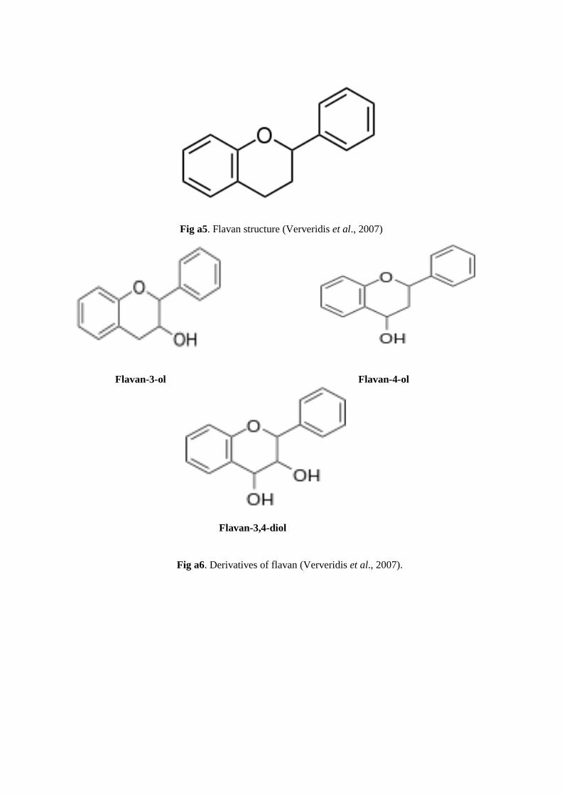
Fig a5. Flavan structure (Ververidis et al., 2007)
Flavan-3-ol Flavan-4-ol
Flavan-3,4-diol
Fig a6. Derivatives of flavan (Ververidis et al., 2007).
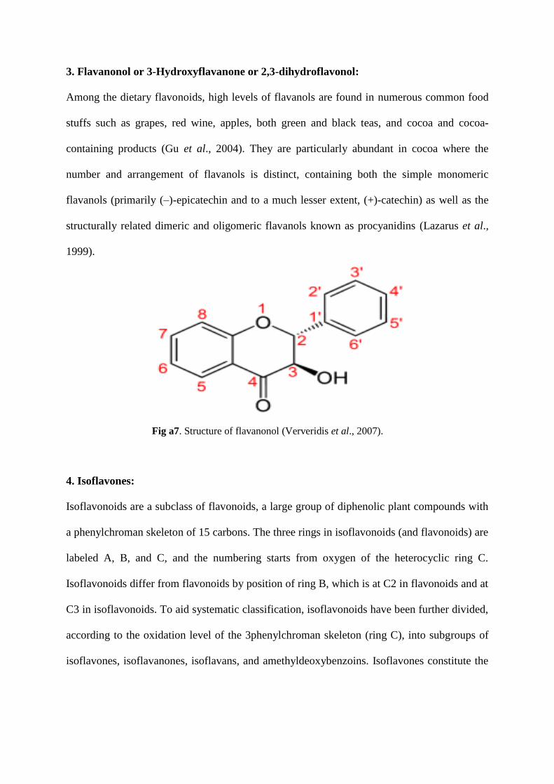
3. Flavanonol or 3-Hydroxyflavanone or 2,3-dihydroflavonol:
Among the dietary flavonoids, high levels of flavanols are found in numerous common food
stuffs such as grapes, red wine, apples, both green and black teas, and cocoa and cocoa-
containing products (Gu et al., 2004). They are particularly abundant in cocoa where the
number and arrangement of flavanols is distinct, containing both the simple monomeric
flavanols (primarily (–)-epicatechin and to a much lesser extent, (+)-catechin) as well as the
structurally related dimeric and oligomeric flavanols known as procyanidins (Lazarus et al.,
1999).
Fig a7. Structure of flavanonol (Ververidis et al., 2007).
4. Isoflavones:
Isoflavonoids are a subclass of flavonoids, a large group of diphenolic plant compounds with
a phenylchroman skeleton of 15 carbons. The three rings in isoflavonoids (and flavonoids) are
labeled A, B, and C, and the numbering starts from oxygen of the heterocyclic ring C.
Isoflavonoids differ from flavonoids by position of ring B, which is at C2 in flavonoids and at
C3 in isoflavonoids. To aid systematic classification, isoflavonoids have been further divided,
according to the oxidation level of the 3phenylchroman skeleton (ring C), into subgroups of
isoflavones, isoflavanones, isoflavans, and amethyldeoxybenzoins. Isoflavones constitute the
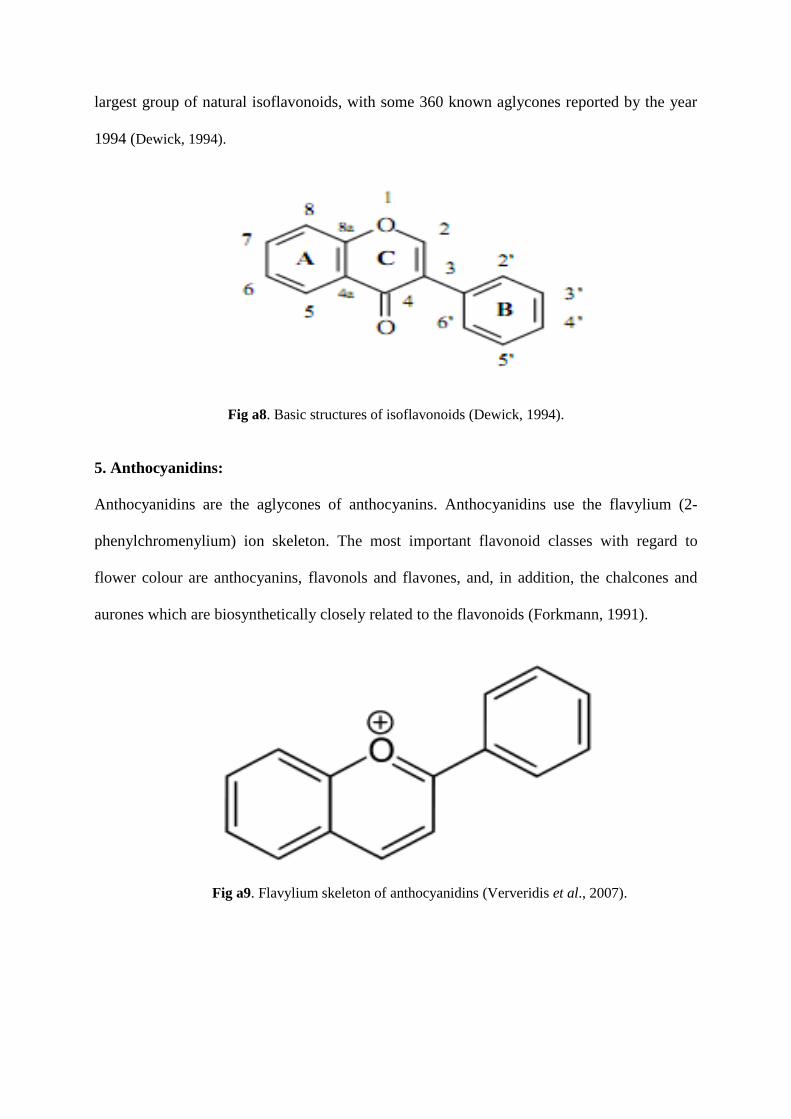
largest group of natural isoflavonoids, with some 360 known aglycones reported by the year
1994 (Dewick, 1994).
Fig a8. Basic structures of isoflavonoids (Dewick, 1994).
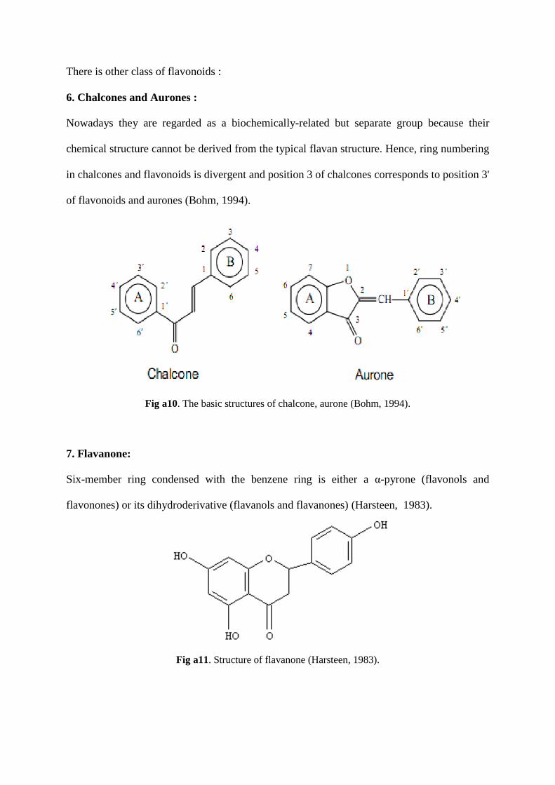
5. Anthocyanidins:
Anthocyanidins are the aglycones of anthocyanins. Anthocyanidins use the flavylium (2-
phenylchromenylium) ion skeleton. The most important flavonoid classes with regard to
flower colour are anthocyanins, flavonols and flavones, and, in addition, the chalcones and
aurones which are biosynthetically closely related to the flavonoids (Forkmann, 1991).
Fig a9. Flavylium skeleton of anthocyanidins (Ververidis et al., 2007).
There is other class of flavonoids :
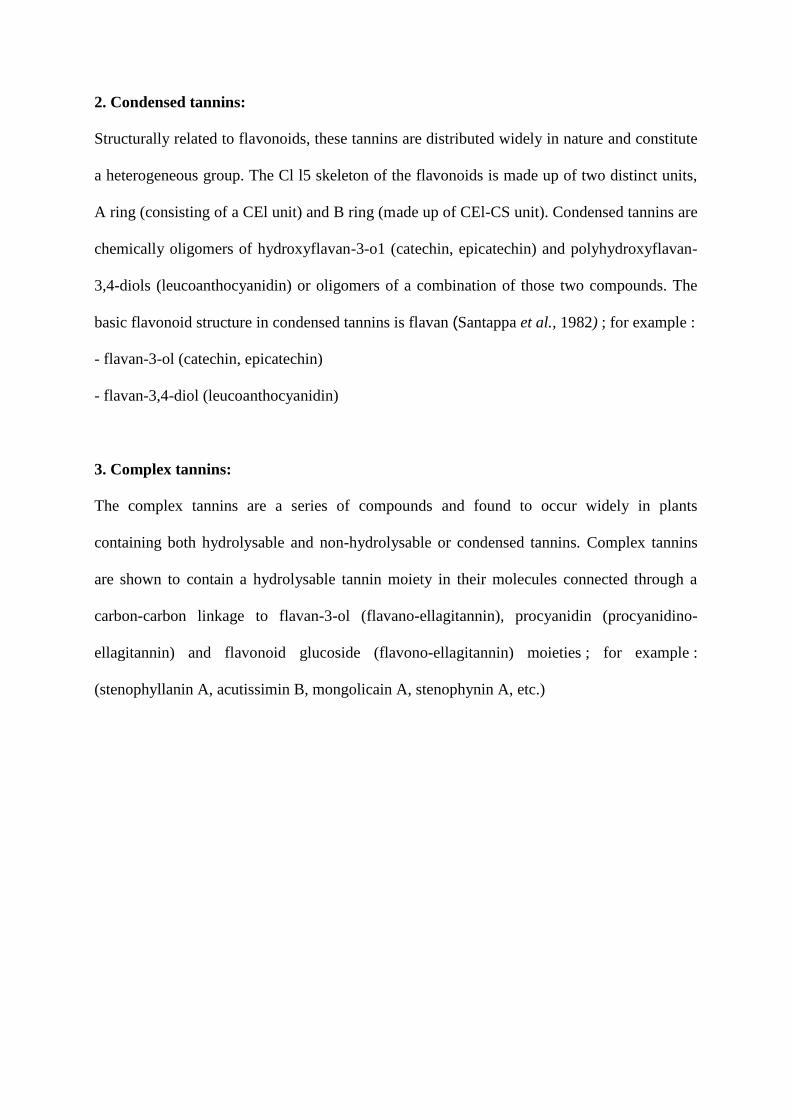
6. Chalcones and Aurones :
Nowadays they are regarded as a biochemically-related but separate group because their
chemical structure cannot be derived from the typical flavan structure. Hence, ring numbering
in chalcones and flavonoids is divergent and position 3 of chalcones corresponds to position 3'
of flavonoids and aurones (Bohm, 1994).
Fig a10. The basic structures of chalcone, aurone (Bohm, 1994).
7. Flavanone:
Six-member ring condensed with the benzene ring is either a α-pyrone (flavonols and
flavonones) or its dihydroderivative (flavanols and flavanones) (Harsteen, 1983).
Fig a11. Structure of flavanone (Harsteen, 1983).

I.6.3. Tannins:
I.6.3.1. Definition :
Tannins (commonly referred to as tannic acid) are water-soluble polyphenols that are present
in many plant foods. They have been reported to be responsible for decreases in feed intake,
growth rate, feed efficiency, net metabolizable energy, and protein digestibility in
experimental animals. Therefore, foods rich in tannins are considered to be of low nutritional
value. However, recent findings indicate that the major effect of tannins was not due to their
inhibition on food consumption or digestion but rather the decreased efficiency in converting
the absorbed nutrients to new body substances.
I.6.3.2. Classification:
1. Hydrolysable tannins:
These are based on esters of phenol carboxylic acids (gallic acid) with a central carbohydrate
core for example :
- gallotannins (gallic acid, quinic acid, tannic acid)
- ellagitannins (ellagic acid, castalagin, vescalagin, etc.)
- hydrolysable tannin oligomers (agrimoniin, rugosin D)
- caffeic acid derivatives (chlorogenic acid, caffeetannin, dicaffeoylquinic acid, rosmarinic
acid).
2. Condensed tannins:
Structurally related to flavonoids, these tannins are distributed widely in nature and constitute
a heterogeneous group. The Cl l5 skeleton of the flavonoids is made up of two distinct units,
A ring (consisting of a CEl unit) and B ring (made up of CEl-CS unit). Condensed tannins are
chemically oligomers of hydroxyflavan-3-o1 (catechin, epicatechin) and polyhydroxyflavan-
3,4-diols (leucoanthocyanidin) or oligomers of a combination of those two compounds. The
basic flavonoid structure in condensed tannins is flavan (Santappa et al., 1982) ; for example :
- flavan-3-ol (catechin, epicatechin)
- flavan-3,4-diol (leucoanthocyanidin)
3. Complex tannins:
The complex tannins are a series of compounds and found to occur widely in plants
containing both hydrolysable and non-hydrolysable or condensed tannins. Complex tannins
are shown to contain a hydrolysable tannin moiety in their molecules connected through a
carbon-carbon linkage to flavan-3-ol (flavano-ellagitannin), procyanidin (procyanidino-
ellagitannin) and flavonoid glucoside (flavono-ellagitannin) moieties ; for example :
(stenophyllanin A, acutissimin B, mongolicain A, stenophynin A, etc.)
II. Herbal therapy:
Phytomedicine, also called herbal therapy is an important branch of complementary and
alternative medicine and is in fact a traditional therapeutic system which takes advantage of
herbal plants to prevent and cure maladies and improve general health (Givens et al., 2006).
Plants are important source of active natural products which differ widely in terms of
structure and biological properties. Many herbs have been used for a long time for claimed
health benefits. They are sold as tablets, capsules, powders, teas, extracts and fresh or dried
plants. However, some can cause health problems, some are not effective and some may
interact with other drugs you are taking.
II.1. Mentha pulegium L.:
II.1.1. Monograph of Mentha pulegium L.:
Scientific name: Mentha pulegium L. ( Lamiaceae)
French vernacular names: Pennyroyal, Pouliot.
Vernacular name: Feliou, Afilgou, Felgou, Moursal, Tamarsa.
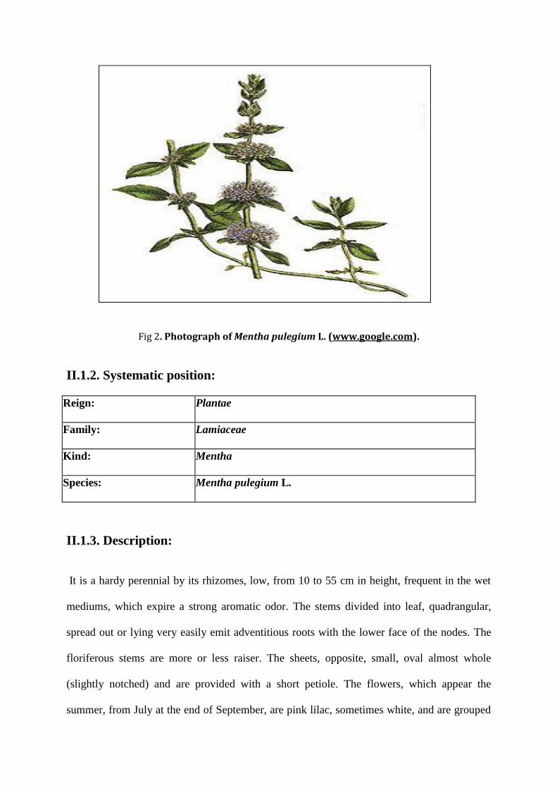
Fig 2. Photograph of Mentha pulegium L. (www.google.com).
II.1.2. Systematic position:
Reign: Plantae
Family: Lamiaceae
Kind: Mentha
Species: Mentha pulegium L.
II.1.3. Description:
It is a hardy perennial by its rhizomes, low, from 10 to 55 cm in height, frequent in the wet
mediums, which expire a strong aromatic odor. The stems divided into leaf, quadrangular,
spread out or lying very easily emit adventitious roots with the lower face of the nodes. The
floriferous stems are more or less raiser. The sheets, opposite, small, oval almost whole
(slightly notched) and are provided with a short petiole. The flowers, which appear the
summer, from July at the end of September, are pink lilac, sometimes white, and are grouped
with the armpit of the sheets in clusters (false verticils) spread out along the stem. The fruits
are akenes.
II.1.4. Botanical description:
The leaves of Pennyroyal are generally small, ovate, slightly serrate, slightly hairy, and
opposite. For the record, the leaf of the non-creeping pennyroyal can be up to 3cm or 1.5 in
long and may be entire rather than slightly toothed. The color depends on the variety and
whether wild or cultivar. The small flowers are produced in distinctive, dense whorls (similar
to corn or fieldmint and gingermint in bloom). The tight, axillary clusters appear in July-
August with colors ranging from reddish -purple to lilac. There are few flowering stems on
the prostate form; they lie on top of what appears to be "a dense green turf". Seed is light
brown, very small and oval.
II.1.5. Origin and distribution:
Pennyroyal (Mentha pulegium) is an aromatic Perennial and is common wild or garden plant.
It is a spontaneous species in the whole of Europe, found in wet grounds around the Med and
the west of Asia (from Chypre to Turkmenistan) and the north of Africa (from Morocco to
Egypt). In France, this plant is very common up to 1800 m of altitude.
Mentha pulegium L among the vegetables recommended in capitulary De Villis with the
Middle Ages.
II.1.6. Traditional medicinal uses:
Iranian peaple usually uses the plant Mentha pulegium L. against the infectious diseases and
finds to be effective against these problems without any scientific base to explain this action.
The increase in resistance to antibiotics of the pathogenic agents associated the infectious
diseases as well as undesirable side effects of antibiotics suggested the use of oil of Mentha
pulegium L. like antibiotic or of replacement. An Additional uses of this plant is well
regarded as an insect repellent, for both humans and pets. However, additional research is
necessary to evaluate the practical values of therapeutic application (www.google.com).
II.1.7. Medicinal uses:
A good digestive tonic, it stimulates digestive juices, relieves flatulence and colic; a good
remedy for headaches and for minor respiratory infections helping to keep fever and
congestion in check; a powerful stimulant to the uterine muscle encouraging menstruation;
externally it can be used to relieve chine and rheumatic conditions including gout
(www.google.com).
II.1.8. Chemical constituents:
Ingredients of Mentha pulegium L. were subjected to a certain number of studies which
showed a difference of its commissar according to the area of culture and it have some
variations in the components of various countries. El-Ghorab (2006) was noted that the oil of
Mentha pulegium L. coming from Egypt contains pulegone (43,5%), piperitone (12,2%); from
Tunisia (Mkaddem et al, 2007), pulegone(8%), isomenthone (11,3%). These studies showed
three chimiotypes of Mentha pulegium L with the following major components oils (1)
pulegone, (2) piperitenone and/or piperitone and (3) isomenthone/neoisomenthol (Topalov
and Dimitrov, 1969; Cook et al, 2007). Although the air part of flowering of this plant is
usually used because of its disinfectants and pharmaceutical properties.
II.2. Camomille romaine:
II.2.1. Monograph of Matricaria chamomilla L.:
Scientific name: Chamaemelum nobile L.
French vernacular names: Camomile roman, Camomile noble, Anthemis noble, Anthemis
odorous, Camomile of Anjou.
Algerian name vernacular: Habak, Babounj.
II.2.2. Systematic position:
Reign: Plantae
Family: Asteraceae
Kind: Chamaemelum
Species: Matricaria chamomilla L.
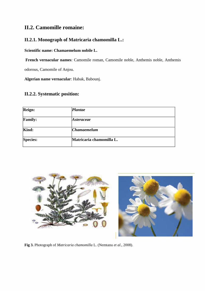
Fig 3. Photograph of Matricaria chamomilla L. (Nemtanu et al., 2008).
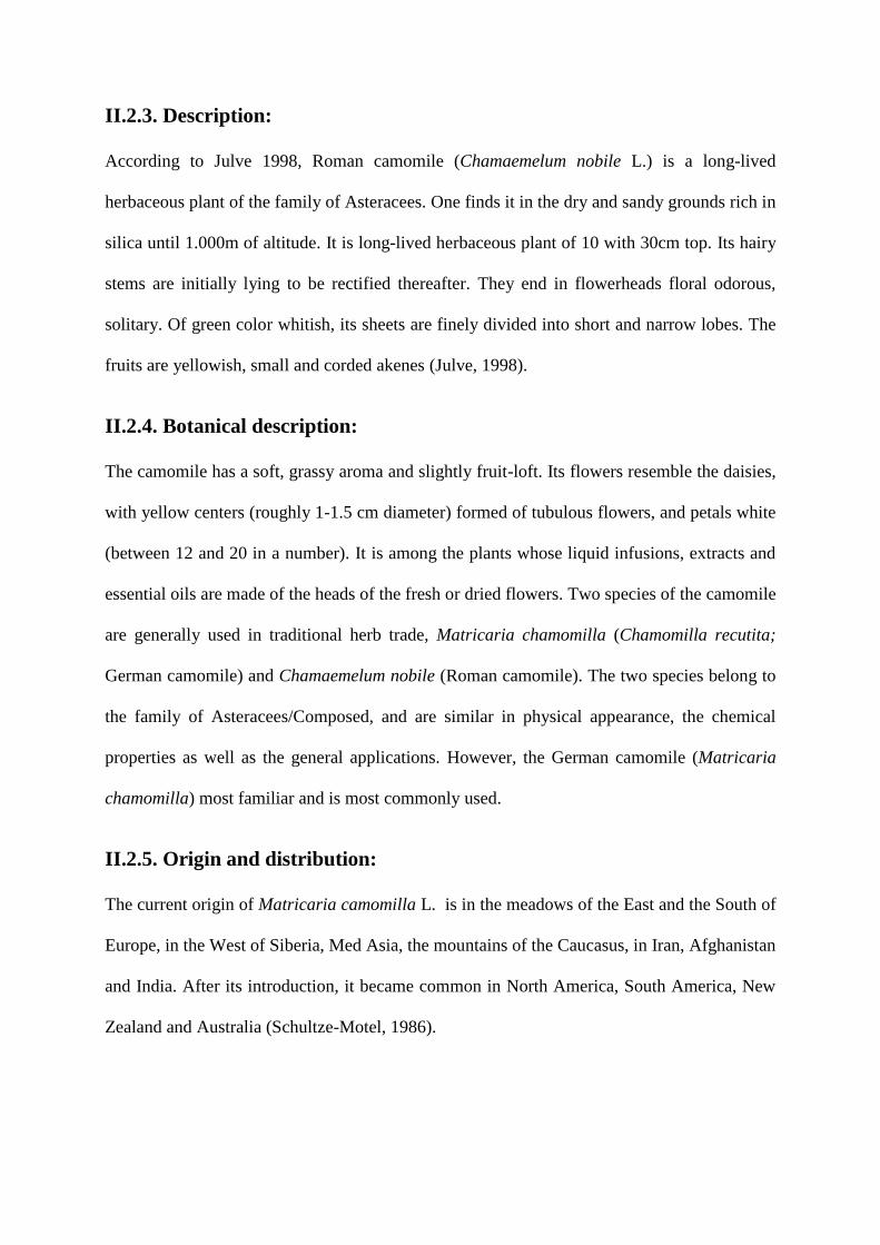
II.2.3. Description:
According to Julve 1998, Roman camomile (Chamaemelum nobile L.) is a long-lived
herbaceous plant of the family of Asteracees. One finds it in the dry and sandy grounds rich in
silica until 1.000m of altitude. It is long-lived herbaceous plant of 10 with 30cm top. Its hairy
stems are initially lying to be rectified thereafter. They end in flowerheads floral odorous,
solitary. Of green color whitish, its sheets are finely divided into short and narrow lobes. The
fruits are yellowish, small and corded akenes (Julve, 1998).
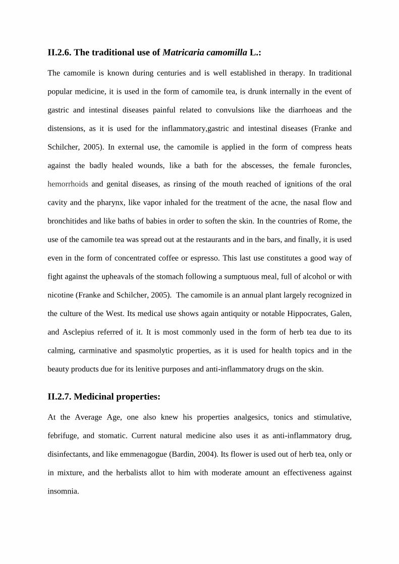
II.2.4. Botanical description:
The camomile has a soft, grassy aroma and slightly fruit-loft. Its flowers resemble the daisies,
with yellow centers (roughly 1-1.5 cm diameter) formed of tubulous flowers, and petals white
(between 12 and 20 in a number). It is among the plants whose liquid infusions, extracts and
essential oils are made of the heads of the fresh or dried flowers. Two species of the camomile
are generally used in traditional herb trade, Matricaria chamomilla (Chamomilla recutita;
German camomile) and Chamaemelum nobile (Roman camomile). The two species belong to
the family of Asteracees/Composed, and are similar in physical appearance, the chemical
properties as well as the general applications. However, the German camomile (Matricaria
chamomilla) most familiar and is most commonly used.
II.2.5. Origin and distribution:
The current origin of Matricaria camomilla L. is in the meadows of the East and the South of
Europe, in the West of Siberia, Med Asia, the mountains of the Caucasus, in Iran, Afghanistan
and India. After its introduction, it became common in North America, South America, New
Zealand and Australia (Schultze-Motel, 1986).
II.2.6. The traditional use of Matricaria camomilla L.:
The camomile is known during centuries and is well established in therapy. In traditional
popular medicine, it is used in the form of camomile tea, is drunk internally in the event of
gastric and intestinal diseases painful related to convulsions like the diarrhoeas and the
distensions, as it is used for the inflammatory,gastric and intestinal diseases (Franke and
Schilcher, 2005). In external use, the camomile is applied in the form of compress heats
against the badly healed wounds, like a bath for the abscesses, the female furoncles,
hemorrhoids and genital diseases, as rinsing of the mouth reached of ignitions of the oral
cavity and the pharynx, like vapor inhaled for the treatment of the acne, the nasal flow and
bronchitides and like baths of babies in order to soften the skin. In the countries of Rome, the
use of the camomile tea was spread out at the restaurants and in the bars, and finally, it is used
even in the form of concentrated coffee or espresso. This last use constitutes a good way of
fight against the upheavals of the stomach following a sumptuous meal, full of alcohol or with
nicotine (Franke and Schilcher, 2005). The camomile is an annual plant largely recognized in
the culture of the West. Its medical use shows again antiquity or notable Hippocrates, Galen,
and Asclepius referred of it. It is most commonly used in the form of herb tea due to its
calming, carminative and spasmolytic properties, as it is used for health topics and in the
beauty products due for its lenitive purposes and anti-inflammatory drugs on the skin.
II.2.7. Medicinal properties:
At the Average Age, one also knew his properties analgesics, tonics and stimulative,
febrifuge, and stomatic. Current natural medicine also uses it as anti-inflammatory drug,
disinfectants, and like emmenagogue (Bardin, 2004). Its flower is used out of herb tea, only or
in mixture, and the herbalists allot to him with moderate amount an effectiveness against
insomnia.
Currently, the camomile is used in a general way to treat all the disorders where the spasm
occupies a significant place. In particular, in the case of functional digestive disorders:
difficult digestions (painful digestive spasms) or of dysmenorrhoea. According to Julve
(1998) one prepares compress and drops with 20 gr. of flowers for one liter of ebullient water,
to look after the conjunctivites and the ignitions of the eyelids. Furoncles, whitlow and the
suppuration of the wounds and to relieve certain aches (Bardin, 2004). In beauty care it is
always present in lotions, creams, shampoos (Julve, 1998).
II.2.8. Chemical constituents:
More than 120 components were identified in the flowers of the camomile (Pino, 2002). The
flowers of the German camomile contain 0,24 to 2% of volatile oil of blue color.The majority
of the secondary components of Mr. chamo-milla belong to three various chemical classes:
sesquiterpenes, coumarins and flavonoides (McKay et al., 2006). The two major components
of oil essence are sesquiterpenes (-)-αbisabolole andα-farnesene of which the percentage is
0.4%. The polyphenoles constitute also most of this plant, represented by coumarins and the
flavonoides. Coumarins: herniarine, umbelliferone, and the esculetine cover 0,1% of the total
components. The major flavonoides are the apigenine, the luteoline and quercetin accounting
for 16.8, 1.9 and 9.9% respectively of total flavonoides (Kato et al., 2008). These coumarins
and flavonoides are hot water soluble, and their quantities obtained by frequent herb tea
consumption are not negligible (Kato et al., 2008). Other components of the oil of the
camomile include: (-)-alpha-bisabolole oxide A and B, (-)-alpha-bisabolone oxide A, the
spiroetheres (cis- and trans in-yn-dicycloethere), cadinene, furfurale, spathulenole, and
proazulene (matricarine and matricine). The chamazulene is also one of the major
components of the plant and is formed of the matricine during the distillation of oil. Thus, the
output depends on the origin and the age of the flowers. The camomile contains also more
than 8% of flavone glycosides (apigénine 7-glycoside and its 6' acetyl derivative) and of
favonoles (luteoline glucosides, quercetin glycosides, and isohame-tine); more than 10%
mucilage of polysaccharides and more than 0.3% of choline. Finally, tannins constitute only
half of 1% of the components of the camomile. The structures of the most significant
components of Mr. chamomilla are represented in the following figure.
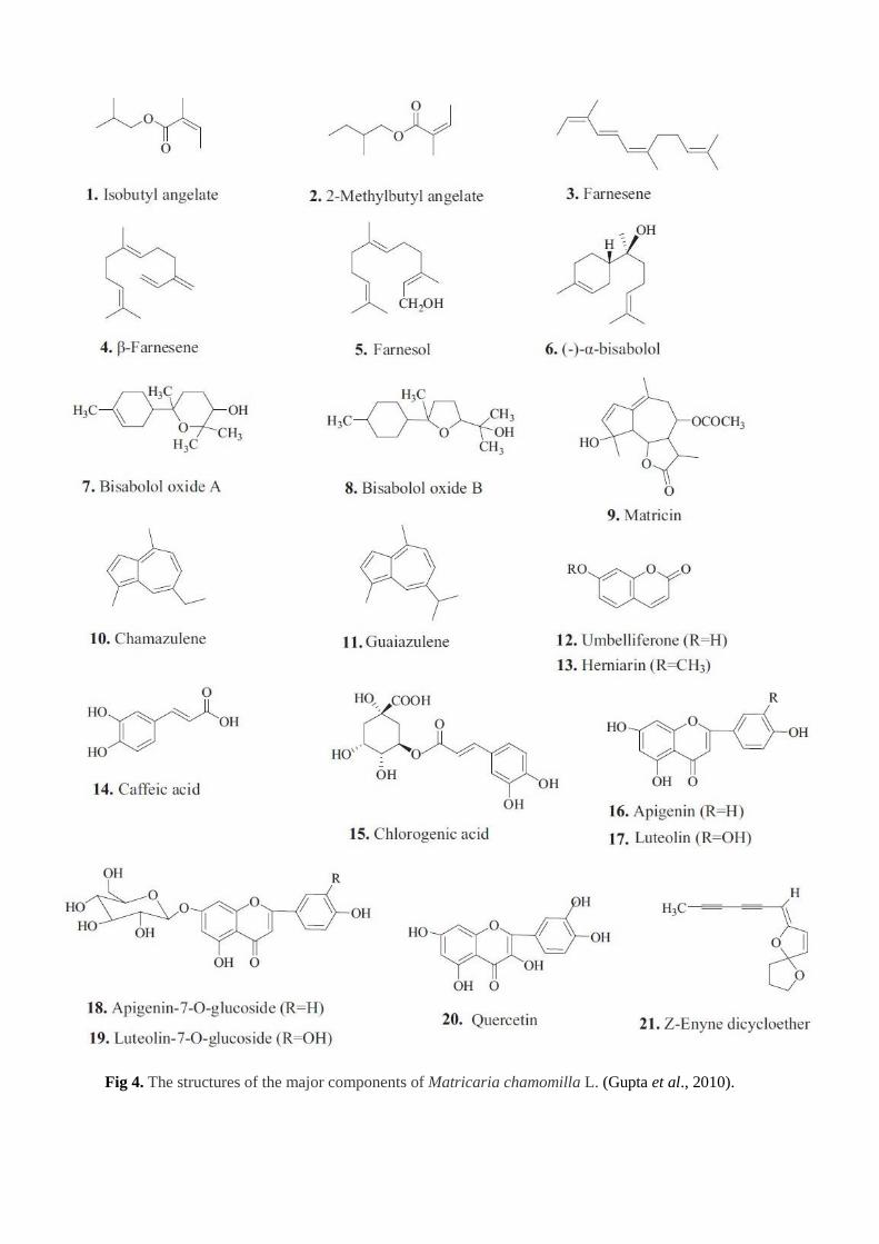
Fig 4. The structures of the major components of Matricaria chamomilla L. (Gupta et al., 2010).
Chapter 2: MATERIALS AND
METHODS
I. MATERIAL:
I.1. Plant material:
Mentha pulegium L. leaves were collected in September,2011 from the capital of Algeria and
the flowers of Matricaria chamomilla L. were collected in the end of May and the beguining
of June from Res-El-Oued. BBA. The two plants were identified by professor Pr. Laouer
Hocine from the Faculty of Sciences. Department of Ecology. University Ferhat Abbass,
Setif, Algeria. The leaves and the flowers were separated from the other parts and dried at
room temperature.
The plant samples were air dried in shadow and finely powdered in a rotating knife grinder.
The powder was sieved through a 1mm mesh to remove large fragments. Each plant powder
was then used for the extraction procedure.
I.2. Chemicals:
Methanol (MeOH) , Hexan , Ethyl acetate , Chloroform , Tannic acid, aluminium trichloride
(AlCl3) , the various polyphenols (Gallic acid, Quercetin and Rutin) ; Folin ; 2,2-diphenyl-1-
picryl-hydrazyl (DPPH), Butylated hydroxytoluene (BHT) , Tween 40 , β carotene , Linoleic
acid and Carbonate.
The various products used were purchased from Aldrich and Sigma.
II. METHODS:
II.1. Choice of solvent:
The extraction of polyphenols from Mentha pulegium L. leaves and Matricaria
chamomilla L. flowers were performed by methanol-distilled water. The
extraction yields was calculated for each sample extracted with solvent system as
the percentage of weight of resulting powder to the weight of extracted material.
Total polyphenols in all extracts were detemnined.
II.1.1. Polyphenols extraction procedures :
100g of Matricaria chamomilla L. flowers or the leaves of Mentha pulegium L. are macerated
to the 1 liter methanol/ water (85% methanol), the mixture is subjected to an agitation (700
tours/minute) during three days (72h) at ambient temperature. The whole is filtered thereafter
on funnel (N03) by the cotton and the filter paper of Wattman, the solvent (methanol) is
eliminated from the filtrate by rotary evaporation in Rotavapor (BÜCHI). The extraction is
remade for the second time (maceration with 50% methanol, followed by an agitation during
4heurs). The second filtrate is mixed with the first. The extract obtained is of a sunk brown
color, it is regarded as being the rough extract of the leaves or the flowers of the two plants.
50 ml of this extract are dried using a drying oven in order to determine their dry matter value
as well as the output of extraction. Volume remaining will be split later on.
Fractionation is carried out according to the method of (Gilani et al, 2001), after
modifications, by using solvents with increasing polarity. The rough extract is initially mixed
with the hexan (1 V/V), the mixture is let elutriate, and the higher organic phase is recovered.
The extraction is remade several times until the solvent (hexan) becomes transparent. The
hexane is evaporated thereafter and the resulting extract is regarded as being the fraction of
hexan. The residual aqueous phase is subjected to another extraction by chloroform, and
finally by the ethyl acetate while following the same stages as the first extraction by hexan.
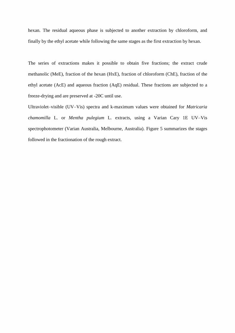
The series of extractions makes it possible to obtain five fractions; the extract crude
methanolic (MeE), fraction of the hexan (HxE), fraction of chloroform (ChE), fraction of the
ethyl acetate (AcE) and aqueous fraction (AqE) residual. These fractions are subjected to a
freeze-drying and are preserved at -20C until use.
Ultraviolet–visible (UV–Vis) spectra and k-maximum values were obtained for Matricaria
chamomilla L. or Mentha pulegium L. extracts, using a Varian Cary 1E UV–Vis
spectrophotometer (Varian Australia, Melbourne, Australia). Figure 5 summarizes the stages
followed in the fractionation of the rough extract.
methanolic extract
Aqueous extract Chloroformic extract Ethyl acetate extract
crushing -Extaraction with methanol 85%
-Conservation at 4C/72h - Filtration
-Extraction (methanol 50%) -Filtration
-Filtration (filter paper) -Evaporation
-Washing (Hexan)
Extraction (chloroform)
Extraction(ethyl acetate)
- freeze-drying
Fig 5. Schematic diagram represents the process of extraction.
jd Plant material (g)
broyat
filtrate sediment
filtrate sediment
Combined filtrate
organic phase= waxes, lipids,
chlorophyl
Sediment Filtrate
aqueous phase= methanolic extract
organic phase aqueous phase
evaporation
aqueous phase
organic phase
II.2.2. Dosage of the metabolites in plants extracts:
II.2.2.1 Determination of total polyphenols:
In order to measure phenolic compounds in plant extracts (the water, chloroform, methanolic
and ethyl acetate extracs) at different occasions, we used the Folin–Ciocalteu assay. The
reagent of Folin–Ciocalteu consists of a mixture of acid phosphotungstic and
phosphomolybdic acid. During oxidation, it is reduced to a mixture of blue oxide. The color
produced is proportional to the amount of polyphenols present in the extract analyzed
(kassemi, 2006).
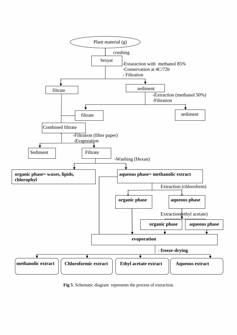
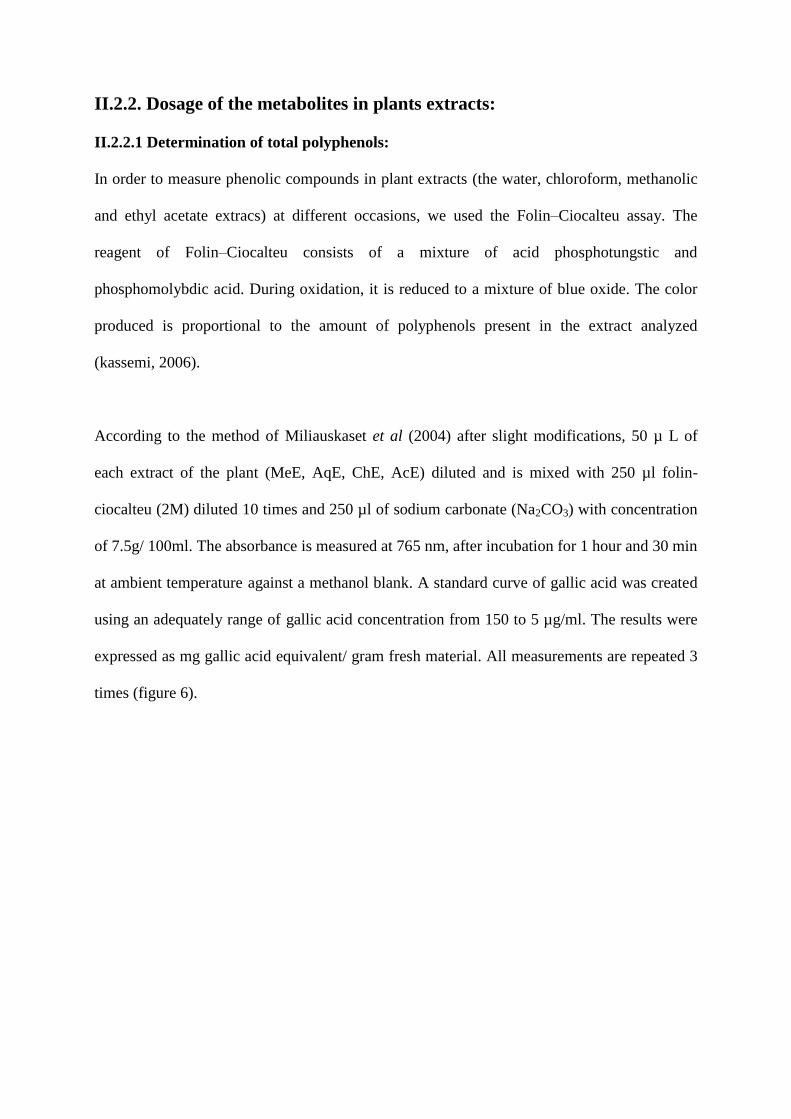
According to the method of Miliauskaset et al (2004) after slight modifications, 50 µ L of
each extract of the plant (MeE, AqE, ChE, AcE) diluted and is mixed with 250 µl folin-
ciocalteu (2M) diluted 10 times and 250 µl of sodium carbonate (Na2CO3) with concentration
of 7.5g/ 100ml. The absorbance is measured at 765 nm, after incubation for 1 hour and 30 min
at ambient temperature against a methanol blank. A standard curve of gallic acid was created
using an adequately range of gallic acid concentration from 150 to 5 µg/ml. The results were
expressed as mg gallic acid equivalent/ gram fresh material. All measurements are repeated 3
times (figure 6).
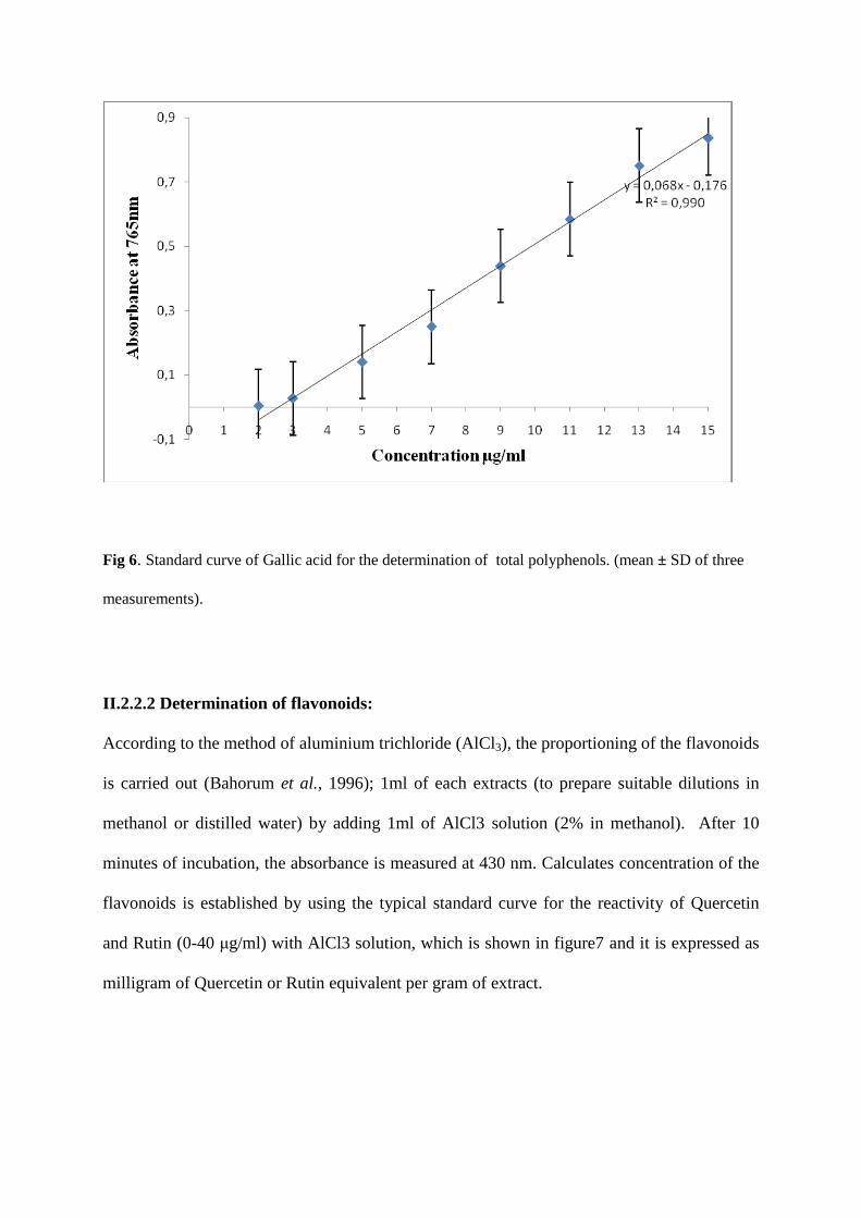
Fig 6. Standard curve of Gallic acid for the determination of total polyphenols. (mean ± SD of three
measurements).
II.2.2.2 Determination of flavonoids:
According to the method of aluminium trichloride (AlCl3), the proportioning of the flavonoids
is carried out (Bahorum et al., 1996); 1ml of each extracts (to prepare suitable dilutions in
methanol or distilled water) by adding 1ml of AlCl3 solution (2% in methanol). After 10
minutes of incubation, the absorbance is measured at 430 nm. Calculates concentration of the
flavonoids is established by using the typical standard curve for the reactivity of Quercetin
and Rutin (0-40 μg/ml) with AlCl3 solution, which is shown in figure7 and it is expressed as
milligram of Quercetin or Rutin equivalent per gram of extract.
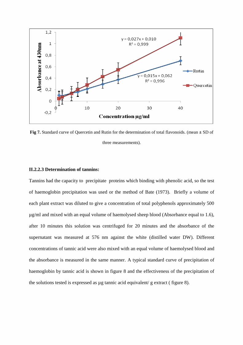
Fig 7. Standard curve of Quercetin and Rutin for the determination of total flavonoids. (mean ± SD of
three measurements).
II.2.2.3 Determination of tannins:
Tannins had the capacity to precipitate proteins which binding with phenolic acid, so the test
of haemoglobin precipitation was used or the method of Bate (1973). Briefly a volume of
each plant extract was diluted to give a concentration of total polyphenols approximately 500
µg/ml and mixed with an equal volume of haemolysed sheep blood (Absorbance equal to 1.6),
after 10 minutes this solution was centrifuged for 20 minutes and the absorbance of the
supernatant was measured at 576 nm against the white (distilled water DW). Different
concentrations of tannic acid were also mixed with an equal volume of haemolysed blood and
the absorbance is measured in the same manner. A typical standard curve of precipitation of
haemoglobin by tannic acid is shown in figure 8 and the effectiveness of the precipitation of
the solutions tested is expressed as µg tannic acid equivalent/ g extract ( figure 8).
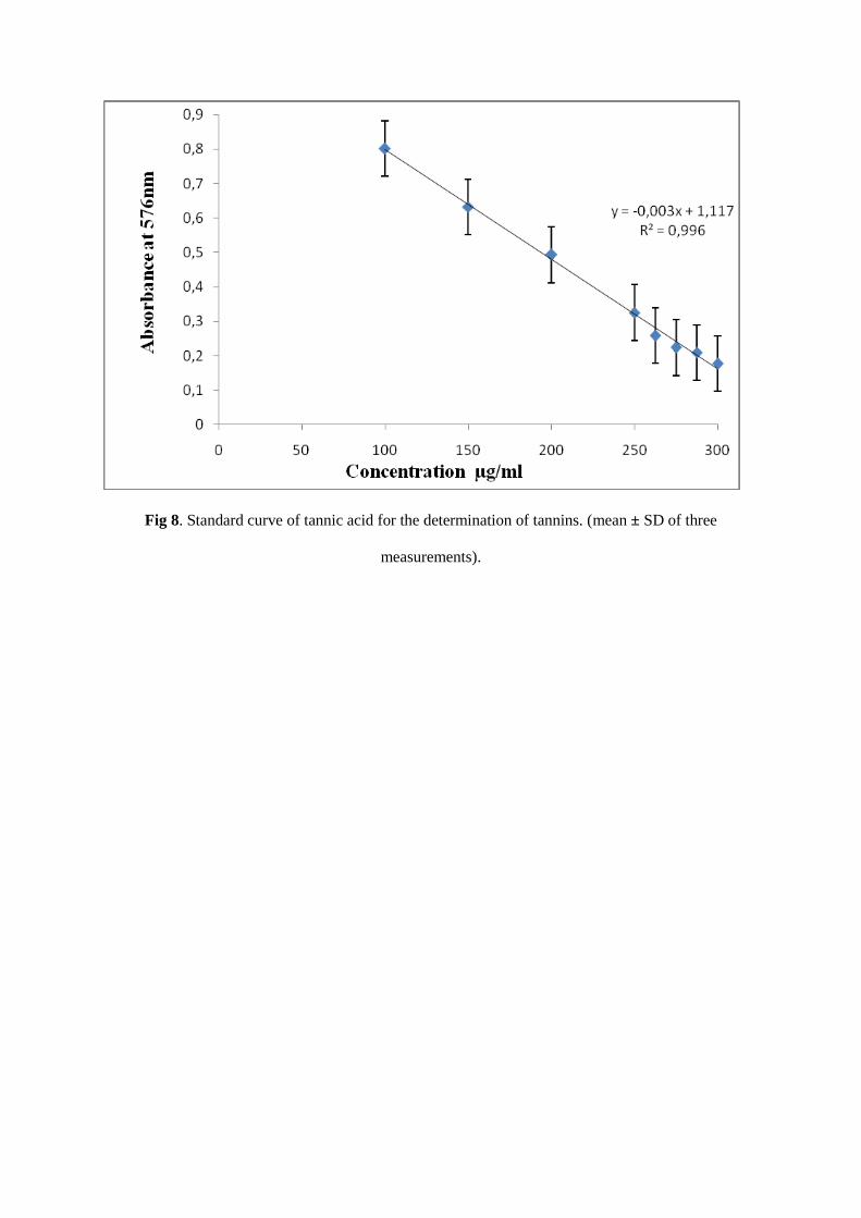
Fig 8. Standard curve of tannic acid for the determination of tannins. (mean ± SD of three
measurements).
II.2.3. Determination of the antioxidant activity of plant extracts:
There is an increasing interest in antioxidants, particularly in those intended to prevent the
presumed deleterious effects of free radicals in the human body, and to prevent the distruction
of fats and other constituents of foodstuffs. In both cases, there is a preference for
antioxidants from natural rather than from synthetic sources (Abdalla and Roozen, 1999).
There is therefore a parallel increase in the use of methods for estimating the efficiency of
such substances as antioxidants (Sa′ nchez-Moreno, 2002; Schwarz et al., 2001).
The antioxydant activity is a complex process which can occur by the means of several
mechanisms. Because of its complexity more than one test must be carried out during the
evaluation of the antioxydant activity of the pure or extracted compounds (Aruoma, 2003).
One such method that is currently popular is based upon the use of the stable free radical
diphenylpicrylhydrazyl (DPPH).
II.2.3.1 DPPH radical scavenging activity of plant extract:
The antioxidant capacity of our extracts which is expressed by the donation of an electron or a
hydrogen atom to radical free 2,2'-diphenyl-1-picrylhydrazyl (DPPH), as a reagent, was
measured by a spectrophotometric (Burits et al, 2000).
The experiment was carried out according to the method described by (Güllüce et al., 2003).
50μl of various concentrations of the extracts is added to 5ml solution of the DPPH of
concentration 0,004%. After 30 minutes of incubation at ambient temperature and in the
darkness, the absorbance is read with a wavelength at 517nm.
Negative control is represented by the methanolic solution of the DPPH and the positive
control is represented by the BHT.
The antioxidant activity, which expresses the capacities to trap the free radical one is
estimated by the percentage of discolouration of the DPPH in solution in methanol (Inhibition
% or I%) according to the formula:
Inhibition % = (ABS control - ABS test) х 100 / ABS control.
Where:
ABS control: Absorbance of control at the wavelength 517nm;
ABS test: Absorbance of the sample at the wavelength 517nm.
The Value IC50 is defined as being the concentration of the substrate which causes the loss of
50% of the activity of the DPPH (color), or, it is the concentration of the sample required to
give a reduction of 50% of the absorbance of the solution controls to constitute methanol and
DPPH. The values of IC50 were calculated by the linear regression where the X-coordinate is
represented by the concentration of the compounds tested and ordered by (I %) the percentage
of inhibition (Mensor et al., 2001).
The concentrations of the extracts in the reactional medium lie between 0,1-1 mg/ml, 3-12
mg/ml, 3-0,03 mg/ml and 0,1-4 mg/ml for AqE, ChE, AcE, and MeE respectively for
Matricaria chamomilla L. while Mentha pulegium L. were betweenn 10-0,25 mg/ml, 30-0,5
mg/ml, 5-0,025 mg/ml for AqE, ChE and (AcE, MeE) respectively.
II.2.3.1. β-carotene/ linoleic acid assay:
In this test, the antioxydant capacity of the extracts is given by measuring the inhibition of the
decomposition oxydative of β-carotene (discolouration) by the products of oxidation of the
linoleic acid according to the method described by (Kartal et al, 2007). The emulsion of β-
carotene/ linoleic acid is prepared by solubilization of β-carotene 0,5mg in 1ml of chloroform,
25µl of the linoleic acid and 200mg of Tween 40 are added, chloroform is completely
evaporated with the rotavapor, thereafter 100ml of distilled water saturated with oxygen are
added, the resulting emulsion is agitated vigorously. 350µl of solution of extracts or
antioxydants of reference (BHT) solubilized in methanol (2mg/ml) is added with 2,5ml with
the preceding emulsion.
The kinetics of discolouration of the emulsion in presence and absence of antioxidant
(negative control in which the sample is replaced by 350µl methanol and distilled water) is
followed to 490 nm with intervals of regular times during 48 heurs (after : 1heure, 2h, 3h, 4h,
6h, 24h, and 48h) of incubation at ambient temperature and in the darkness.
The percentage of inhibition of the extracts antioxidant is measured as follows:
AA% = ABS test / ABS BHT ×100
AA%: Percentage of the antioxidant activity;
ABS test: Absorbance in the presence of the extract (test);
ABS BHT: Absorbance in the presence of positive control BHT.
Chapter 3: RESULTS AND
DISCUSSION
III.1. Preparation of extracts from plants:
The different extract of Matricaria chamomilla L. and Mentha pulegium L. were obtained
following the extraction method described by (Markham, 1982). This method is based on the
degree of solubility of polyphenols in organic solvents. It takes place in four stages:
1)- solubilization of polyphenols in methanol.
2)- defatting of the extract by adding hexan.
3)- the addition of chloroform to obtain the flavonoid aglycons.
4)- the addition of ethyl acetate to obtain the glucoflavonoids.
This method allowed us to obtain five different fractions yield an extract variable to another
(tables 1, 2). The yield was calculated on the total weight of the crushed flowers or leaves of
Matricaria chamomilla L. and Mentha pulegium L. respectively.
III.1.1. Extracts from Mentha pulegium L.:
In the leaves of Mentha pulegium L, the ME had the higher yield (14,4%) and for other
extracts were lower than these values in this order: AqE (13,872%), AcE (4,324%), ChE
(1,012%) and HxE (0,46%) as shown in table 1.
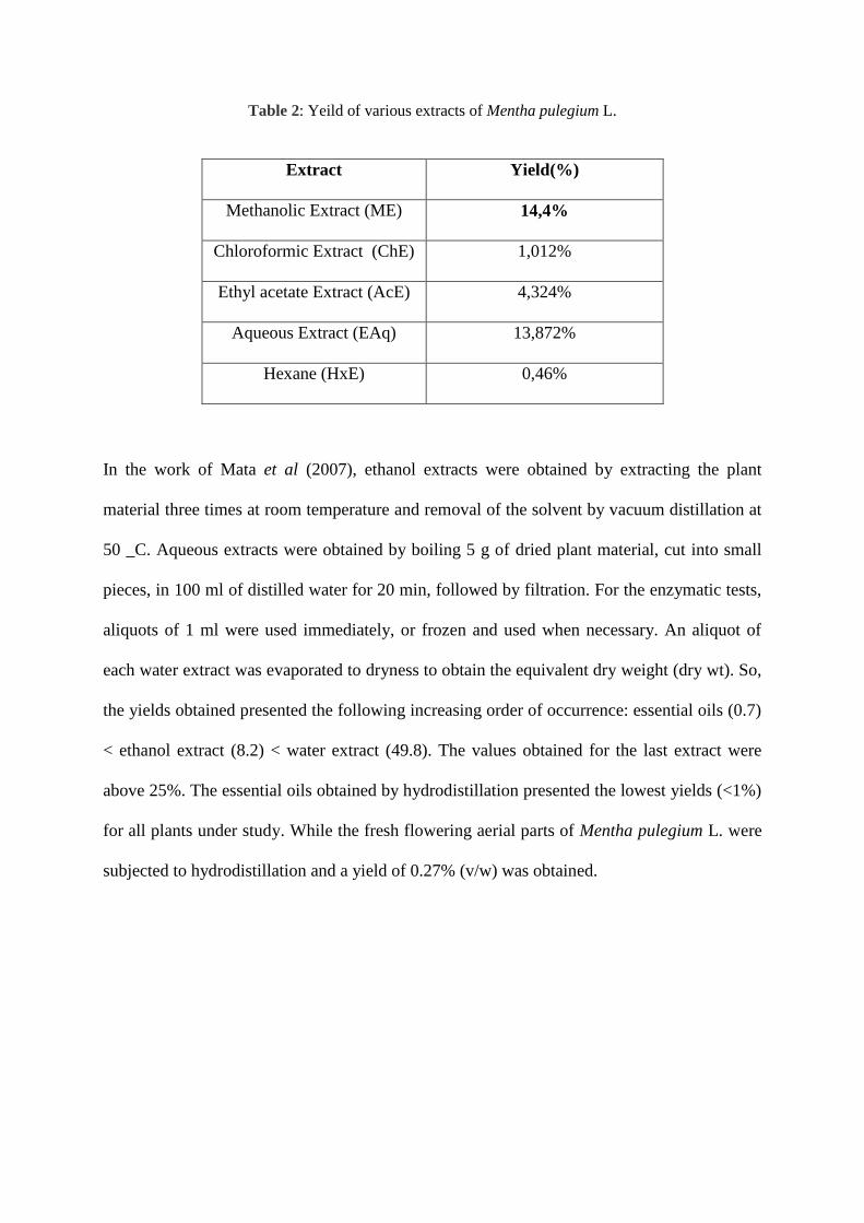
Table 2: Yeild of various extracts of Mentha pulegium L.
In the work of Mata et al (2007), ethanol extracts were obtained by extracting the plant
material three times at room temperature and removal of the solvent by vacuum distillation at
50 _C. Aqueous extracts were obtained by boiling 5 g of dried plant material, cut into small
pieces, in 100 ml of distilled water for 20 min, followed by filtration. For the enzymatic tests,
aliquots of 1 ml were used immediately, or frozen and used when necessary. An aliquot of
each water extract was evaporated to dryness to obtain the equivalent dry weight (dry wt). So,
the yields obtained presented the following increasing order of occurrence: essential oils (0.7)
< ethanol extract (8.2) < water extract (49.8). The values obtained for the last extract were
above 25%. The essential oils obtained by hydrodistillation presented the lowest yields (<1%)
for all plants under study. While the fresh flowering aerial parts of Mentha pulegium L. were
subjected to hydrodistillation and a yield of 0.27% (v/w) was obtained.
Extract Yield(%)
Methanolic Extract (ME) 14,4%
Chloroformic Extract (ChE) 1,012%
Ethyl acetate Extract (AcE) 4,324%
Aqueous Extract (EAq) 13,872%
Hexane (HxE) 0,46%
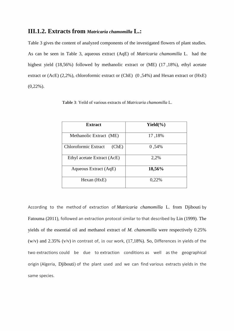
III.1.2. Extracts from Matricaria chamomilla L.:
Table 3 gives the content of analyzed components of the investigated flowers of plant studies.
As can be seen in Table 3, aqueous extract (AqE) of Matricaria chamomilla L. had the
highest yield (18,56%) followed by methanolic extract or (ME) (17 ,18%), ethyl acetate
extract or (AcE) (2,2%), chloroformic extract or (ChE) (0 ,54%) and Hexan extract or (HxE)
(0,22%).
Table 3: Yeild of various extracts of Matricaria chamomilla L.
According to the method of extraction of Matricaria chamomilla L. from Djibouti by
Fatouma (2011), followed an extraction protocol similar to that described by Lin (1999). The
yields of the essential oil and methanol extract of M. chamomilla were respectively 0.25%
(w/v) and 2.35% (v/v) in contrast of, in our work, (17,18%). So, Differences in yields of the
two extractions could be due to extraction conditions as well as the geographical
origin (Algeria, Djibouti) of the plant used and we can find various extracts yields in the
same species.
Extract Yield(%)
Methanolic Extract (ME) 17 ,18%
Chloroformic Extract (ChE) 0 ,54%
Ethyl acetate Extract (AcE) 2,2%
Aqueous Extract (AqE) 18,56%
Hexan (HxE) 0,22%
III.2. Determination of total polyphenols, flavonoids and tannins
in plants extracts:
III.2.1. Determination of total polyphenols, flavonoids and tannins
in Mentha pulegium L. extracts:
Based on the absorbance value of the plant extracts solution reacting with Folin-Ciocalteu
phenol reagent and compaired with the absorbance values of standard solutions of gallic acid,
total phenolics content of the plant extracts was estimated in this order: AcE (191,99±0,016
µg GAE/g of extract)> ME (183,45±0,125 µg GAE/g of extract)> ChE (119,73±0,036 µg
GAE/g of extract)> AqE (88,84±0,112 µg GAE/g of extract). This values indicate that each
milligram of the plant extracts contains phenolic compounds equivalent to about 191,99;
183,45; 119,73; 88,84 µg of pue gallic acid respectively.
As can be seen in the table 4, AcE had the higher contents of tannins (265,33±0,030 µg
TAE/gE ) followed by ChE (209±0,017 µg TAE/gE) then ME (149,33±0,0046 µgTAE/gE)
and the AqE (137,22±0,029 µg TAE/gE) with lower content.
In the AlCl3 method, the flavonoid results measured either by: the Quercetin equivalent
which as the following order: AcE(110,37±0,023 µg QE/gE)> ME (59,87±0,005 µg QE/gE)>
ChE (19,50±0,013 µg QE/gE) > AqE (1,19±0,004 µg QE/gE) or the Rutin equivalent which
revealed that the extracts had: AcE (151,11±0,023 µg RE/gE), ME (60,92±0,005 µg RE/gE),
AqE (1,41±0,004 µg RE/gE) and ChE (0,37±0,013 µg RE/gE).
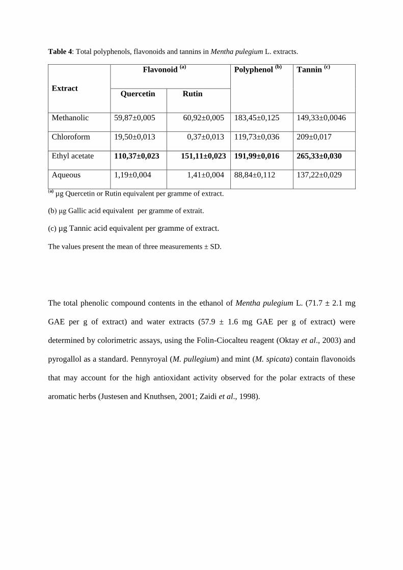
Table 4: Total polyphenols, flavonoids and tannins in Mentha pulegium L. extracts.
Extract
Flavonoid (a)
Polyphenol (b)
Tannin (c)
Quercetin Rutin
Methanolic 59,87±0,005 60,92±0,005 183,45±0,125 149,33±0,0046
Chloroform 19,50±0,013 0,37±0,013 119,73±0,036 209±0,017
Ethyl acetate 110,37±0,023 151,11±0,023 191,99±0,016 265,33±0,030
Aqueous 1,19±0,004 1,41±0,004 88,84±0,112 137,22±0,029
(a) µg Quercetin or Rutin equivalent per gramme of extract.
(b) μg Gallic acid equivalent per gramme of extrait.
(c) µg Tannic acid equivalent per gramme of extract.
The values present the mean of three measurements ± SD.
The total phenolic compound contents in the ethanol of Mentha pulegium L. (71.7 ± 2.1 mg
GAE per g of extract) and water extracts (57.9 ± 1.6 mg GAE per g of extract) were
determined by colorimetric assays, using the Folin-Ciocalteu reagent (Oktay et al., 2003) and
pyrogallol as a standard. Pennyroyal (M. pullegium) and mint (M. spicata) contain flavonoids
that may account for the high antioxidant activity observed for the polar extracts of these
aromatic herbs (Justesen and Knuthsen, 2001; Zaidi et al., 1998).
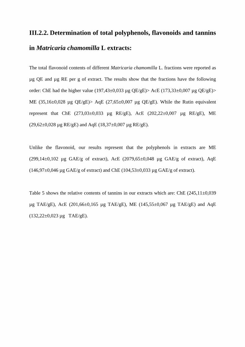
III.2.2. Determination of total polyphenols, flavonoids and tannins
in Matricaria chamomilla L extracts:
The total flavonoid contents of different Matricaria chamomilla L. fractions were reported as
µg QE and µg RE per g of extract. The results show that the fractions have the following
order: ChE had the higher value (197,43±0,033 µg QE/gE)> AcE (173,33±0,007 µg QE/gE)>
ME (35,16±0,028 µg QE/gE)> AqE (27,65±0,007 µg QE/gE). While the Rutin equivalent
represent that ChE (273,03±0,033 µg RE/gE), AcE (202,22±0,007 µg RE/gE), ME
(29,62±0,028 µg RE/gE) and AqE (18,37±0,007 µg RE/gE).
Unlike the flavonoid, our results represent that the polyphenols in extracts are ME
(299,14±0,102 µg GAE/g of extract), AcE (2079,65±0,048 µg GAE/g of extract), AqE
(146,97±0,046 µg GAE/g of extract) and ChE (104,53±0,033 µg GAE/g of extract).
Table 5 shows the relative contents of tannins in our extracts which are: ChE (245,11±0,039
µg TAE/gE), AcE (201,66±0,165 µg TAE/gE), ME (145,55±0,067 µg TAE/gE) and AqE
(132,22±0,023 µg TAE/gE).
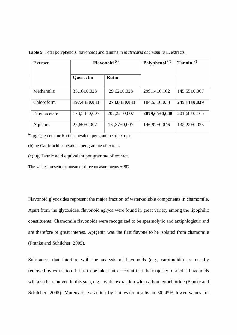
Table 5: Total polyphenols, flavonoids and tannins in Matricaria chamomilla L. extracts.
Extract Flavonoid (a)
Polyphenol (b)
Tannin (c)
Quercetin Rutin
Methanolic 35,16±0,028 29,62±0,028 299,14±0,102 145,55±0,067
Chloroform 197,43±0,033 273,03±0,033 104,53±0,033 245,11±0,039
Ethyl acetate 173,33±0,007 202,22±0,007 2079,65±0,048 201,66±0,165
Aqueous 27,65±0,007 18 18 ,37±0,007 146,97±0,046 132,22±0,023
(a) µg Quercetin or Rutin equivalent per gramme of extract.
(b) μg Gallic acid equivalent per gramme of extrait.
(c) µg Tannic acid equivalent per gramme of extract.
The values present the mean of three measurements ± SD.
Flavonoid glycosides represent the major fraction of water-soluble components in chamomile.
Apart from the glycosides, flavonoid aglyca were found in great variety among the lipophilic
constituents. Chamomile flavonoids were recognized to be spasmolytic and antiphlogistic and
are therefore of great interest. Apigenin was the first flavone to be isolated from chamomile
(Franke and Schilcher, 2005).
Substances that interfere with the analysis of flavonoids (e.g., carotinoids) are usually
removed by extraction. It has to be taken into account that the majority of apolar flavonoids
will also be removed in this step, e.g., by the extraction with carbon tetrachloride (Franke and
Schilcher, 2005). Moreover, extraction by hot water results in 30–45% lower values for
flavonoids compared to methanol extraction. It is therefore not possible to avoid the co-
extraction of chlorophyll and related substances by using water.
The photometric determination of flavonoids has many advantages, although the absolute
values are actually about 20–30% higher. The absolute content of flavonoids ranged between
1.0 and 2.5% in a study of 102 commercially available plants and determination according to
References (Franke and Schilcher, 2005) and (Christ and Müller, 1960). Twelve samples of
material of different origin cultivated by Schilcher showed values between 0.3 and 2.96%
(Schilcher, 1987).
III.3. Antioxidant activity:
III.3.1. Test of DPPH:
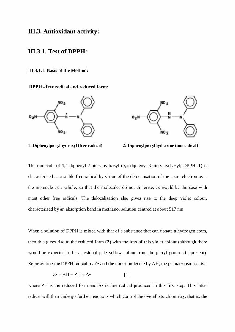
III.3.1.1. Basis of the Method:
DPPH - free radical and reduced form:
1: Diphenylpicrylhydrazyl (free radical) 2: Diphenylpicrylhydrazine (nonradical)
The molecule of 1,1-diphenyl-2-picrylhydrazyl (α,α-diphenyl-β-picrylhydrazyl; DPPH: 1) is
characterised as a stable free radical by virtue of the delocalisation of the spare electron over
the molecule as a whole, so that the molecules do not dimerise, as would be the case with
most other free radicals. The delocalisation also gives rise to the deep violet colour,
characterised by an absorption band in methanol solution centred at about 517 nm.
When a solution of DPPH is mixed with that of a substance that can donate a hydrogen atom,
then this gives rise to the reduced form (2) with the loss of this violet colour (although there
would be expected to be a residual pale yellow colour from the picryl group still present).
Representing the DPPH radical by Z• and the donor molecule by AH, the primary reaction is:
Z• + AH = ZH + A• [1]
where ZH is the reduced form and A• is free radical produced in this first step. This latter
radical will then undergo further reactions which control the overall stoichiometry, that is, the
number of molecules of DPPH reduced (decolorised) by one molecule of the reductant. The
reaction [1] is therefore intended to provide the link with the reactions taking place in an
oxidising system, such as the autoxidation of a lipid or other unsaturated substance; the DPPH
molecule Z• is thus intended to represent the free radicals formed in the system whose activity
is to be suppressed by the substance AH.
The parameter EC50 (“efficient concentration” value)
One parameter that has been introduced recently for the interpretation of the results from the
DPPH method, is the “efficient concentration” or EC50 value (otherwise called the IC50
value). This is defined as the concentration of substrate that causes 50% loss of the DPPH
activity (colour).
III.3.1.2. DPPH scavenging of extracts of Mentha pulegium L:
The antioxidant activity profiles obtained show that extracts of plants had a dose-dependent
antioxidant activity, the IC50 of each of the different extracts were determined (table6).
The DPPH free radical method determined the antiradical power of antioxidants. Regarding
the IC50 values, all the extracts and the commercial standards (BHT, Gallic acid, Quercetin,
Rutin) depleted the initial DPPH concentration by 50% within 1h. The lower of IC50 value is
the higher of free radical scavenging activity of a sample. The free radical scavenging
activities of all extracts of Mentha pulegium L. were in this order: ethyl acetate > methanolic
> chloroform > aqueous (Table 6). The ethyl acetate extract, which contained the most tannin,
had the highest free radical scavenging activity. All of the extracts had higher IC50 values
compared to Gallic acid, Quercetin, Rutin and BHT. When we compared to BHT, the ethyl
acetate extract and methanolic extract, Rutin, GA, Quercetin did not show any significant
differences (P>0,005). While the chloroformic extract and aqueous extract were significant
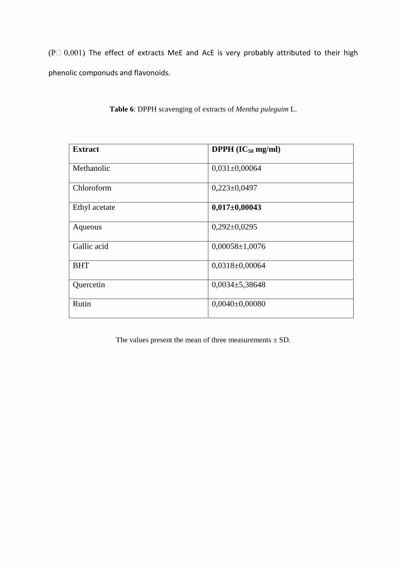
(P˂0,001). The effect of extracts MeE and AcE is very probably attributed to their high
phenolic componuds and flavonoids.
Table 6: DPPH scavenging of extracts of Mentha puleguim L.
Extract DPPH (IC50 mg/ml)
Methanolic 0,031±0,00064
Chloroform 0,223±0,0497
Ethyl acetate 0,017±0,00043
Aqueous 0,292±0,0295
Gallic acid 0,00058±1,0076
BHT 0,0318±0,00064
Quercetin 0,0034±5,38648
Rutin 0,0040±0,00080
The values present the mean of three measurements ± SD.
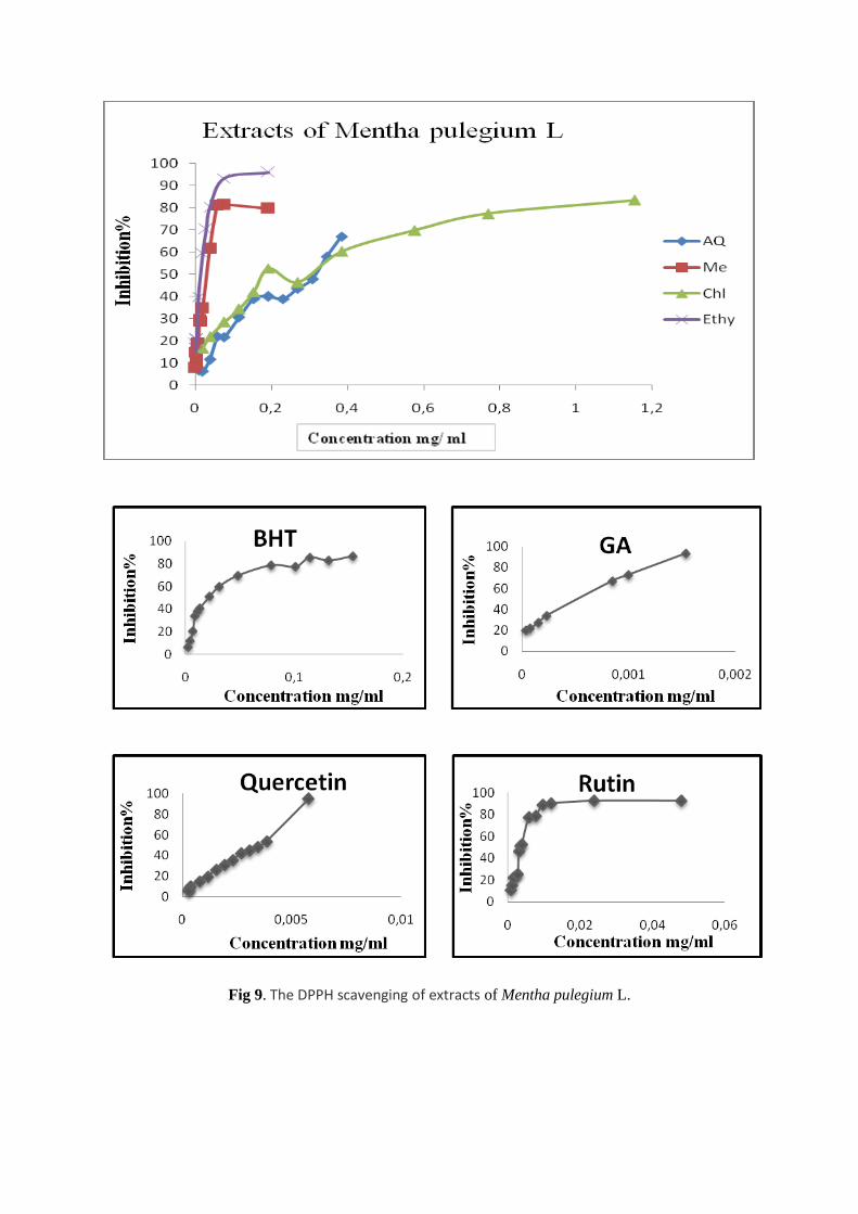
Fig 9. The DPPH scavenging of extracts of Mentha pulegium L.
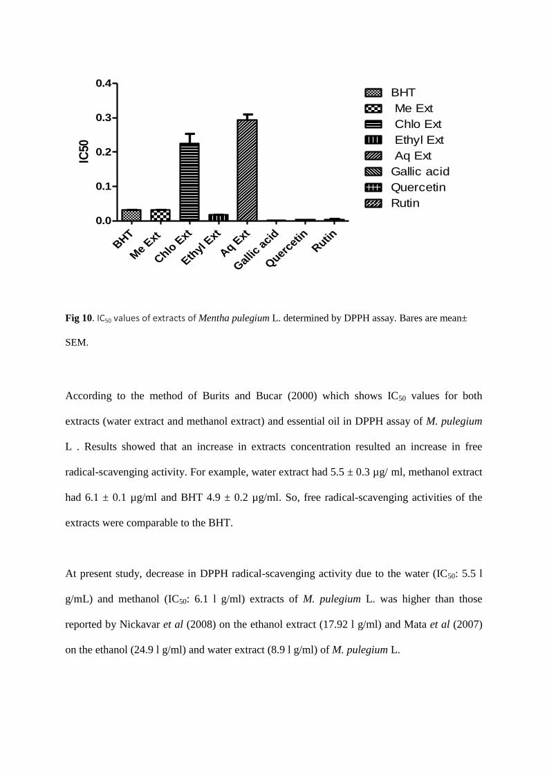
BHT
Me E
xt
Chlo
Ext
Eth
yl Ext
Aq E
xt
Gal
lic a
cid
Quer
cetin
Rutin
0.0
0.1
0.2
0.3
0.4BHT
Me Ext
Chlo Ext
Ethyl Ext
Aq Ext
Gallic acid
Quercetin
Rutin
IC50
Fig 10. IC50 values of extracts of Mentha pulegium L. determined by DPPH assay. Bares are mean±
SEM.
According to the method of Burits and Bucar (2000) which shows IC50 values for both
extracts (water extract and methanol extract) and essential oil in DPPH assay of M. pulegium
L . Results showed that an increase in extracts concentration resulted an increase in free
radical-scavenging activity. For example, water extract had 5.5 ± 0.3 µg/ ml, methanol extract
had 6.1 ± 0.1 µg/ml and BHT 4.9 ± 0.2 µg/ml. So, free radical-scavenging activities of the
extracts were comparable to the BHT.
At present study, decrease in DPPH radical-scavenging activity due to the water (IC50: 5.5 l
g/mL) and methanol (IC50: 6.1 l g/ml) extracts of M. pulegium L. was higher than those
reported by Nickavar et al (2008) on the ethanol extract (17.92 l g/ml) and Mata et al (2007)
on the ethanol (24.9 l g/ml) and water extract (8.9 l g/ml) of M. pulegium L.
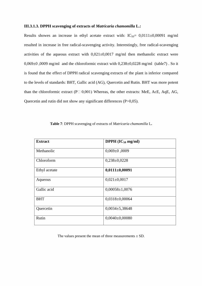
III.3.1.3. DPPH scavenging of extracts of Matricaria chamomilla L.:
Results showes an increase in ethyl acetate extract with: IC50= 0,0111±0,00091 mg/ml
resulted in increase in free radical-scavenging activity. Interestingly, free radical-scavenging
activities of the aqueous extract with 0,021±0,0017 mg/ml then methanolic extract were
0,069±0 ,0009 mg/ml and the chloroformic extract with 0,238±0,0228 mg/ml (table7) . So it
is found that the effect of DPPH radical scavenging extracts of the plant is inferior compared
to the levels of standards: BHT, Gallic acid (AG), Quercetin and Rutin. BHT was more potent
than the chloroformic extract (P˂0,001). Whereas, the other extracts: MeE, AcE, AqE, AG,
Quercetin and rutin did not show any significant differences (P>0,05).
Table 7: DPPH scavenging of extracts of Matricaria chamomilla L.
Extract DPPH (IC50 mg/ml)
Methanolic 0,069±0 ,0009
Chloroform 0,238±0,0228
Ethyl acetate 0,0111±0,00091
Aqueous 0,021±0,0017
Gallic acid 0,00058±1,0076
BHT 0,0318±0,00064
Quercetin 0,0034±5,38648
Rutin 0,0040±0,00080
The values present the mean of three measurements ± SD.
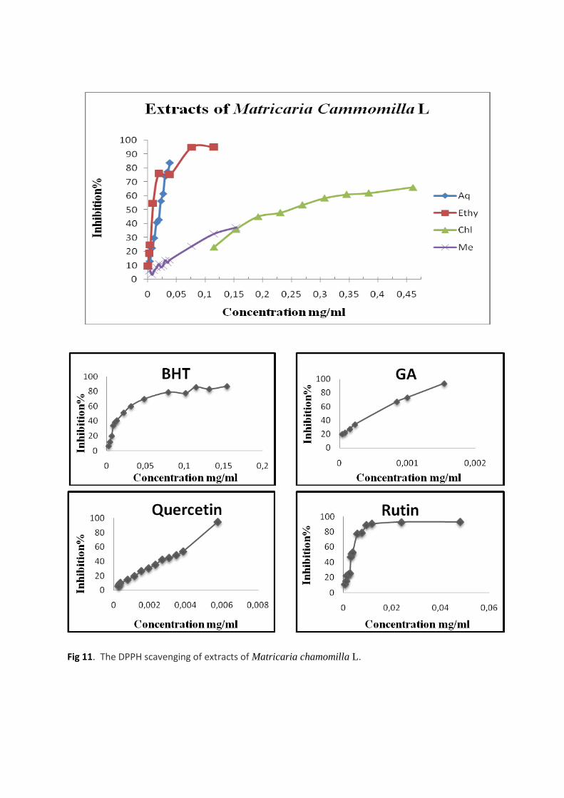
Fig 11. The DPPH scavenging of extracts of Matricaria chamomilla L.
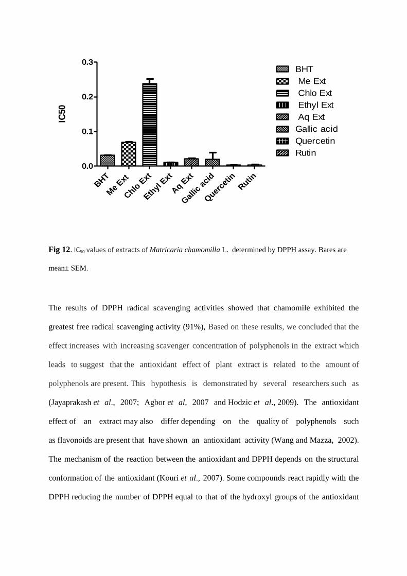
BHT
Me
Ext
Chlo
Ext
Eth
yl E
xt
Aq E
xt
Gal
lic a
cid
Quer
cetin
Rutin
0.0
0.1
0.2
0.3BHT
Me Ext
Chlo Ext
Ethyl Ext
Aq Ext
Gallic acid
Quercetin
Rutin
IC50
Fig 12. IC50 values of extracts of Matricaria chamomilla L. determined by DPPH assay. Bares are
mean± SEM.
The results of DPPH radical scavenging activities showed that chamomile exhibited the
greatest free radical scavenging activity (91%), Based on these results, we concluded that the
effect increases with increasing scavenger concentration of polyphenols in the extract which
leads to suggest that the antioxidant effect of plant extract is related to the amount of
polyphenols are present. This hypothesis is demonstrated by several researchers such as
(Jayaprakash et al., 2007; Agbor et al, 2007 and Hodzic et al., 2009). The antioxidant
effect of an extract may also differ depending on the quality of polyphenols such
as flavonoids are present that have shown an antioxidant activity (Wang and Mazza, 2002).
The mechanism of the reaction between the antioxidant and DPPH depends on the structural
conformation of the antioxidant (Kouri et al., 2007). Some compounds react rapidly with the
DPPH reducing the number of DPPH equal to that of the hydroxyl groups of the antioxidant
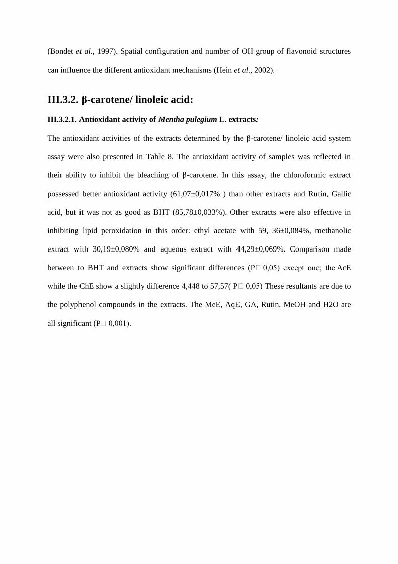
(Bondet et al., 1997). Spatial configuration and number of OH group of flavonoid structures
can influence the different antioxidant mechanisms (Hein et al., 2002).
III.3.2. β-carotene/ linoleic acid:
III.3.2.1. Antioxidant activity of Mentha pulegium L. extracts:
The antioxidant activities of the extracts determined by the β-carotene/ linoleic acid system
assay were also presented in Table 8. The antioxidant activity of samples was reflected in
their ability to inhibit the bleaching of β-carotene. In this assay, the chloroformic extract
possessed better antioxidant activity (61,07±0,017% ) than other extracts and Rutin, Gallic
acid, but it was not as good as BHT (85,78±0,033%). Other extracts were also effective in
inhibiting lipid peroxidation in this order: ethyl acetate with 59, 36±0,084%, methanolic
extract with 30,19±0,080% and aqueous extract with 44,29±0,069%. Comparison made
between to BHT and extracts show significant differences (P˂0,05) except one; the AcE
while the ChE show a slightly difference 4,448 to 57,57( P˂0,05). These resultants are due to
the polyphenol compounds in the extracts. The MeE, AqE, GA, Rutin, MeOH and H2O are
all significant (P˂0,001).
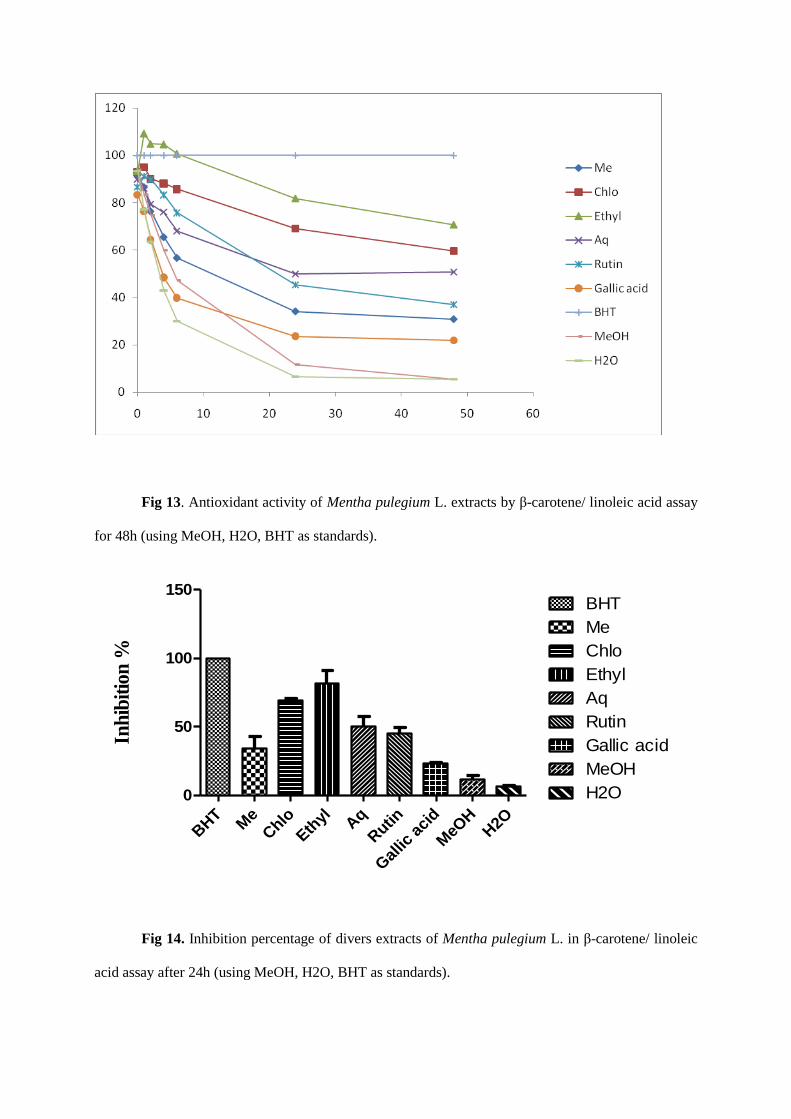
Fig 13. Antioxidant activity of Mentha pulegium L. extracts by β-carotene/ linoleic acid assay
for 48h (using MeOH, H2O, BHT as standards).
BHT
Me
Chlo
Ethyl A
q
Rutin
Gal
lic a
cid
MeO
HH2O
0
50
100
150BHT
Me
Chlo
Ethyl
Aq
Rutin
Gallic acid
MeOH
H2O
Inh
ibit
ion
%
Fig 14. Inhibition percentage of divers extracts of Mentha pulegium L. in β-carotene/ linoleic
acid assay after 24h (using MeOH, H2O, BHT as standards).
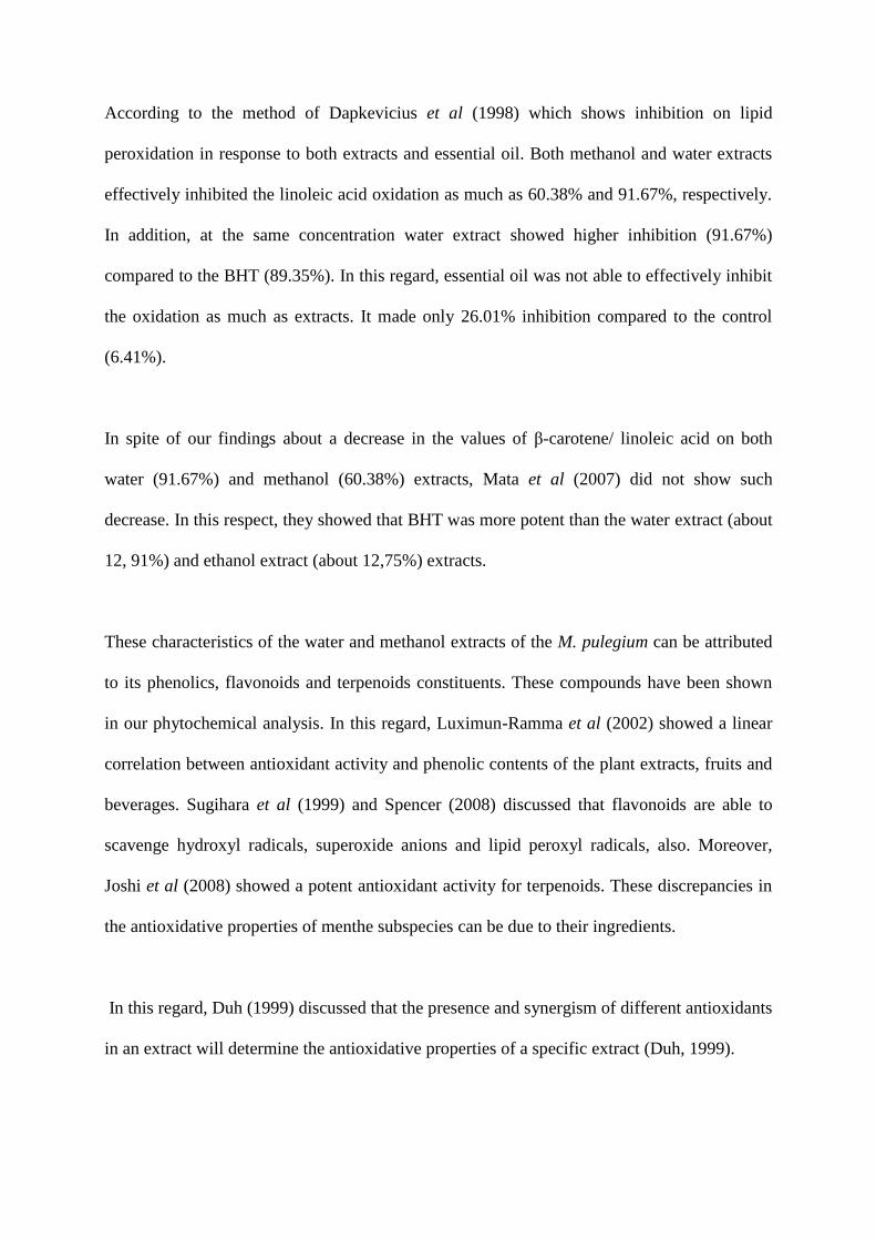
According to the method of Dapkevicius et al (1998) which shows inhibition on lipid
peroxidation in response to both extracts and essential oil. Both methanol and water extracts
effectively inhibited the linoleic acid oxidation as much as 60.38% and 91.67%, respectively.
In addition, at the same concentration water extract showed higher inhibition (91.67%)
compared to the BHT (89.35%). In this regard, essential oil was not able to effectively inhibit
the oxidation as much as extracts. It made only 26.01% inhibition compared to the control
(6.41%).
In spite of our findings about a decrease in the values of β-carotene/ linoleic acid on both
water (91.67%) and methanol (60.38%) extracts, Mata et al (2007) did not show such
decrease. In this respect, they showed that BHT was more potent than the water extract (about
12, 91%) and ethanol extract (about 12,75%) extracts.
These characteristics of the water and methanol extracts of the M. pulegium can be attributed
to its phenolics, flavonoids and terpenoids constituents. These compounds have been shown
in our phytochemical analysis. In this regard, Luximun-Ramma et al (2002) showed a linear
correlation between antioxidant activity and phenolic contents of the plant extracts, fruits and
beverages. Sugihara et al (1999) and Spencer (2008) discussed that flavonoids are able to
scavenge hydroxyl radicals, superoxide anions and lipid peroxyl radicals, also. Moreover,
Joshi et al (2008) showed a potent antioxidant activity for terpenoids. These discrepancies in
the antioxidative properties of menthe subspecies can be due to their ingredients.
In this regard, Duh (1999) discussed that the presence and synergism of different antioxidants
in an extract will determine the antioxidative properties of a specific extract (Duh, 1999).
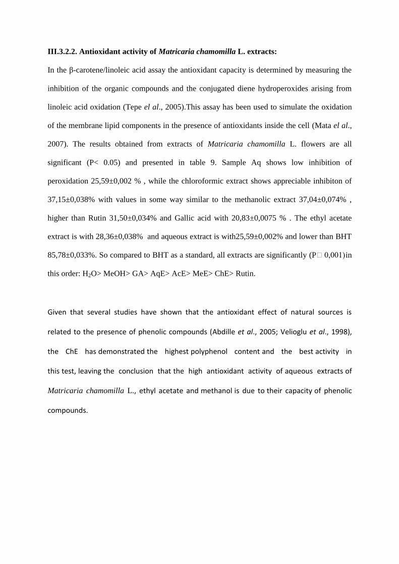
III.3.2.2. Antioxidant activity of Matricaria chamomilla L. extracts:
In the β-carotene/linoleic acid assay the antioxidant capacity is determined by measuring the
inhibition of the organic compounds and the conjugated diene hydroperoxides arising from
linoleic acid oxidation (Tepe el al., 2005).This assay has been used to simulate the oxidation
of the membrane lipid components in the presence of antioxidants inside the cell (Mata el al.,
2007). The results obtained from extracts of Matricaria chamomilla L. flowers are all
significant (P< 0.05) and presented in table 9. Sample Aq shows low inhibition of
peroxidation 25,59±0,002 % , while the chloroformic extract shows appreciable inhibiton of
37,15±0,038% with values in some way similar to the methanolic extract 37,04±0,074% ,
higher than Rutin 31,50±0,034% and Gallic acid with 20,83±0,0075 % . The ethyl acetate
extract is with 28,36±0,038% and aqueous extract is with25,59±0,002% and lower than BHT
85,78±0,033%. So compared to BHT as a standard, all extracts are significantly (P˂0,001) in
this order: H2O> MeOH> GA> AqE> AcE> MeE> ChE> Rutin.
Given that several studies have shown that the antioxidant effect of natural sources is
related to the presence of phenolic compounds (Abdille et al., 2005; Velioglu et al., 1998),
the ChE has demonstrated the highest polyphenol content and the best activity in
this test, leaving the conclusion that the high antioxidant activity of aqueous extracts of
Matricaria chamomilla L., ethyl acetate and methanol is due to their capacity of phenolic
compounds.
Fig 15. Antioxidant activity of Matricaria chamomilla L. extracts in β-carotene/ linoleic acid
assay for 48h (using MeOH, H2O, BHT as standards).
BHT
Me
Chlo
Ethyl A
q
Rutin
Gal
lic a
cid
MeO
HH2O
0
50
100
150BHT
Me
Chlo
Ethyl
Aq
Rutin
Gallic acid
MeOH
H2O
Inh
ibit
ion
%
Fig 16. Inhibition percentage of divers extracts of Matricaria chamomilla L. in β-carotene/
linoleic acid assay after 24h (using MeOH, H2O, BHT as standards).

CONCLUSION
CONCLUSION:
We have focused on plants belonging to several different families from Algeria (Lamiaceae,
Asteraceae) to understand their therapeutic uses and their potential antioxidant activities. The
two plants are widely used in the country as aromatic herbs in cooking, and also for their
medicinal properties in folk medicine, may act as important supplements of antioxidants in
the diet, especially in the cooking of some dishes of poor nutritional value. But unfortunately,
most of the species that are claimed to contain potent antioxidant activity have not been
studied in vivo. Screening with in vitro assays has little meaning if there is no clear evidence
of the effectiveness of the extracts in vivo. Therefore, further in vivo studies of these species
are required, and a systematic investigation of these antioxidant rich species is needed before
they can be used in the food processing industry and as preventive medicine.
Overall, it could be concluded that plants bear a potent antioxidant activity at a concentration
level which is related to constituents. Their constituents scavenge free radicals and exert a
protective effect against oxidative damage induced to cellular macromolecules because it has
shown the presence of number of polyphenols, which are related to their chemical structure
and may be responsible of the antioxidant activities. Further studies on the isolation of these
compounds are in progress.
So all plants exhibited moderate to good antioxidant activity, at least in one of the extract
tested, being the more active, the ethyl acetate for Mentha pulegium L. and chloroformic
extract for Matricaria camomilla L. In general, as effective as a known standard, BHT, in
scavenging free radicals.
Plants showing simultaneous polyphenols contents and antioxidant activity capacity could be
considered as food, having some function besides their traditional value, which makes them
promising candidates for more detailed in vitro and in vivo studies because of the different
experimental methods used in various studies.
REFERENCES
References:
Abdalla AE and Roozen JP., 1999. Effect of plant extracts on the oxidative stability of sunflower oil
and emulsion. Food chemistry. 64, 323-329.
Abdille MDH, Singh RP, Jayaprakasha GK and Jena BS., 2005. Antioxidant activity of the
extracts from Dillenia indica fruits. Food chemistry. 90, 891-896.
Agbor GA, Kuate D and Oben JE., 2007. Medicinal plants can be good source of antioxidants: case
study in Cameroon. Pakistan journal of biological sciences. 10, 537-544.
Arouma OI., 1993. Free radicals and food. Chemistry in britain. 29, 210-4.
Aruoma OL., 1998. Free radicals, oxidative stress and antioxidants in human health and disease.
Journal of the american oil chemistry. 75, 199-212.
Aruoma OI., 2003. Methodological considerations for characterizing potential antioxidant actions of
bioactive components in plant foods. Mutation research. 523-524, 9-20.
Bahorum T, Gressier B, Trotin F, Brunete C, Dine T, Vasseur J, Gazin JC, Pinkas M, Luycky,
M and Gazin M., 1996. Oxygen species scavenging activity of phenolic extracts from hawthorn fresh
plant organs and pharmaceutical preparation. Arzneimittel-Forsch. 46, 1086-1089.
Bardin JM., 2004. Illustrated Dictionary of the medicinal plants. ED, Brodard & Taupin. France. p:
312.
Bate-Smith E C., 1973. Haemanalysis of tannins, the concept of relative astringency. Phytochemistry.
12, 907-912.
Bhatia S, Shukla R, Madhu SV, Gambhir JK and Prabhu KM., 2003. Antioxidant status, lipid
peroxidation and NO end products in patients of type 2 diabetes mellitus with nephropathy. Clinical
biochemistry. 36, 557-562.
Bodamyali T, Stevens CR, Blake DR and Winyard PG., 2000. Reactive oxygen/nitrogen species
and acute inflammation: a physiological process. In: Winyard PG, Blake DR, Evans CH, editors. Free
radicals and inflammation. Basel, Switzerland: Birkhauser. pp: 11-9.
Bohm B., 1994. The minor flavonoids. Flavonoids. 387, 440.
Bondet V, Williams WB and Berset C., 1997. Kinetic and mechanism of antioxidant activity using
the DPPH free radical method. Lebensmittel-Wissenschaft und Technologie. 30, 609-615.
Bradley DE, Min DB., 1992. Singlet oxygen oxidation of foods. (Review) Food science and nutrition.
31, 211-236.
Bruneton J., 1993. Composés phénoliques: Shikimate-acétates. In: “Pharmacognosie: Phytochimie;
Plantes médicinales”. Technique et Documentation- Lavoisier (Paris). Chap.3, pp: 199-383.
Burits M and Bucar F., 2000. Antioxidant activity of Nigella sativa essential oil. Phytotherapy
research. 14, 323-328.
Chen C, Zhou J, Xu H, Jiang Y and Zhu G., 1997. Effect of selenium supplementation on mice
infected with LP-BM5 MuLV, a murine AIDS model. Biological trace of element research. 59, 187-
93.
Chen F, Lu Y and Demers LM., 1998. Role of hydroxyl radical in silicainduced NF-kappa B
activation in macrophages. Annals of clinical laboratory science. 28, 1-13.
Christ B and Müller KH., 1960. Zur serienmäßigen Bestimmung des Gehaltes an Flavonol-Derivaten
in Drogen. Archiv der Pharmazie. 293, 1033-1042.
Cook CM, Maloupa E, Kokkini S and Lanaras T., 2007. Differences between the inflorescence,
leaf and stem essential oils of wild Mentha pulegium plants from Zakynthos Greece. Journal of
essential oil research. 19, 239-244.
Dapkevicius A, Venskutonis R, Van Beek, TA and Linssen, PH., 1998. Antioxidant activity of
extracts obtained by different isolation procedures from some aromatic herbs grown in Lithuania.
Journal of the science of food and agriculture. 77, 140-146.
Davies KJA., 1987. Protein damage and degradation by oxygen radicals. I General aspects. Journal of
biological chemistry. 162, 9895-9901.
DeWaart FG, Portengen L, Doekes G, Verwaal CJ and Kok FJ., 1997. Effect of 3 months vitamin
E supplementation on indices of the cellular and humoral immune response in elderly subjects. The
British journal of nutrition. 78, 761-4.
Dewick P M., 1994. Isoflavonoids. In The Flavonoids: Advances in research since 1986, Harborne J
B. Ed. Chapman and Hall Ltd, London. p: 676.
Duh PD., 1999. Antioxidant activity of water extract of four Harng Jyur (Chrysanthemum morifolum
Ramat) varieties in soybean oil emulsion. Food chemistry. 66, 471-476.
El-Ghorab AH., 2006. The chemical composition of Mentha pulegium L. essential oil from Egypt and
its antioxidant activity. Journal of essential oil bearing plant. 9, 183-195.
Farr SB and Kogoma T., 1991. Oxidative stress responses in Escherichia coli and Salmonella
typhimurium. Microbiology review. 55, 561-585.
Fatouma M. Abdoul-Latif, Nabil Mohamed, Prosper Edou, Adwa A Ali, Samatar O Djama,
Louis-Clément Obame, Ismael H N Bassolé and Mamoudou H Dicko., 2011. Antimicrobial and
antioxidant activities of essential oil and methanol extract of Matricaria chamomilla L. from Djibouti.
Journal of medicinal plants research. Vol. 5(9), 1512-1517.
Forkmann G., 1991. Flavonoids as flower pigments: the formation of the natural spectrum and its
extension by genetic engineering (Review). Plant breeding. 100, 1-20.
Franke R. and Schilcher H., 2005. Chamomile: Industrial Profiles (Medicinal and Aromatic Plants -
Industrial Profiles), CRC Press, Taylor & Francis Group, USA, 1st edition. 29, 1118.
Frankel EN., 1985. Chemistry of free radical and singlet oxidation of lipids. Progress in lipid
research. 23, 197-221.
Galan P, Preziosi P and Monget AL., 1997. Effects of trace elements and/or vitamin supplementation
on vitamin and mineral status, free radical metabolism and immunological markers in elderly long
hospitalized subjects. International journal of vitamin in nutrition research. 67, 450-60.
Gilani AH, Aziz N, Khurram IM, Chaudary KS and Iqbal A., 2001. Bronchodilator, spasmolytic
and calcium antagonist activities of Nigella sativa seeds (Kalonji). Journal of the pakistan medical
association. 51, 115-120.
Givens JA, K Reeds and A Touwaide., 2006. Visualizing Medieval Medicine and Natural History.
1200-1550. Great Britain: Ashgate Publishing. p: 189.
Grassmann J, Hippeli S and Elstner E F., 2002. Plant‟s defence and its benefits for animals and
medicine: role of phenolics and terpenoids in avoiding oxygen stress. Plant physiology and
biochemistry. 40, 471-478.
Grisham MB., 1992. Reactive metabolites of oxygen and nitrogen in biology and medicine. Reactive
oxygen metabolites. Chemistry and medical consequences. 5 (2), 227-23.
Gu L, Kelm MA and Hammerstone JF., 2004. Concentrations of proanthocyanidins in common
foods and estimations of normal consumption. The journal of nutrition. 134, 613-617.
Gulcin I, Oktay M, Kirecci E and Kufrevioglu OI., 2003. Screening of antioxidant and
antimicrobial activities of anise (Pimpinella anisum L.) seed extracts. Food chemistry. 83, 371-382.
Güllüce M, Sokmen M, Daferera D, Agar G, Ozkan H, Kartal N, Polissiou M, Sokmen A and
Gupta V, Mittal P, Bansal P, Khokra SL andKaushik D., 2010. Pharmacological potential of
Matricaria recutita-A review. International journal of pharmacology science of drug research. 2, 12-6.
http://en.wikipedia.org/wiki/Pennyroyal.
Ha HC, Sirisoma NS, Kuppusamy P, Zweier JL, Woster PM and Halliwell B., 1990. How to
characterize a biological antioxidant. Free radical research communications. 9, 1-32.
Halliwell B., 1999. Vitamin C. poison, prophylactic or panacea? Trends in Biochemical sciences. 24,
255-9.
Halliwell B and Gutteridge JMC., 1984. Oxygen toxicity, oxygen radicals, transition metals and
disease. Biochemistry journal. 219, 1-14.
Harborne JB., in: Cody V, Middleton E and Harborne JB., 1986. (Eds), Plant Flavonoids in
Biology and Medicine. Alan R. Liss: New York. pp: 15-24.
Harsteen B., 1983. Flavonoids. A class of natural products of high pharmacological potency.
Biachemisry and pharmacology. 32, 1141-8.
Hein KE, Tagliaferro AR and Bobilya DJ., 2002. Flavonoid antioxidants: chemistry metabolism and
structure-activity relationships. Journal of nutritional biochemistry. 13, 572-584.
Herrmann K., 1989. Occurrence and content of hydroxycinnamic and hydroxybenzoic acid
compounds in foods. Critical reviews in food science and nutrition. 28, 315-347.
Hodzic Z, Pasalic H, Memisevic A, Srabovic M, Saletovic M and Poljakovic M., 2009. The
Influence of total phenols content on antioxidant capacity in the whole grain extracts. Scientific
research. 28 (3), 471-477.
Huang MT, Ho CT and Lee CY., 1992. (Eds). Phenolic compounds in foods and their effects on
human health. II: Antioxidants and cancer prevention. American Chemical Society, Washington DC.
ACS Symposium Series. Vol. No. 507.
Jayaprakasha GK and Patil BS., 2007. In vitro evaluation of the antioxidant activities in fruit
extracts from citron and blood orange. Food chemistry. 101, 410-418.
Joshi S, Chanotiya CS, Aganwal G, Prakash D, Pant AK and Mathela CS., 2008. Terpenoid
compositions and antioxidant and antimicrobial properties of the rhizome essential oils of different
Hedychium species. Chemical biodiversity. 5, 299-309.
Julve PH., 1998.Mentha pulegium L. Base flora. Botanical index, ecological and chorologic index
Flora of France.Version: April 23, 2004.
Justesen U and Knuthsen P., 2001. Composition of flavonoids in fresh herbs and calculation of
flavonoid intake by use of herbs in traditional Danish dishes. Food chemistry, 73, 245-250.
Kagaya K, Miyakawa Y, Watanabe K and Fukazawa Y., 1992. Antigenic role of stress-induced
catalase of Salmonella typhimurium in cell mediated immunity. Infection and immunity. 60, 1820-5.
Kaouass M, Deloyer P, Gouders I, Peulen O and Dandrifosse G., 1997. Role of interleukin-1 beta,
interleukin-6 and TNF-alpha in intestinal maturation induced by dietary spermine in rats. Endocrine.
6, 187-94.
Kartal N, Sokmen M, Tepe B, Daferera D, Polissiou M and Sokmen A., 2007. Investigation of the
antioxidant properties of Ferula orientalis L. using a suitable extraction procedure. Food chemistry.
100, 584–589.
Kato A, Minoshima Y, Yamamoto J, Adachi I, Awatson A and Nash RJ., 2008. Protective Effects
of Dietary Chamomile Tea on Diabetic Complications. J. Agric. Food Chemistry. 56, 8206-8211.
Kawanishi S, Hiraku Y, Murata M and Oikawa S., 2002. The role of metals in site-specific DNA
damage with reference to carcinogenesis. Free radical biology & medicine. 32, 822-32.
Kessemi N., 2006. Relation entre un insecte phytophage et sa principale plante hôte: cas de la bruche
du haricot (Acanthoscelides obtectus ), (Coleoptera Bruchidae).Thèse de Magistère de l‟université
Abou Bakr Belkaid de Tlemcen. Algérie.
Kouri G, Tsimogiannis D, Bardouki H, Oreopoulou V., 2007. Extraction and analysis of
antioxidant components from Origanum dictamnus. Innovative food science and emerging
technologies. 8, 155-162.
Lazarus SA, Hammerstone JF and Schmitz HH., 1999. Chocolate contains additional flavonoids
not found in tea. Lancet. 354, 1825.
Lefer DJ and Granger DN., 2000. Oxidative stress and cardiac disease. The american journal of
medecine. 109, 315-323.
Linn S., 1998. DNA damage by iron and hydrogen peroxide in vitro and in vivo. Drug metabolism
review. 30, 313-26.
Lin X, Lee GG, Casale ES, Shih JCH., 1999. Purification and characteri-zation of a keratinase from
feather degradating Bacillus lichenformis Strain. Applied and environmental microbiology. 5, 3271-
3275.
Lloyd RV, Hanna PM and Mason RP., 1997. The origin of the hydroxyl radical oxygen in the
Fenton reaction. Free radical biology & medicine. 22, 885-8.
Luximun-Ramma A, Bahorun T, Soobrattee MA and AruomaI OI., 2002. Antioxidant activities
of phenolic, proanthocyanidin, and flavonoid components in extracts of Cassia fistula. Journal of
agricultural and food chemistry. 50, 5042-5047.
Maddipati KR and Marnett LJ., 1987. Characterization of the major hydroperoxide- reducing
activity of human plasma. Purification and properties of a selenium-dependent glutathione peroxidase.
Journal of biological chemistry. 262, 17398-403.
Markham KR., 1982. Techniques of flavonoid identification (Chapter 1 and 2). London: Academic
press. 1-113.
Mata AT, Proenc C¸ Ferreira AR, Serralheiro MLM, Nogueira JMF and Arauj MEM., 2007.
Antioxidant and anti- acetylcholinesterase activities of five plants used as Portuguese food spices.
Food chemistry. 103, 778-786.
Mata AT, ProençaC, Ferreira AR, Serralheiro MLM, Nogueira JMF and Araújo MEM., 2007.
Antioxidant and anti- acetylcholinesterase activities of five plants used as Portuguese food spices.
Food chemistry. 103, 778.
Mates JM, Gomez CP and Blanca M., 2000. Chemical and biological activity of free radical
scavengers in allergic diseases. Clinica chimica acta. 296, 1-15.
Matkowski A., 2008. Plant in vitro culture for the production of antioxidants - A Review.
Biotechnology advances. 26(6), 548-560.
McKay DL and Blumberg JB., 2006. A review of the bioactivity and potential health benefits of
chamomile tea (Matricaria recutita L). Phytoterapy research. 20, 519-530.
Melov S, Schneider JA and Day BJ et al., 1998. A novel neurological phenotype in mice lacking
mitochondrial manganese superoxide dismutase. Nature genetic. 18, 159-63.
Mensor LL, Menezes FS, Leitāo GG, Reine AS, Santos TC, Coube CS and Leitāo SG., 2001.
Screening of Brazilian plant extracts for antioxidant isoactivity by the use of DPPH free radical
method. Phy-totherapy research. 15, 127-130.
Middleton E., 1984. The flavonoids. Trends in Biochemical sciences. 5, 335-8.
Mkaddem M, Bousaid M and Ben Fadhel N., 2007. Variability of volatiles in Tunisian Mentha
pulegium L (Lamiaceae). Journal of essential oil research. 19, 211-215.
Moon BH, Lee JHAhn and LimY., 2006. Complete assignment of 1H and 13C NMR data of
dihyroxyflavone derivatives. Magnetic resonance in chemistry. 44, 99-101.
Nappi AJ and Vass E., 1998. Hydroxyl radical formation via iron-mediated Fenton Chemistry is
inhibited by methylated catechols. Biochimical and biophysical Acta. 1425, 159-67.
Nemtanu MR, Kikuchi IS, de Jesus Andreoli Pinto T, Mazilu E, Setnic S, Bucur M, Duliu OG,
Meltzer V and Elena Pincu E., 2008. Electron beam irradiation of Matricaria chamomilla L. for
microbial decontamination. Nuclear Instruments and Methods in Physics Research B. 266, 2520–2523.
Nickavar B, Alinaghi A and Kamalinejad M., 2008. Evaluation of the antioxidant properties of five
Mentha species. Iranian journal of pharmaceutical research. 7, 203-209.
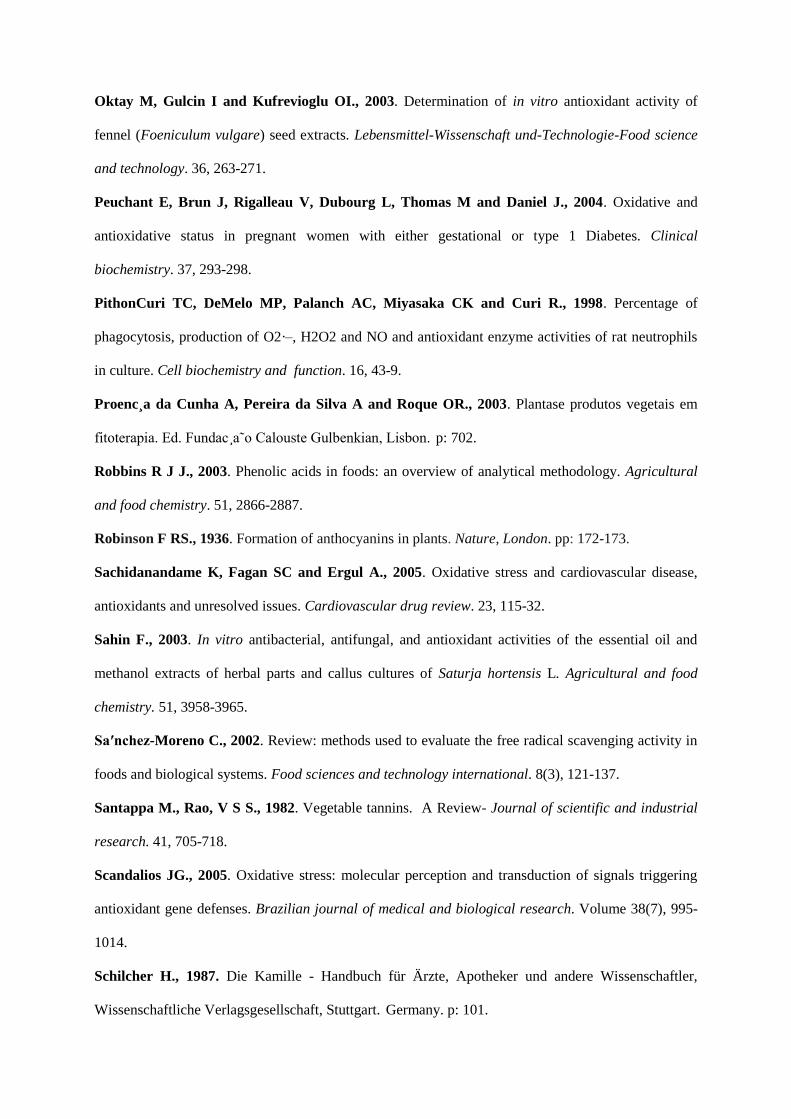
Oktay M, Gulcin I and Kufrevioglu OI., 2003. Determination of in vitro antioxidant activity of
fennel (Foeniculum vulgare) seed extracts. Lebensmittel-Wissenschaft und-Technologie-Food science
and technology. 36, 263-271.
Peuchant E, Brun J, Rigalleau V, Dubourg L, Thomas M and Daniel J., 2004. Oxidative and
antioxidative status in pregnant women with either gestational or type 1 Diabetes. Clinical
biochemistry. 37, 293-298.
PithonCuri TC, DeMelo MP, Palanch AC, Miyasaka CK and Curi R., 1998. Percentage of
phagocytosis, production of O2·–, H2O2 and NO and antioxidant enzyme activities of rat neutrophils
in culture. Cell biochemistry and function. 16, 43-9.
Proenc¸a da Cunha A, Pereira da Silva A and Roque OR., 2003. Plantase produtos vegetais em
fitoterapia. Ed. Fundac¸a˜o Calouste Gulbenkian, Lisbon. p: 702.
Robbins R J J., 2003. Phenolic acids in foods: an overview of analytical methodology. Agricultural
and food chemistry. 51, 2866-2887.
Robinson F RS., 1936. Formation of anthocyanins in plants. Nature, London. pp: 172-173.
Sachidanandame K, Fagan SC and Ergul A., 2005. Oxidative stress and cardiovascular disease,
antioxidants and unresolved issues. Cardiovascular drug review. 23, 115-32.
Sahin F., 2003. In vitro antibacterial, antifungal, and antioxidant activities of the essential oil and
methanol extracts of herbal parts and callus cultures of Saturja hortensis L. Agricultural and food
chemistry. 51, 3958-3965.
Sa′nchez-Moreno C., 2002. Review: methods used to evaluate the free radical scavenging activity in
foods and biological systems. Food sciences and technology international. 8(3), 121-137.
Santappa M., Rao, V S S., 1982. Vegetable tannins. A Review- Journal of scientific and industrial
research. 41, 705-718.
Scandalios JG., 2005. Oxidative stress: molecular perception and transduction of signals triggering
antioxidant gene defenses. Brazilian journal of medical and biological research. Volume 38(7), 995-
1014.
Schilcher H., 1987. Die Kamille - Handbuch für Ärzte, Apotheker und andere Wissenschaftler,
Wissenschaftliche Verlagsgesellschaft, Stuttgart. Germany. p: 101.
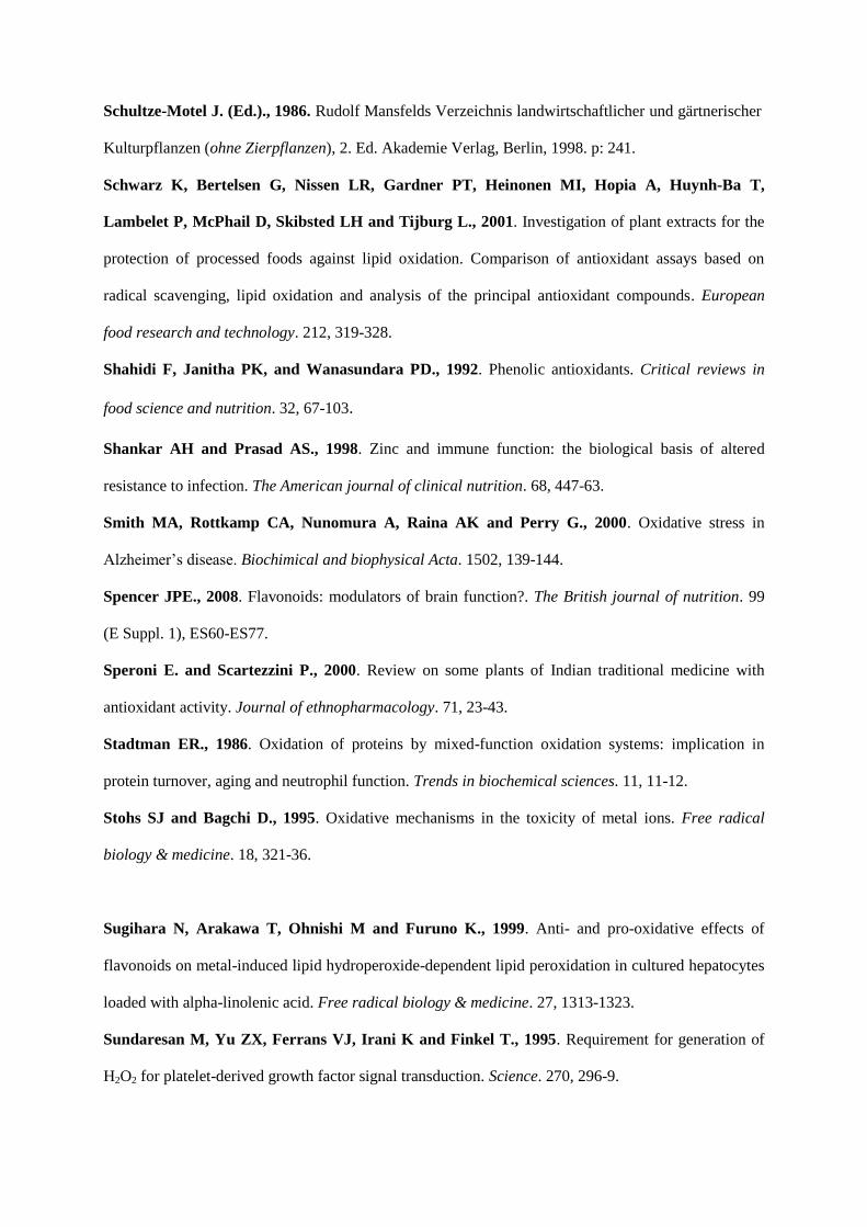
Schultze-Motel J. (Ed.)., 1986. Rudolf Mansfelds Verzeichnis landwirtschaftlicher und gärtnerischer
Kulturpflanzen (ohne Zierpflanzen), 2. Ed. Akademie Verlag, Berlin, 1998. p: 241.
Schwarz K, Bertelsen G, Nissen LR, Gardner PT, Heinonen MI, Hopia A, Huynh-Ba T,
Lambelet P, McPhail D, Skibsted LH and Tijburg L., 2001. Investigation of plant extracts for the
protection of processed foods against lipid oxidation. Comparison of antioxidant assays based on
radical scavenging, lipid oxidation and analysis of the principal antioxidant compounds. European
food research and technology. 212, 319-328.
Shahidi F, Janitha PK, and Wanasundara PD., 1992. Phenolic antioxidants. Critical reviews in
food science and nutrition. 32, 67-103.
Shankar AH and Prasad AS., 1998. Zinc and immune function: the biological basis of altered
resistance to infection. The American journal of clinical nutrition. 68, 447-63.
Smith MA, Rottkamp CA, Nunomura A, Raina AK and Perry G., 2000. Oxidative stress in
Alzheimer‟s disease. Biochimical and biophysical Acta. 1502, 139-144.
Spencer JPE., 2008. Flavonoids: modulators of brain function?. The British journal of nutrition. 99
(E Suppl. 1), ES60-ES77.
Speroni E. and Scartezzini P., 2000. Review on some plants of Indian traditional medicine with
antioxidant activity. Journal of ethnopharmacology. 71, 23-43.
Stadtman ER., 1986. Oxidation of proteins by mixed-function oxidation systems: implication in
protein turnover, aging and neutrophil function. Trends in biochemical sciences. 11, 11-12.
Stohs SJ and Bagchi D., 1995. Oxidative mechanisms in the toxicity of metal ions. Free radical
biology & medicine. 18, 321-36.
Sugihara N, Arakawa T, Ohnishi M and Furuno K., 1999. Anti- and pro-oxidative effects of
flavonoids on metal-induced lipid hydroperoxide-dependent lipid peroxidation in cultured hepatocytes
loaded with alpha-linolenic acid. Free radical biology & medicine. 27, 1313-1323.
Sundaresan M, Yu ZX, Ferrans VJ, Irani K and Finkel T., 1995. Requirement for generation of
H2O2 for platelet-derived growth factor signal transduction. Science. 270, 296-9.
Tepe K, Daferera D, Sokmen A, Sokmen M, Polissiou M., 2005. Antimicrobial and antioxidative
activity of essential oil and various extracts of Cyclotrichium origanifolium (Labill.). Journal of food
energy. 69, 335-342
Topalov V and Dimitrov S., 1969. Studies on the content and quality of essential oil from some
peppermint species from Bulgarian Flora. Plant Science. 6, 77-83.
Tsai KJ, Hung IJ, Chow CK, Stern A, Chao SS and Chin DTY., 1998. Impared production of nitric
oxide, superoxide and hydrogen peroxide in glucose 6-phosphate dehydrogenase-deficient ranulocytes.
FEBS Letter. 436, 411-4.
Underhill E, Woo Watkin J E and Neish A C., 1957. Biosynthesis of quercetin in buckwheat. (part
1). Canadian journal of biochemistry and physiology. 35, 219-228.
Velioglu YS, Mazza G, Gao L and Oomah BD., 1998. Antioxidant activity and total phenolics in
selected fruits, vegetables and grain products. Journal of agricultural and food chemistry. 46, 4113-
4117.
Ververidis Filippos F, Trantas Emmanouil, Douglas Carl, Vollmer Guenter, Kretzschmar Georg
and Panopoulos Nickolas., October 2007. "Biotechnology of flavonoids and other phenylpropanoid-
derived natural products. Part I: Chemical diversity, impacts on plant biology and human health".
Biotechnology journal. 2 (10), 1214-34.
Wang J and Mazza G., 2002. Effects of anthocyanins and other phenolic compounds on the
production of tumor necrosis factor α in LPS/IFN-γ-activated RAW 264.7 macrophages. Journal of
agricultural and food chemistry. 50, 4183-4189.
Ward JF, Evans JW, Limoli CL and Calabro-Jones PM., 1987. Radiation and hydrogen peroxide
induced free radical damage to DNA. The brtish journal of cancer. 55, 105-12.
Watkin J E, Heish A C., 1960. Biosynthesis of quercetin in buckwheat (part 111). Canadian journal
of biochemistry and physiology. 38, 559-567.
www.floralimages.co.uk/images/mentha_pulegium
Zaidi F, Voirin B, Jay M and Viricel MR., 1998. Free flavonoid aglycones from leaves of Mentha
pulegium and Mentha suaveolens (Labiaceae). Phytochemistry. 48, 991-994.