Embed Size (px)
Citation preview
Phytochemistry Reviews (2004) 3: 371–379
Monitoring defensive responses in macroalgae – limitations andperspectives
© Springer 2005
S.L. La Barre1,∗, F. Weinberger1, N. Kervarec2 & P. Potin1
1UMR 7139 CNRS/GOEMAR/UPMC, Végétaux Marins et Biomolécules, B.P. 74, Station Biologique, 29682 Ro-scoff Cedex, France; 2Laboratoire de Résonance Magnétique Nucléaire, Faculté des Sciences, B.P. 809 Universitéde Bretagne Occidentale, 29285 Brest Cedex, France; ∗Author for correspondence (Phone: +33 (1) 9829 2360;Fax: +33 (1) 9829 2324; E-mail: [email protected])
Key words: elicitor, macrophytic algae, mass spectrometry, metabolomics, nuclear magnetic resonancespectroscopy
Abstract
As part of an ongoing research program aiming at monitoring molecular changes in the tissues and metabolitetrafficking in the hydrosphere of algae subjected to chemical stresses, we are discussing the various analyticaltechniques that have been employed to characterize, and sometimes to quantity these metabolites. High-fieldmultinuclear and solid-state nuclear magnetic resonance (NMR) spectroscopies are powerful tools for metabolitecharacterization from extracts and in vivo, but quantification and kinetic aspects show some limitations. ModernMS (mass spectrometry) is extremely useful for fingerprinting samples against databases and when dealing withvery low concentrations of metabolites, the limitations being set by the type of chromatographic separation andmode of detection coupled with the mass spectrometer. Regarding chemical communication, optimization in termsof resolution and efficiency of hydrosphere chemical analysis can theoretically be achieved in a system whichintegrates (i) a multiparametric incubation chamber, (ii) a gasphase or a liquid-phase separation system and (iii)mass spectrometer(s) equipped with one or two detectors responding to the analytical and quantitative needs. Thistext reviews some of the techniques that have been employed in various types of plant metabolic studies, whichmay serve as a basis towards an integrative analytical strategy directly applicable to the metabolomics of selectedmarine macrophytes.
Abbreviations: APCI – Atmospheric Pressure Chemical Ionization; cITP – Capillary Isotachophoresis (cITP);DAD – Diode Array Detector; ECD – Electron Capture Detection; EI-MS – Electron-Impact Mass Spectroscopy;ESI – Electro-Spray Ionization; GC – Gas Chromatography; 1H NMR – Proton Nuclear Magnetic Resonance;HPLC – High-Performance Liquid Chromatography; HR-MAS – High-Resolution Magic Angle Spin; HVOC –Halogenated Volatile Organic Compound; HXO – Hypohalous Acid; ICP-MS – Inductively Coupled Plasma MassSpectrometry; LC-MS – Liquid Chromatography coupled with Mass Spectroscopy; LMWHC – Low-MolecularWeight Halocarbon; MAA – Mycosporin-like Amino Acids; MIP-AED – Microwave-Induced Plasma AtomicEmission Detection; MS – Mass Spectroscopy; MS/MS – refers to two tandem-coupled mass spectrometers; NMR– Nuclear Magnetic Resonance; 31P NMR – Phosphorus Nuclear Magnetic Resonance; PAR – PhotosyntheticActive Radiation; SBSE – Stir Bar Sorptive Extraction; SIM – Single Ion Monitoring; SPME – Solid-Phase MicroExtraction; UV – Ultra-Violet; UVR – Ultra-Violet Radiation; VHC – Volatile HaloCarbon; VHOC – VolatileHalogenated Organic Compounds; X- – Halide Ions.
Introduction
Marine algae are continuously being challenged bymicro-organisms, including viruses, bacteria and other
algae as well as grazers, all of which may causewounds and diseases. In addition, underwater compet-ition for settlement and epiphytism may affect growthby reducing light availability or by increasing hy-
372
drodynamic drag. The combination of competitionand predation pressures represents a constant threat tothe fitness of macrophytic algae throughout their life,equivalent to that encountered by sessile autotrophs interrestrial ecosystems.
Macroalgae normally grow in shallow or eulittoralenvironments where the hydrodynamic regime (cur-rents, tides, turbidity) is fast-changing. In additionto biotic stress, they may have to adjust to rapid anddramatic physical or chemical changes, e.g., UV ra-diation (UVR), high Photosynthetic Active Radiation(PAR), desiccation, salinity and nutrient limitation. Tosurvive in such a seemingly hostile and unpredictableenvironment, marine plants obviously had to evolvesurvival strategies, under the form of so-called escaperesponses e.g. habitat refuges, species association,rapid growth etc., reviewed by Biber (2002), in com-bination with the production of chemical deterrentsand growth inhibitors, UV screens or osmolytes, eachrepresenting a metabolic investment on behalf of theindividual.
A rapid response likely to represent a chemical de-fence against biotic stress in seaweeds is the emissionof halogenated volatile organic compounds (HVOC’s).An increased production of such iodinated, brom-inated or chlorinated carbon skeletons is associatedwith oxidative stress from various origins, whethercaused by carbon deprivation, high light intensity ormechanical injuries (Mtolera et al., 1996). HVOC’sbiogenesis involves vanadium-haloperoxidases, whichcatalyse the oxidation of halides (X−) into hypohal-ous acids (HXOs). As highly electrophilic agents,hypohalous acids can react with a variety of organiccompounds and as such are considered as potent nat-ural biocides (Wever et al., 1991). In brown algae,phlorotannins are induced by exposure to UV radiationand in response to grazing (Cronin and Hay, 1996;Pavia and Toth, 2000; Toth and Pavia, 2000). Anotherclass of secondary metabolites, the mycosporine-likeamino acids (MAAs) may be viewed as antioxid-ants in the rhodophytes (Dunlap and Shick, 1998).MAAs are synthesised via the shikimic acid path-way, a metabolic route which leads to the productionof the higher plant antibiotics known as phytoalex-ins (Sommsich and Halbrock, 1998), and shikimicacid may be a central element of defence activa-tion in these algae. In higher plants low molecularweight hydrocarbons (LMWHCs) such as isoprene(2-methyl-1,3-butadiene) and ethene are potent sig-naling molecules which are synthesised during en-vironmental stress or upon grazing (Kimmerer and
Kozlowski, 1982; Abeles et al., 1992; Kesselmeierand Staudt, 1999). In marine brown algae LMWHCsare so far recognized as sexual pheromones (Pohnertand Boland, 2002) or herbivore deterrents (Hay et al.,1998; Schnitzler et al., 2001). These compounds mayhelp to ward off infectious or fouling microorganisms(Hay, 1996) and they are very useful for broadcastingwarning messages, i.e. informing the population of thepresence of pathogens or grazers. Recent bioassay-guided separations of crude bioactive algal extractshave proved the power and usefulness of such ap-proaches to decipher the function of algal metabolitesPrevious investigations of the role of seaweed chemic-als against herbivores in fucoid brown algae assumedthat phlorotannins (Ragan et al., 1986) act as anti-feedants (see Hay and Fenical, 1988). However, thedeterrence of Fucus vesiculosus chemical extracts wasrecently reinvestigated using herbivore bioassays toguide chemical investigations (Deal et al., 2003). Al-though crude extracts from F. vesiculosus stronglydeterred feeding by the sea urchin Arbacia punctu-lata, phlorotannins from this extract did not affectfeeding at 2-fold or 4-fold natural concentration bydry mass. Feeding deterrence was instead due to: (1)one polar galactolipid in the ethyl acetate soluble ex-tract and (2) at least one non-phenolic compound inthe water-soluble extract. Given the high polarity ofthese chemical deterrents, they could co-occur withphlorotannins and confound bioassays performed onpolar extracts before separation. The hypothesis thatmany marine plants use chemical defences selectivelyagainst microbial pathogens, epiphytes, and sapro-phytes was recently tested using a similar approach.Lobophorolide [1], a polycyclic macrolide with sub-µM antifungal activity, appears to function as an an-tifungal chemical defence, protecting the brown algaLobophora variegata from at least one pathogenic andone saprophytic fungus, while being inactive against athrausochytrid fungus, a pathogenic bacterium as wellas against herbivorous fishes (Kubanek et al., 2003).
Most researchers studying environmental or ge-netic effects on secondary metabolite production tendto focus on a particular class of compounds for prac-tical reasons. Analyzing several groups of secondarycompounds with different chemical properties is time-consuming and often requires the use of individualextraction procedures and chromatographic methods.This functional diversity stems from the fact thatmany different secondary metabolic pathways are co-ordinately regulated by biotic elicitors and environ-mental stresses, as now evidenced by the abundant lit-
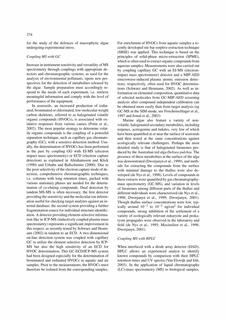
373
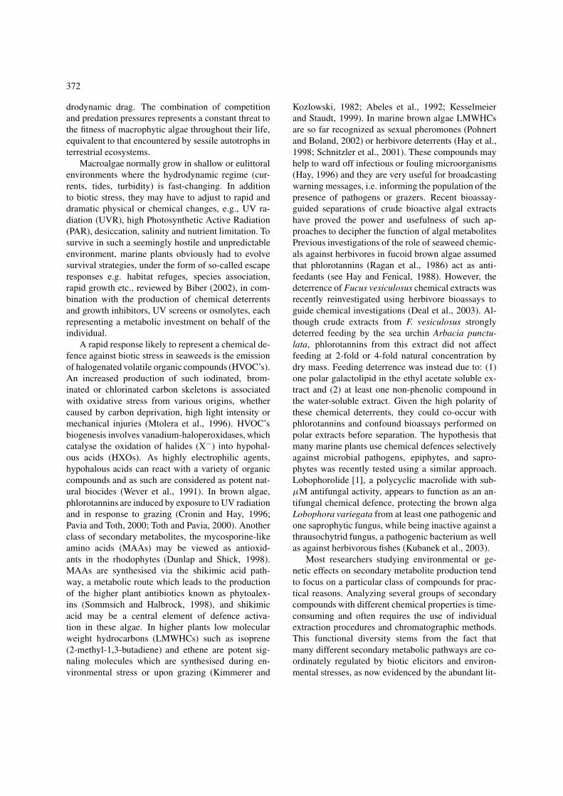
Figure 1. Diagram representing a circulating incubation chamber for the time-lapse analysis of compounds released in the medium, and furtheranalyses of algal samples (1) Time-lapse analysis of compounds trapped on SPME fibers (2) Analysis of algal tissues and of their extracts(3) Alternative analysis of seawater samples collected in sealed vials and processed using a cold purge and trap system. (boldface): analyticalmethods of choice for the metabolic profiling and the chemical characterization
erature in terrestrial plants, (Sommsich and Halbrock,1998; Scheel, 1998).
When prompted, sessile marine organisms releasecommunication (lato sensu) metabolites as bundles orsequentially in their immediate hydrosphere. The ana-lysis of these metabolites, together with that obtainedclassically from tissue extracts, is essential for a com-plete ‘metabolomic picture’. It follows that differentsampling procedures must be followed when operat-ing in vivo and in vitro (i.e. between molecules highlydiluted in seawater and metabolites concentrated in anorganic extract). This also concerns to some extent theseparation steps where appropriate chromatographicequipment must be used for online treatment, and ofcourse, the choice of detection sensitivity.
Here we briefly review recent methodological de-velopment s in the chemical ecology and biochemistryof marine algae, from which we can begin to out-line an integrated methodology for a broad-spectrummetabolite fingerprinting of induced compounds, withoptimal interactivity between the extraction and sep-aration (HPLC, GC) steps, and the pertinent spectro-scopic analysis and profiling of selected metabolites.
Mass spectrometry analysis
The recent developments of mass spectrometry inbiology were initially motivated by medical researchtargeted towards high throughput screening as wellas key analytical applications, e.g. understanding
cellular processes and the discovery of disease bio-markers. The use of mass spectrometry (MS) as theprincipal method for the detection of bioactive mo-lecules presents an alternative way of performing nat-ural products screening, especially in the absence ofspecific biomarkers (review by Van Elswijk and Irth,2003). In particular, the need for dereplication ofstructures at initial stages of the analysis of ‘unknown’natural samples, calls for outstandingly discriminat-ing technology. In this regard, LC-MS, thanks to theuse of appropriate interfaces e.g. electrospray (ES) oratmospheric pressure chemical ionization (APCI) isnow capable of analyzing a tremendous range of mo-lecules of all sizes. Database search using the nominalmolecular mass of a compound then becomes highlyefficient (Jemal et al., 2003).
Ecotoxicological applications are presently tar-geted towards analysis of anthropogenic wastes andtheir fate in the atmosphere, water and soil, withclimatic and toxicology concerns stimulating state-of-the-art technological advances allowing detectionand identification of metabolites at picomolar levels.In this respect, the modularity of MS systems, andthe miniaturization of most components often allowson-site analysis.
Metabolomic studies, in which the molecular rep-ertoire of a sample serves as a basis to the elucidationof biosynthetic processes as well as for the descriptionof osmolyte trafficking, coud indeed benefit from allthese technological advances, as proposed hereunder
374
for the study of the defences of macrophytic algaeundergoing experimental stress.
Coupling MS with GC
Increase in instrument sensitivity and versatility of MSspectrometry through couplings with appropriate de-tectors and chromatographic systems, as used for theanalysis of environmental pollutants, opens new per-spectives for the detection of metabolites released bythe algae. Sample preparation must accordingly re-spond to the needs of each experiment, i.e. retrievemeaningful information and comply with the level ofperformance of the equipment.
In seaweeds, an increased production of iodin-ated, brominated or chlorinated, low-molecular weightcarbon skeletons, referred to as halogenated volatileorganic compounds (HVOCs), is associated with ox-idative responses from various causes (Potin et al.,2002). The most popular strategy to determine volat-ile organic compounds is the coupling of a powerfulseparation technique, such as capillary gas chromato-graphy (GC), with a sensitive detection method. Usu-ally, the determination of HVOCs has been performedin the past by coupling GG with EI-MS (electronimpact mass spectrometry) or ECD (electron capturedetection) as explained in Abrahamsson and Klick(1990) and Urhahn and Ballschmiter (2000). Due tothe poor selectivity of the electron capture mode of de-tection, comprehensive chromatographic techniques,i.e. columns with long retention times, packed withvarious stationary phases are needed for the determ-ination of co-eluting compounds. Dual detection bytandem MS-MS is often necessary, the first detectorproviding the sensitivity and the molecular ion inform-ation useful for checking target analytes against an in-ternal database, the second system providing a furtherfragmentation source for individual structure identific-ation. A detector providing element-selective informa-tion like in ICP-MS (inductively coupled plasma massspectrometry) represents a significant improvement inthis respect, as recently tested by Schwarz and Heum-ann (2002) in tandem to an ECD. A two-dimensionalon-line detection system was coupled with capillaryGC to utilize the element selective detection by ICP-MS but also the high sensitivity of an ECD forHVOC determination. This GC-ECD/ICP-MS systemhad been designed especially for the determination ofbrominated and iodinated HVOCs in aquatic and airsamples. Prior to the measurements, the HVOCs musttherefore be isolated from the corresponding samples.
For enrichment of HVOCs from aquatic samples a re-cently developed stir bar sorptive extraction technique(SBSE) was applied. This technique is based on theprinciples of solid-phase micro-extraction (SPME),which is often used to extract organic compounds fromaqueous samples. Measurements were also carried outby coupling capillary GC with an EI-MS (electron-impact mass spectrometer) detector and a MIP-AED(microwave-induced plasma atomic emission detec-tion), respectively, often used for HVOC determina-tions (Schwarz and Heumann, 2002). As well as in-formation on elemental composition, quantitative dataof selected molecules from GC-MIP-AED screeninganalysis after compound independent calibration canbe obtained more easily than from target analysis (egGC-MS in the SIM-mode, see Frischenschlager et al.,1997 and Jemal et al., 2003)
Marine algae also feature a variety of non-volatile, halogenated secondary metabolites, includingterpenes, acetogenins and indoles, very few of whichhave been quantified at or near the surface of seaweedsand then tested at the same concentrations againstecologically relevant challengers. Perhaps the mostdetailed study is that of halogenated furanones pro-duced by the Australian red alga Delisea pulchra. Thepresence of these metabolites at the surface of the algawas demonstrated (Dworjanyn et al., 1999), and meth-ods for extracting the compounds from the surfacewith minimal damage to the thallus were also de-veloped (de Nys et al., 1998). Levels of compounds inthese extracts were quantified by gas chromatography-mass spectrometry (GC-MS), and variation in levelsof furanones among different parts of the thallus anddifferent individuals were characterized (de Nys et al.,1998; Dworjanyn et al., 1999; Dworjanyn, 2001).Though thallus surface concentrations were low, typ-ically around 10−1 to 10−2 ng/cm2 for individualcompounds, strong inhibition of the settlement of avariety of ecologically relevant eukaryote and proka-ryote propagules were observed in the laboratory andfield (de Nys et al., 1995; Maximilien et al., 1998;Dworjanyn, 2001).
Coupling MS with HPLC
When interfaced with a diode array detector (DAD),HPLC allows an experienced analyst to identifyknown compounds by comparison with their HPLCretention times and UV spectra (Van Elswijk and Irth,2003). In the application of liquid chromatography(LC)-mass spectrometry (MS) to biological samples,
375
electrospray ionization (ESI) has shown to desolv-ate and ionise ‘fragile’ species e.g. protein, peptides,nucleic acids and bioactive macrolides. The interestsof this technique are non-destructive soft ionizationand suitability for compounds with a wide range ofmolecular masses. However, few applications to theanalysis of algal metabolites have been reported.
MAAs are one of nature’s sunscreens, with about20 structurally distinct MAAs presently identified inmarine organisms (Dunlap and Shick, 1998). Thecommonly used method for MAA detection and quan-tification is separation by high-performance liquidchromatography (HPLC) followed by chromophoredetection at characteristic UV wavelengths (gener-ally ∼310 and ∼340 nm) or obtaining entire UV,λ scans via diode array detection (DAD, see Dun-lap et al., 1986). A common difficulty has been thelack of commercially available MAA standards, thepreparation of which is costly and time consuming.Therefore, much of the characterization of MAAs hasbeen accomplished using the distinctive nature of theirabsorption spectra. While such spectral methods aregenerally suitable, difficulties arise in the identifica-tion and quantification of MAAs for which standardsare not available or in the examination of MAAsaltered by dietary, chemical, or bacterial degradation.In addition, there are instances where closely relatedcompounds share similar absorption maxima and anisocratic HPLC retention time (e.g., shinorine [λmax =334 nm, RT= 3.5 min] and porphyra-334 [λmax =334 nm, RT= 3.7 min]), which further complicates theidentification process. Lastly, characterization of un-known UV-absorbing compounds is initially limited toobtaining the retention time and wavelength maxima,without additional information relating to their struc-tural features and classification. Recently, a mass spec-tral approach was applied to MAA characterizationutilizing liquid chromatography coupled with electro-spray ionization mass spectrometry (LC/MS) (White-head and Hedges, 2002). Quantitative mass spectralanalysis of MAAs with respect to a L-tyramine hy-drochloride internal standard was found to have aprecision of better than 3% and a detection limit ofapprox. 2.0 pg. Overall, this method adds anotherlevel in MAA elucidation by providing the molecularweight and/or fragmentation pattern of each constitu-ent. Using a data matrix of retention time, absorp-tion maximum, and molecular weight, the identity ofthe MAAs can be unambiguously established. Massspectrometry also provides a means to better charac-
terize novel MAAs and MAA degradation products ingeneral.
Activated defences, i. e., which consist in therapid conversion of defensive precursor into harm-ful molecules following cell damage, are also foundin both macro- and micro-algae. (Cetrulo and Hay,2000; Potin et al., 2002). A rapid transformation ofan acetylated terpene has been found in the greenalga Caulerpa taxifolia. This alga reacts upon wound-ing with the rapid, esterase-mediated transformationof its major secondary metabolite caulerpenyne intohighly reactive 1,4-dialdehydes (Jung and Pohnert,2001). In LC/APCI-MS measurements, a complexseries of closely related hydrazones bearing one ortwo derivatised aldehyde groups could be assigned bytheir characteristic fragmentation pattern. Since thesetransformations occur immediately after wounding orcomplete tissue disruption, a de novo expression of therequired esterases is highly unlikely. It was suggestedinstead that the disruption of an existing intracellularsegregation leads to the transformation of the terpen-oid substrates after getting into contact with pre-storedesterases.
The demonstration that fatty acid oxidative path-ways are involved in the immunity of marine algaewas lacking until very recently. Following the oxidat-ive burst in C. crispus (Bouarab et al., 1999), variouslipid hydroperoxides are produced, which were iden-tified by LC/ACPI-MS. They were not only derivedfrom arachidonic acid, the ‘animal-type’ eicosanoicfatty acid already well documented in red algae (Ger-wick, 1999), but also from linolenic and linoleic acids,precursors common in higher plants (Bouarab et al.,2004).
Nuclear magnetic resonance spectroscopy
Classical spectroscopic methods have been applied tothe identification, and occasionally to the quantifica-tion of metabolites produced or destroyed as a result ofan experimental event inflicted to a plant, in particularthe action of a chemical elicitor, i.e. an extra-cellularsignal mimicking attack by an aggressor. This meth-odology has been applied to metabolic studies ofmacrophytic algae undergoing various experimentalstresses.
Progress in solid-state NMR spectroscopy now al-lows chemical fingerprinting of tissues without theneed to extract the metabolites, with severe limitationswhen it comes to quantification. NMR spectroscopy is
376
the choice method for the study of such metabolites,in vitro from solvent extracts, and in vivo from cellsuspensions or from ‘intact’ tissues (Ratcliffe, 1994;Ratcliffe and Sachard-Hill, 2001). Soluble metaboliteprofiles can be established and compared before andafter an experimental perturbation capable of indu-cing a metabolic stress, for example by presenting anelicitor. Whereas 1H NMR profiles can get quite com-plex with many overlapping signals, detection of othernuclei may sometimes prove very useful in specific de-tections. 31P NMR is an attractive method, in combin-ation with 13C NMR, for detecting changes in phos-phorylated compounds, organic and inorganic, whichare observed in aqueous extracts during the first stagesof the perception of an elicitor (Bligny et al., 2001).We attempted to evaluate early metabolic changes fol-lowing the elicitation of young thalli of Laminariadigitata by oligoguluronate fractions. Phosphorylatedfractions were prepared using a protocol designedfor plant cell cultures (Pugin et al., 1977), but theyield was too low to obtain useful NMR information.The same problems arose with 31P NMR experimentson the red alga Gracilaria conferta exposed to oli-goagar fractions (unpublished results). Whereas 1HNMR profiles on neutralized perchloric acid extractsproved quite informative, peak-shifting was signific-ant between corresponding 31P NMR spectra, evenwith minimal differences in pH during the bufferingprocess. Care must be taken in the preparation andconditioning of the extracts, so that i) sufficient ma-terial is available for phosphorus-proton correlationstudies and ii) pH-related drifting of chemical shifts iskept minimal. In vivo (solid-state) 31P NMR is said todiscriminate between metabolites from different sub-cellular compartments, allowing comparisons betweenvacuolar and cytoplasmic contents.
Under HR-MAS (High Resolution Magic AngleSpin) NMR, it is possible to analyse minute amountsof intact plant material using both 1D and 2D spectraof low molecular mass compounds in solution insidethe organisms, as well as spectra of structural poly-mers (Defernez and Colquhoun, 2003). We have beenable to observe significant profile differences in bothsoluble and insoluble material between i) holdfast, ii)stipe iii) meristematic and iv) thallus region of thebrown kelp Laminaria digitata.
The HR-MAS technique is also suited to the mon-itoring the accumulation or disappearance of variousclasses of metabolites or of their intermediates fol-lowing stress, provided identically treated samples arereplaced between runs. The usefulness of HR-MAS
NMR as an analytical tool, both in the study of thecontent of low-molecular-weight compounds in sea-weeds, as well as in the study of metabolism was firstexemplified in the red alga Gracilariopsis lemanei-formis (Broberg et al., 1998). Some metabolites, suchas the major heterosides digeneaside and isofloridos-ide, other components such as isethionic acid andthe amino acids taurine and citrulline were identifiedin situ, using HR-MAS NMR, and some metabolicevents, in the same red alga, following a change insalinity, were monitored using the same technique(Broberg et al., 1998). In our laboratory, using HR-MAS, we compared the 1H NMR metabolic profilesof living tissues of the brown kelp Laminaria digitatainfested by the endophytic parasite Laminarionemaelsbetiae to unexposed material, and observed theemergence of new proton signals in the δ = 2−4 ppmregion, presumably corresponding to the biosynthesisof defence compounds (study under way).
Ecotoxicologists are now able to detect traceamounts of known or novel pollutants, and to quantifythem in complex mixtures from highly pollutedsamples, the new challenge being to characterize thecontaminant-degradation products at the molecularlevel (Cardoza et al., 2003). This is achieved onlineby optimizing the separation of analytes by capil-lary isotachophoresis (cITP), using high-field magnets(800 MHz), and by complementing this system withHPLC-MS/MS analysis, while high-field NMR sys-tems are rarely accessible, which may pose a problemwith sample conservation.
Monitoring algal defence responses: towards anintegrated approach?
Chemical elicitors are capable of selectively inducingthe production and emission of chemically aggressiveand of bioactive metabolites, yet there is no evidenceto date of their implication in the tissue storage of tox-ins in macrophytic algae. The complete metabolomic‘scan’ of a model e.g. a young brown kelp Laminariafollowing elicitation by oligoguluronates (Potin et al.,1999; Küpper et al., 2001) would require both thetime-lapse analysis of compounds released in the me-dium, and that of the tissue themselves, against samefrom naive samples.
Incubation and sampling devices
Any in vivo experiment involving metabolic kineticsor biosynthesis monitoring on cell cultures or whole
377
organisms ideally requires an incubation chamber op-erating under a constant physico-chemical environ-ment (Fig. 1, this paper). Macrophytic algae can bekept well alive under a reduced volume of seawaterunlike many marine invertebrates, provided mediumchanges can be made regularly. The size of a cham-ber should be sufficient to accommodate a seaweedsample around 10–20 g, in a water volume not ex-ceeding 100 ml. At least two chambers (experimentaland control) are needed. Circulation must be stoppedduring coincubation (about 30 min) with the elicitor(usually 0.01% concentration for oligoguluronates),but medium sampling can be made in circulating orin still conditions, depending on whether metabolitebuilt-up is necessary (e.g., for HVOC) or not for ana-lysis. In over-confined medium, the consequence isthat the alga may react to increased concentrations ofits own excreta, or to CO2 or nutrient deprivation, inaddition to the elicitor. The purged volume may bepartially or totally retrieved using sealed ampoules orSPME fibers or other such traps, for subsequent sep-aration and spectroscopic analysis using one of themethods described above. Under circulating condi-tions, a cup-like collector, through which interfacialwater may be drawn by a peristaltic pump through asolid-phase trapping device can be positioned againstalgal tissue. Automation of sampling procedure isonly necessary for online analysis, but if samplingis done manually, great care must be taken to avoidcontamination of medium samples. This certainly ap-plies if algal internal fluids are sampled by e.g. SPME.Whether an arithmetic or a geometric time scale is tobe used for sampling must be determined experiment-ally, knowing that (i) the release of allelochemicals isunlikely to be qualitatively nor quantitatively uniformthroughout the duration of the experiment, up to 48 hrsor more, and ii) that some metabolites may absorbedby the tissues at the same time.
Separation, detection and analysis
Complete metabolic profiles of water-borne metabol-ites that are likely to be up or down regulated followingan elicitation or an infestation, are not easily ob-tainable by mass spectroscopy. The co-occurrence ofpolymers with small metabolites that mayor may notrequire positive or negative ionization by chemical de-rivatization for detection, supposes that there is nosingle mass spectroscopy method that is amenable togeneral metabolic profile monitoring. Thus MS meta-bolic profiles must be limited to the range of molecules
and fragments defined by the detection limits of a par-ticular MS system with its chromatographic couplings.Bearing this in mind, semi-automated systems link-ing one or a series of algal incubation chambers to adedicated analytical suite should provide almost real-time monitoring of metabolites trafficking followingexperimental stress. The simplest illustration is thatof volatile organic species in seawater analyzed onGC MS/MS suites (discussed earlier), provided timeintervals between samples allow complete processing.
Since very high field magnets are not easily access-ible, (800 MHz and up), the use of NMR spectroscopyis more or less confined to biological samples ortheir extracts, and excludes external medium analysis.Going back to the Laminaria incubation chamber, ametabolic profile by e.g. HR-MAS NMR of tissuesamples can be run concurrently in order to completethe ‘metabolomic picture’.
Alternatively, NMR spectra can be continuouslyrecorded on living biological material, in especiallydesigned non-rotating incubation cells (in 25-mmNMR tubes), which are fed with appropriately oxy-genated nutrient media. Such systems are particularlyuseful with long acquisition times, provided spec-tral complexity and peak shifting remain manageable(Roby et al., 1987) using 31P NMR on plant cell cul-tures). Such arrangements are not compatible withHR-MAS-NMR, in which small solid samples arerotated at up to 15,000 Hz. Thanks to reduced ac-quisition times, however, this technique appears bettersuited to one-time sample analysis.
Conclusions
NMR and MS recent developments allow chemicalfingerprinting in liquid and solid samples, and it be-comes gradually possible to monitor biosynthetic pro-cesses in a variety of sources, with detection levelsaccurate down to nanomolar or even picomolar level.Lengthy extraction and separation procedures, oftendetrimental to the sample, appear no longer necessary.
When introducing experimental stresses such as aneliciting agent to a living sample, in order to bettercharacterize chemical responses, it is now technicallypossible to design fully integrated incubators whichallow microsampling of internal or external fluids,automated or not, with little or no damage to the or-ganism, and to couple this system to the appropriateanalytical equipment.
378
We now have the possibility of experimentallymimicking attacks by pathogens in marine algae andof exploring the nature and the transduction pathwaysof the induced defences (Bouarab et al., 1999, 2004;Potin et al., 1999; Weinberger et al., 1999, 2001;Küpper et al., 2001). Establishing whether the keypoints underlined above actually are involved in chem-ical defence against pathogens and grazers can be nowstudied by elicitation with oligosaccharides followedby monitoring the induced responses and by assays ofacquired resistance. Combining investigations in bothlaboratory cultures and in the field will give inform-ation on the threshold concentrations for defensiveactivity and on the biotope levels under stressed ornormal conditions and contribute major insights in theregulation mechanisms that control the environmentallevel of these metabolites. Systematic investigation ofgene expression in the presence or absence of signalswill identify genes involved in these defences. In thiscontext analytical chemists will be asked to providerapid screens of many different classes of metabolites.When chemical or gene defence markers are avail-able, the search will be open for waterborne, distancecommunication molecules.
References
Abeles FB, Morgan PW & Saltveit ME Jr (1992) Ethylene in PlantBiology, Academic Press, New York/Boston.
Abrahamsson K & Klick S (1990) Determination of biogenic andanthropogenic volatile halocarbons in sea water by liquid-liquidextraction and capillary gas chromatography. J. Chrom. 513: 39–45.
Biber PD (2002) The effects of environmental stressors on thedynamics of three functional groups of algae in Thalassia tes-tudinum habitats of Biscayne Bay, Florida: a modelling ap-proach. PhD. dissertation, University of Miami, Coral GablesFL, 350 p.
Bligny R, Gout E, Aubert S, Van Der Rest B & Douce R (2001)Utilisation de la RMN du 13C et du 31P pour les analyses méta-boliques in vivo et in vitro chez les cellules et les tissus végétaux.Ecole Thématique de Biologie Végétale du CNRS (pp. 1–8).
Bouarab K, Adas F, Gaquerel E, Kloareg B, Salaun J-P & Potin P(2004) The innate immunity of a marine red alga involves oxyli-pins from both the eicosanoid and octadecanoid pathways. PlantPhysiol., 135: 1838–1848.
Bouarab K, Potin P, Correa J & Kloareg B (1999) Sulfated oligosac-charides mediate the interaction between a marine red alga andits green algal pathogenic endophyte. Plant Cell 11: 1635–1650.
Broberg A, Kenne L & Pedersén M (1998) In situ identification ofmajor metabolites in the red alga Gracilariopsis lemaneiformisusing high-resolution magic angle spinning nuclear magneticresonance spectroscopy. Planta 206: 300–307.
Cardoza LA, Almeida VK, Carr A, Larive CK & Graham D (2003)Separations coupled with NMR detection. Trends Analyt. Chem.22: 766–775.
Cetrulo GL & Hay ME (2000) Activated chemical defenses intropical versus temperate seaweeds, Mar. Ecol. Prog. Ser. 207:243–253.
Cronin G & Hay M E (1996) Induction of seaweed chemicaldefences by amphipod grazing. Ecology 77: 2287–2301.
De Hoffmann E, Charette J & Stroobant V (1999) Quantitativeinformation. In : Spectrometrie de masse, De Hoffmann E,Charette J & Stroobant V (eds) ch. 5 (pp. 162–173), Dunod,Paris.
De Nys R, Steinberg PO, Willemsen P, Dworjanyn SA, Gabalish CL& King RJ (1995) Broad spectrum effects of secondary meta-bolites from the red alga Delisea pulchra in antifouling assays.Biofouling 8: 259–271.
De Nys R, Dworjanyn SA & Steinberg PD (1998) A new method-ology for determining surface concentrations of marine naturalproducts on seaweeds. Mar. Ecol. Prog. Ser. 162: 79–87.
Deal Michael S, Hay ME, Wilson D & Fenical W (2003) Galactol-ipids rather than phlorotannins as herbivore deterrents in thebrown seaweed Fucus vesiculosus. Oecologia 136: 107–114.
Defernez M & Colquhoun IJ (2003) Factors affecting the ro-bustness of metabolite fingerprinting using 1H NMR spectra.Phytochemistry 62: 1009–1017.
Dunlap WC, Chalker BE & Oliver JK, (1986) Bathymetric adapta-tions of reef-building corals at Davies Reef, Great Barrier Reef,Australia: III. UV-B absorbing compounds. J. Exp. Mar. Biol.Ecol. 104: 239–248.
Dunlap WC & Shick JM (1998) Ultraviolet radiation-absorbingmycosporine-like amino acids in coral reef organisms: A bio-chemical and environmental perspective. J. Phycol. 34: 418–430.
Dworjanyn SA (2001) Chemically mediated antifouling and thecost of producing secondary metabolites in a marine alga. PhDThesis. University of New South Wales, Sydney.
Dworjanyn SA, De Nys R & Steinberg PD (1999) Localisation andsurface quantification of secondary metabolites in the red algaDelisea pulchra. Mar. Biol. 133: 727–36.
Frischenschlager H, Mittermayr C, Peck M, Rosenberg E &Grasserbauer M (1997) The potential of gas chromatographywith microwave-induced plasma atomic emission detection (GC-MIP-AED) as a complementary analytical technique in environ-mental screening analysis of aqueous samples. Anal. Bioanal.Chem. 359: 213–221.
Gerwick WH (1999) Eicosanoids in nonmammals In: Comprehens-ive Natural Products Chemistry, Vol. 1. Edited by Sankawa U,Barton DHR, Nakanishi K, Meth-Cohn New York: Elsevier,pp 207–254.
Hay M & Fenical W (1988) Marine plant-herbivore interactions: theecology of chemical defense. Ann. Rev. Ecol. Syst. 19: 111–145.
Hay ME (1996) Marine chemical ecology: what’s known and what’snext? J. Exp. Mar. Biol. Ecol. 200: 103–134.
Hay ME, Piel J, Boland W & Schnitzler I (1998) Seaweed sexpheromones and their degradation products frequently sup-press amphipod feeding but rarely suppress sea urchin feeding.Chemoecology 8: 91–98.
Jemal M, Ouyang Z, Zhao W, Zhu M & Wu WW (2003) A strategyfor metabolite identification using triple-quadrupole mass spec-trometry with enhanced resolution and accurate mass capability.Rapid Commun. Mass Spectrom. 17: 2732–2740.
Jung V & Pohnert G (2001) Rapid wound-activated transformationof the green algal defensive metabolite caulerpenyne Tetrahedron57: 7169–7172.
Kesselmeier J & Staudt M (1999) Biogenic volatile organic com-pounds (VOC): an overview on emission, physiology and eco-logy. J. Atmos. Chem. 33: 23–88.
379
Kimmerer TW & Kozlowski TT (1982) Ethylene, ethane, acetalde-hyde and ethanol produced by plants under stress. Plant Physiol.69: 840–847.
Kubanek et al. (2003) Seaweed resistance to microbial attack: Atargeted chemical defense against marine fungi. Proc. Nat. Acad.Sci. USA 100: 6916–6921.
Küpper FC, Kloareg B, Guern J & Potin P (2001) Oligoguluronateselicit an oxidative burst in brown algal kelp, Laminaria digitata.Plant Physiol. 125: 278–291.
Maximilien R, De Nys R, Holmstrom C, Gram L, Givskov M, CrassK, Kjelleberg S & Steinberg PD (1998) Chemical mediation ofbacterial surface colonisation by secondary metabolites from thered alga Delisea pulchra. Aquat. Microb. Ecol. 15: 233–246.
Mtolera MSP, Collen J, Pedersen M, Ekdahl A, Abrahamson K &Semesi AK (1996) Stress-induced production of volatile halo-genated organic compounds in Eucheuma denticulatum (Rhodo-phyta) caused by elevated pH and high light intensities. Eur. J.Phycol. 31: 89–95.
Pavia H & Toth G (2000) Inducible chemical resistance in the brownseaweed Ascophyllum nodosum. Ecology 81: 3212–3225.
Pohnert G & Boland W (2002) The oxylipin chemistry of attractionand defense in brown algae and diatoms. Nat. Prod. Rep. 19:108–122.
Potin P, Bouarab K, Küpper FC & Kloareg B (1999) Oligosac-charide recognition signals and defence reactions in marineplant-microbe interactions. Curr. Opin. Microbiol. 2: 276–283.
Potin P, Bouarab K, Salaün JP, Pohnert G & Kloareg B (2002) Bioticinteractions of marine algae. Curr. Opin. Plant Bioi. 5: 1–10.
Pugin A, Frachisse JM , Tavernier E, Bligny R, Gout E, Douce R &Guerne J (1997) Early events induced by the elicitor cryptogeinin tobacco cells: involvement of a plasma membrane NADPHoxydase and activation of glycolysis and the pentose phosphatepathway. Plant Cell 9: 2077–2091.
Ragan MA, Glombitza KW (1986): Phlorotannins, brown algalpolyphenols. Prog. Phycol. Res. 4: 129–241.
Ratcliffe GR & Sachard-Hili Y (2001) Probing plant metabolismwith NMR. Ann. Rev. Plant Physiol. Plant Mol. Biol. 52: 499–526.
Ratcliffe GR (1994) In vivo NMR studies of higher plants and al-gae. In: Callow JA (ed) Advances in Botanical Research, Vol. 20(pp. 43–123). Academic Press, London.
Roby C, Martin JB, Bligny R & Douce R (1987) Biochem-ical changes during sucrose deprivation in higher plant cells.Phosphorus-31 nuclear magnetic resonance studies. J. Biol.Chem. 262: 5000–5007.
Scheel, D. (1998) Resistance response physiology and signaltranduction. Curr. Opin. Plant Biol. 1: 305–310.
Schnitzler I, Pohnert G, Hay M & Boland W (2001) Chemical de-fence of brown algae (Dictyopteris spp.) against the herbivorousamphipod Ampithoe longimana, Oecologia 126: 515–521.
Schwartz Heumann (2002) Two-dimensional on-line detection ofbrominated and iodinated volatile organic compounds by ECDand ICP-MS after GC separation. Anal. Bioanal. Chem. 374:212–219.
Sommsich IE & Halbrock K (1998) Pathogen defense in plants: aparadigm of biological complexity. Trends Plant Sci. 3: 86–90.
Toth GB & Pavia H (2000) Water-borne cues induce chemical de-fence in a marine alga Ascophyllum nodosum. Proc. Natl. Acad.Sci. USA 97: 14418–14420.
Urhahn T & Ballschmiter K (2000) Analysis of halogenated C1/C2-trace compounds in marine atmosphere. Fresenius J. Anal.Chem. 366: 365–367.
Van de Velde F, Utsen SH , Usov AI., Rollema HS & Cerezo AS(2002) 1H and 13C high resolution NMR spectroscopy of car-rageenans: application in research and industry. Trends Food Sci.Tech. 13: 73–92.
Van Elswijk DA & Irth H (2003) Analytical tools for the detec-tion and characterization of biologically active compounds fromnature. Phytochem. Rev. 1: 427–439.
Weinberger F, Friedlander M & Hoppe H-G (1999) Oligoagars elicita physiological response in Gracilaria conferta (Rhodophyta). J.Phycol. 35: 747–755.
Weinberger F, Richard C, Kloareg B, Kashman Y, Hoppe HG& Friedlander M (2001) Structure-activity relationships of oli-goagar elicitors towards Gracillaria conferta (Rhodophyta). J.Phycol. 37: 418–426.
Wever R, Tromp MGM, Krenn BE, Marjani A & Van Tol M (1991)Brominating activity of the seaweed Ascophyllum nodosum:impact on the biosphere. Environ. Sci. Technol. 25: 446–449.
Whitehead K & Hedges JI (2002) Analysis of mycosporine-likeamino acids in plankton by liquid chromatography electrosprayionization mass spectrometry. Mar. Chem. 80: 27–39.


![Reciprocal Responses in the Interaction between ... · Reciprocal Responses in the Interaction between Arabidopsis and the Cell-Content-Feeding Chelicerate Herbivore Spider Mite1[W][OPEN]](https://img.pdfslide.fr/doc/110x75/5f07b53e7e708231d41e56c6/reciprocal-responses-in-the-interaction-between-reciprocal-responses-in-the.jpg)