Embed Size (px)
Citation preview
Nanostructured thin films from mixed magnetic Co–Ag clusters
L. Favrea,*, S. Stanescua, V. Dupuisa, E. Bernsteina,T. Epicierb, P. Melinona, A. Pereza
aLaboratoire de Physique de la Matiere Condensee et Nanostructures,
Universite Claude Bernard Lyon 1 and CNRS, 69622 Villeurbanne Cedex, FrancebGroupe d’Etude de Metallurgie Physique et de Physique des Materiaux,
Institut Nationale des Sciences Appliquees de Lyon, 69621 Villeurbanne Cedex, France
Abstract
Nanostructured thin films were prepared from mixed Co–Ag clusters preformed in the gas phase, in order to improve the
magnetic properties previously obtained for pure superparamagnetic Co clusters embedded in miscible matrices. In this paper,
we focus on the morphology of such mixed nanoparticles to confirm a core–shell model. Electron diffraction has shown that a
Co–Ag segregation occurs but preliminary local structure and magnetic results indicate that the Ag-shell around the Co-core is
not perfect, some Co atoms being in direct contact with matrix atoms.
# 2003 Elsevier B.V. All rights reserved.
PACS: 36.40-c; 79.60.Jv; 75.30.Gw
Keywords: Cobalt–silver mixed clusters; Core-shell morphology; Magnetic anisotropy
1. Introduction
Magnetic nanostructured films are of great interest
due to the various fields of potential applications
[1,2], especially for high density magnetic recording
media and spin electronics. Unfortunately, for nano-
size magnetic systems the so-called superparamag-
netic limit due to thermal fluctuations exceeding the
magnetic anisotropy energy (MAE) is reached at low
temperature preventing the use of such small systems
to store information [3]. Therefore, new materials
with high MAE and improved magnetic moments are
needed [2].
Compared with other conventional co-evaporation
techniques (MBE, sputtering, PLD), the low energy
cluster beam deposition (LECBD) enables the synth-
esis of nanostructured films from clusters preformed
in the gas phase. The magnetic properties of free
clusters are adjusted before deposition from the size
and the chemical composition in the case of mixed
clusters. Therefore, tuning the magnetic properties of
such assemblies become as simple as changing the
concentration of the clusters. Thin nanostructured
films from non-interacting to interacting clusters
assemblies can be obtained varying the cluster con-
centration and the nature of the embedding matrix
(conducting or insulating). Moreover, any kind of
cluster/matrix combination can be produced inde-
pendently of their nature and properties (conducting,
isolating, metal, oxide, miscibility, etc).
Applied Surface Science 226 (2004) 265–270
* Corresponding author. Tel.: þ33-4-72-44-80-46;
fax: þ33-4-72-43-15-92.
E-mail address: [email protected] (L. Favre).
0169-4332/$ – see front matter # 2003 Elsevier B.V. All rights reserved.
doi:10.1016/j.apsusc.2003.11.040
Our aim is to obtain clusters with individual
improved magnetic properties. Previous work showed
that interfacial diffusion occurred for Co clusters
embedded in a Nb matrix leading to poor magnetic
properties (low blocking temperature) [4]. In order to
reduce the direct interaction between the magnetic
clusters and the non-magnetic matrices we have cho-
sen to produce and study the properties of mixed Co–
Ag clusters where Ag surface segregation should
occur for thermodynamic reasons: silver and cobalt
being immiscible elements, the lower surface energy
of silver compared to the cobalt one (gðAgÞ ¼1250 mJ/m2 and gðCoÞ ¼ 2550 mJ/m2), the higher
Ag-atomic radius (rðAgÞ ¼ 1:13 A and rðCoÞ ¼0:74 A), should favor surface silver-segregation. This
phenomenon has been already observed in very simi-
lar Ni–Ag clusters [5]. As a consequence, a core–shell
morphology is expected, with a Ag shell protecting the
magnetic Co core.
In this paper, we report on the first structural and
morphological studies of mixed Co–Ag clusters. After
a brief overview of the production method, we will
present successively the obtained results, trying to
answer whether the core–shell morphology is obtained
or not. Finally, we will discuss preliminary magnetic
results and explain them with help of the structural
characterization.
2. Sample preparation
Our nanostructured films are synthesized by an
original co-deposition technique. It consists of a
vaporization laser source (i.e. cluster source), coupled
with an evaporation cell (i.e. electron bombardment
system). Concentration is controlled trough the rela-
tive evaporation rates of the cluster and atomic matrix
beams.
Co–Ag clusters are produced using the LECBD
technique [6] using a laser vaporization source under
vacuum. Briefly, a Nd:YAG laser provides energies up
to 200 mJ/pulse, for a pulse duration of about 4 ns with
a frequency rate fixed at 10 Hz. It is focused on a
target-rod with a 50% Co, 50% Ag atomic composi-
tion. A pure He or mixed He þ Ar continuous gas flow
at 20–45 mbar is injected into the nucleation chamber,
producing an isentropic expansion of the nascent
cluster beam at the exit of a conical nozzle. The
intense supersonic cluster beam is then collimated
by a skimmer in a second vacuum chamber
(2 � 10�4 mbar). Because of the isentropic expansion,
clusters do not fragment upon impact on the substrate.
The matrix is simultaneously evaporated with an
electron gun under ultra high vacuum. Clusters and
matrix beams reach the substrate at room temperature
with a 458 angle in a UHV chamber (base pressure:
2 � 10�9 Torr reaching 2 � 10�8 Torr during evapora-
tion).
3. Structural characterization
Table 1 displays the clusters mean diameter (Dm)
and chemical composition, as a function of several
vaporization source parameters. One equivalent
monolayer of neutral clusters was deposited on a
carbon coated copper grid and subsequently protected
with a thin amorphous carbon or silicon layer (�3 nm)
to perform ex situ transmission electron microscopy
(TEM) observations. The best fits of the TEM size
distributions of supported clusters were obtained with
a log–normal function. Complementary RBS mea-
surements (with 2 M eV a-particles) on 80 nm
thick-films of Co–Ag clusters protected by a thin
amorphous carbon layer on top were performed to
analyze the mean compositions of the supported clus-
ter assemblies.
As it can be observed, by increasing laser power or
introducing Ar in gas flow, it is possible to simulta-
neously increase the mean cluster size and the Co
atomic concentration (see Table 1). Afterwards, we
used in our experiments a mixture of He–Ar gas flow
Table 1
Mean Co–Ag cluster diameter and atomic composition as a
function of vaporization source parameters. Bold line corresponds
to source parameters used for the samples of our studies
Sample Mean diameter
Dm (nm)
Atomic con-
centration (%)
Parameters
Co Ag Gas Laser
power
(W)
Co–Ag (1) 2.7 52 48 He 0.35
Co–Ag (2) 2.9 54 46 He þ Ar 0.35
Co–Ag (3) 3.1 57 43 He þ Ar 0.65
266 L. Favre et al. / Applied Surface Science 226 (2004) 265–270
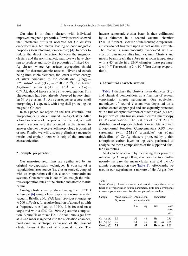
and a laser power fixed at 0.65 W, leading to Co–Ag
clusters with a mean diameter Dm of about 3.1 nm
(�0.1 nm) (�1150 atoms) and a standard deviation
s ¼ 0:40 (see Fig. 1). RBS measurements revealed in
this case an atomic composition of 57% Co–43% Ag
(�2%).
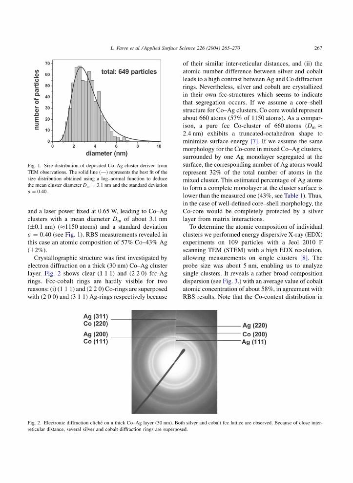
Crystallographic structure was first investigated by
electron diffraction on a thick (30 nm) Co–Ag cluster
layer. Fig. 2 shows clear (1 1 1) and (2 2 0) fcc-Ag
rings. Fcc-cobalt rings are hardly visible for two
reasons: (i) (1 1 1) and (2 2 0) Co-rings are superposed
with (2 0 0) and (3 1 1) Ag-rings respectively because
of their similar inter-reticular distances, and (ii) the
atomic number difference between silver and cobalt
leads to a high contrast between Ag and Co diffraction
rings. Nevertheless, silver and cobalt are crystallized
in their own fcc-structures which seems to indicate
that segregation occurs. If we assume a core–shell
structure for Co–Ag clusters, Co core would represent
about 660 atoms (57% of 1150 atoms). As a compar-
ison, a pure fcc Co-cluster of 660 atoms (Dm �2:4 nm) exhibits a truncated-octahedron shape to
minimize surface energy [7]. If we assume the same
morphology for the Co-core in mixed Co–Ag clusters,
surrounded by one Ag monolayer segregated at the
surface, the corresponding number of Ag atoms would
represent 32% of the total number of atoms in the
mixed cluster. This estimated percentage of Ag atoms
to form a complete monolayer at the cluster surface is
lower than the measured one (43%, see Table 1). Thus,
in the case of well-defined core–shell morphology, the
Co-core would be completely protected by a silver
layer from matrix interactions.
To determine the atomic composition of individual
clusters we performed energy dispersive X-ray (EDX)
experiments on 109 particles with a Jeol 2010 F
scanning TEM (STEM) with a high EDX resolution,
allowing measurements on single clusters [8]. The
probe size was about 5 nm, enabling us to analyze
single clusters. It reveals a rather broad composition
dispersion (see Fig. 3.) with an average value of cobalt
atomic concentration of about 58%, in agreement with
RBS results. Note that the Co-content distribution in
Fig. 1. Size distribution of deposited Co–Ag cluster derived from
TEM observations. The solid line (—) represents the best fit of the
size distribution obtained using a log–normal function to deduce
the mean cluster diameter Dm ¼ 3:1 nm and the standard deviation
s ¼ 0:40.
Fig. 2. Electronic diffraction cliche on a thick Co–Ag layer (30 nm). Both silver and cobalt fcc lattice are observed. Because of close inter-
reticular distance, several silver and cobalt diffraction rings are superposed.
L. Favre et al. / Applied Surface Science 226 (2004) 265–270 267
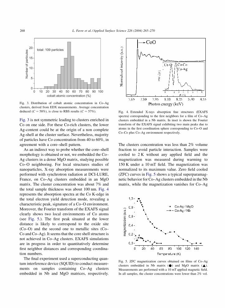
Fig. 3 is not symmetric leading to clusters enriched in
Co on one side. For these Co-rich clusters, the lower
Ag-content could be at the origin of a non complete
Ag-shell at the cluster surface. Nevertheless, majority
of particles have Co concentration from 40 to 60%, in
agreement with a core–shell pattern.
As an indirect way to probe whether the core–shell
morphology is obtained or not, we embedded the Co–
Ag clusters in a dense MgO matrix, studying possible
Co–O neighboring. For local structures studies of
nanoparticles, X-ray absorption measurements were
performed with synchrotron radiation at DCI-LURE,
France, on Co–Ag clusters embedded in an MgO
matrix. The cluster concentration was about 7% and
the total sample thickness was about 100 nm. Fig. 4
represents the absorption spectra at the Co K-edge in
the total electron yield detection mode, revealing a
characteristic peak, signature of a Co–O environment.
Moreover, the Fourier transform of the EXAFS signal
clearly shows two local environments of Co atoms
(see Fig. 5.). The first peak situated at the lower
distance is likely to correspond to the oxide site
(Co–O) and the second one to metallic sites (Co–
Co and Co–Ag). It seems that the core shell structure is
not achieved in Co–Ag clusters. EXAFS simulations
are in progress in order to quantitatively determine
first neighbor distances and corresponding coordina-
tion numbers.
The final experiment used a superconducting quan-
tum interference device (SQUID) to conduct measure-
ments on samples containing Co–Ag clusters
embedded in Nb and MgO matrices, respectively.
The clusters concentration was less than 2% volume
fraction to avoid particle interaction. Samples were
cooled to 2 K without any applied field and the
magnetization was measured during warming to
150 K under a 10 mT field. The magnetization was
normalized to its maximum value. Zero field cooled
(ZFC) curves in Fig. 5 shows a typical superparamag-
netic behavior for Co–Ag clusters embedded in the Nb
matrix, while the magnetization vanishes for Co–Ag
Fig. 3. Distribution of cobalt atomic concentration in Co–Ag
clusters, derived from EDX measurements. Average concentration
deduced (C ¼ 58%), is close to RBS results (C ¼ 57%). Fig. 4. Extended X-rays absorption fine structures (EXAFS
spectra) corresponding to the first neighbors for a film of Co–Ag
clusters embedded in a Nb matrix. In inset is shown the Fourier
transform of the EXAFS signal exhibiting two main peaks due to
atoms in the first coordination sphere corresponding to Co–O and
Co–Co plus Co–Ag environment respectively.
Fig. 5. ZFC magnetization curves obtained on films of Co–Ag
clusters embedded in Nb matrix (*) and MgO matrix (~).
Measurements are performed with a 10 mT applied magnetic field.
In all samples, the cluster concentrations were lower than 2% vol.
268 L. Favre et al. / Applied Surface Science 226 (2004) 265–270

clusters embedded in MgO matrix. Such a strong
reduction of magnetic properties can only be
explained by Co–O bonds for most Co–Ag clusters.
Thus, a core–shell structure is not clearly completely
achieved in Co–Ag clusters. Co atoms are not well
protected from matrix interaction by silver atoms.
4. Discussion
For Co–Ag/Nb films, the maximum magnetization
is achieved at a magnetic blocking temperature TB
above 10 K (Fig. 5). This temperature is proportional
to the magnetic anisotropy-energy of the clusters
which depends on both ‘‘the magnetic diameter’’ of
the particles and their environment. For comparison,
Table 2 shows the blocking temperatures (TB) and the
‘‘magnetic diameters’’ (Dmag) measured for pure Co-
clusters embedded in Nb and Ag matrices [4,9]. In the
first case (Co/Nb, miscible elements), the cluster–
matrix interface is composed of two magnetically-
dead monolayers. Therefore, Dmag is smaller than the
mean cluster diameter Dm determined from TEM-
observations [4] leading to a rather low blocking
temperature (TB � 12 K). A dominant surface-aniso-
tropy effect was observed in this case. On the contrary,
an abrupt interface has been observed for pure Co-
clusters embedded in a non-miscible Ag-matrix. In
this case, Dm ¼ Dmag, leading to a higher TB-value
(�30 K) and a dominant volume-anisotropy energy
[10]. For Co–Ag clusters, the low blocking tempera-
ture reported above could be due to the same effects: a
Co-core (Dmag � 2:3 nm) smaller than the clusters
diameter (Dm ¼ 3 nm), and probably a diffusion of
Nb-atoms in the Co-core to reduce the magnetic
volume since silver atoms segregated at the surface
do not form a perfect protective shell. Further experi-
mentation are in progress to determine the origin of the
magnetic anisotropy in mixed Co–Ag clusters.
5. Conclusion
The preparation of mixed Co–Ag clusters and the
studies of their structure/morphology are reported in
this paper. The mean size of deposited clusters
deduced from TEM observations is about 3.1 nm
and the average atomic-composition deduced from
RBS and EDX is 57% Co–43% Ag when using a
Co0.5 Ag0.5 target mounted in the laser vaporization
cluster source. From thermodynamic considerations,
Ag-atom segregation towards the cluster surface was
expected in such nanosystem leading to a Co–Ag
core–shell morphology and consequently to a protec-
tion of the Co-core by the Ag-surface layer. Electron
diffraction patterns indicate that both cobalt and silver
are crystallized in their own fcc-structure, which is a
first indication that segregation occurs. However,
EXAFS and SQUID-magnetometry measurements
reveal the presence of cobalt-oxygen bonds in the
case of Co–Ag clusters embedded in a MgO matrix.
This could be due to the formation of a non-perfect
core–shell structure and consequently a non-complete
protection of the Co-core by the Ag-segregated sur-
face layer. Consequently, the magnetic properties of
Co–Ag clusters, especially the magnetic blocking
temperature, are not improved with respect to the pure
Co-cluster ones. Further investigations to understand
the origin of the weak magnetic anisotropy in mixed
Co–Ag clusters are in progress as well as molecular
dynamic simulations to confirm the cluster morphol-
ogy. Studies of other systems of interest such as, i.e.
Co–Pt are also in progress.
Acknowledgements
The authors are indebted to O. Boirons, G. Guiraud
and C. Clavier for their continuous and efficient
technical assistances and developments during
LECBD experiments. The authors would like to thank
E. Bonet from the Laboratoire Louis Neel in Grenoble,
France for their collaborations on magnetic measure-
ments. Many thanks also to A. Traverse from the
LURE in Orsay, France for her assistance during
absorption experiments using the synchrotron radia-
tion sources. The authors gratefully acknowledge
support of part of this work from the EC (AMMARE
contract no. G5RD-CT 2001-00478).
Table 2
Magnetic blocking temperatures (TB) and magnetic diameters
(Dmag) for various cluster/matrix systems studied
Cluster/matrix TB (K) Dmag (nm)
Co–Ag 30 3.0
Co/Nb 12 2.3
Co–Ag/Nb 10 2.4
L. Favre et al. / Applied Surface Science 226 (2004) 265–270 269
References
[1] S. Sun, C.B. Murray, D. Weller, L. Folks, A. Moser, Science
287 (2000) 1989.
[2] D.J. Sellmyer, M. Yu, R.D. Kirby, Nanostruct. Mater. 12
(1999) 1021.
[3] J.L. Dormann, Rev. Phys. Appl. 16 (1981) 275.
[4] M. Jamet, V. Dupuis, P. Melinon, G. Guiraud, A. Perez, W.
Wernsdorfer, A. Traverse, B. Baguenard, Phys. Rev. B 62
(2000) 493.
[5] M. Gaudry, E. Cottancin, M. Pellarin, J. Lerme, L. Arnaud,
J.R. Huntzinger, J.L. Vialle, B. Broyer, Phys. Rev. B 67
(2003) 155409.
[6] A. Perez, P. Melinon, V. Dupuis, P. Jensen, B. Prevel, J.
Tuaillon, L. Bardotti, C. Martet, M. Treilleux, M. Broyer, M.
Pellarin, J.L. Vialle, B. Palpant, J. Lerme, J. Phys. D, Appl.
Phys. 30 (1997) 709.
[7] R. Van Hardeveld, F. Hartog, Surf. Sci. 15 (1969) 189.
[8] O. Holderer, T. Epicier, C. Esnouf, G.J. Fuchs, Phys. Chem. B
107 (8) (2003) 1723.
[9] M. Jamet, V. dupuis, C. Thirion, W. Wernsdorfer, P. Melinon,
A. Perez, Phys. Rev. Lett. 86 (2001) 4676.
[10] F. Parent, J. Tuaillon, V. Dupuis, B. Prevel, A. Perez, P.
Melinon, G. Guiraud, F. Parent, L.B. Steren, R. Morel, A.
Barthelemy, A. Fert, Phys. Rev. B 55 (1997) 3683.
270 L. Favre et al. / Applied Surface Science 226 (2004) 265–270