Embed Size (px)
Citation preview
Quasi-isotropic 3-D resolution in two-photon scanning microscopy
Cristina Ibáñez-López+, Genaro Saavedra+, and Gilbert Boyer Laboratoire d’Optique Appliquée, Ecole Polytechnique–Ecole Nationale Supérieure de Techniques Avancées, Centre
de l’Yvette, Chemin de la Hunière, 91761 Palaiseau, France + Permanent address: Departamento de Óptica, Universidad de Valencia, E46100 Burjassot, Spain
[email protected], [email protected], [email protected]
Manuel Martínez-Corral Departamento de Óptica, Universidad de Valencia, E46100 Burjassot, Spain
Abstract: One of the main challenges in three-dimensional microscopy is to overcome the lack of isotropy of the spatial resolution, which results from the axially-elongated shape of the point spread function. Such anisotropy gives rise to images in which significant axially-oriented structures of the sample are not resolved. In this paper we achieve an important improvement in z resolution in two-photon excitation microscopy through spatial modula-tion of the incident beam. Specifically, we demonstrate that the design and implementation of a simple shaded ring performs quasi-isotropic three-dimensional imaging and that the corresponding loss in luminosity can be easily compensated by most available femtosecond lasers. The outcome looks particularly relevant to nano-fabrication and optical manipulation.
© 2005 Optical Society of America
OCIS Codes: (180.5810) Scanning microscopy; (1000.6640) Superresolution; (100.1220) Aper-tures.
References and Links 1. Abbe, E. Arch. Mikrosk. Anat. 9, 413–468 (1873). 2. T. R. M. Sales, "Smallest focal spot," Phys. Rev. Lett. 81, 3844-3847 (1998). 3. J. Miao, T. Ishikawa, B. Johnson, E. K. Anderson, B. Lai and K. O. Hodgson, "High resolution 3D x-ray
diffraction microscopy," Phys. Rev. Lett. 89, 088303 (2002). 4. J. Miao, T. Ohsuna, O. Terasaki, K. O. Hodgson and M. A. O’Keefe, "Atomic resolution three-dimensional
electron diffraction microscopy," Phys. Rev. Lett. 89, 155502 (2002). 5. C. M. Blanca and S. W. Hell, "Axial superresolution with ultrahigh aperture lenses," Opt. Express 10, 893-
898 (2002). 6. J. B. Pawley, ed., Handbook of biological confocal microscopy. Plenum Press, New York, 1995. 7. The term superresolution introduced here is understood in the sense of Rayleigh criterion; i. e., as the nar-
rowness of the PSF or, equivalently, the enhancement of the OTF for frequencies under the cut-off fre-quency.
8. S. Lindek, J. Swoger and E. H. K. Stelzer, "Single-lens theta microscopy: resolution, efficiency and work-ing distance," J. Mod. Opt. 46, 843-858 (1999).
9. B. Bailey, D. L. Farkas, D. Lansing-Taylor and F. Lanni, "Enhancement resolution in fluorescence micros-copy by standing-wave excitation," Science 366, 44-48 (1993).
10. J. Huisken, J. Swoger, F. Del Bene, J. Wittbrodt and E. H. K. Stelzer, "Optical sectioning deep inside live embryos by selective plane illumination microscopy," Science 305, 1007-1009 (2004).
11. M. A. A. Neil, R. Juskaitis, T. Wilson, Z. J. Laczik and V. Sarafis, "Optimized pupil-plane filters for confo-cal microscope point-spread function engineering," Opt. Lett. 25, 245-247 (2000).
12. C. J. R. Sheppard, "Binary optics and confocal imaging," Opt. Lett. 24, 505-506 (1999). 13. M. Martínez-Corral, M. T. Caballero, E. H. K. Stelzer and J. Swoger, "Tailoring the axial shape of the point
spread function using the Toraldo concept," Opt. Express 10, 98-103 (2002). 14. G. Boyer, "New class of axially apodizing filters for confocal scanning microscopy," J. Opt. Soc. Am. A 19,
584-589 (2002). 15. M. Martínez-Corral, C. Ibáñez-López, G. Saavedra and M. T. Caballero, "Axial gain in resolution in optical
sectioning fluorescence microscopy by shaded-ring filters," Opt. Express 11, 1740-1745 (2003). 16. S. S. Sherif and P. Török, "Pupil plane masks for super-resolution in high-numerical-aperture focusing," J.
Mod. Opt. 51, 2007-2019 (2004).
(C) 2005 OSA 8 August 2005 / Vol. 13, No. 16 / OPTICS EXPRESS 6168#7916 - $15.00 US Received 23 June 2005; revised 28 July 2005; accepted 29 July 2005
17. W. Denk, J. H. Strickler and W. W. Webb, "Two-photon laser scanning fluorescence microscopy," Science 248, 73-76 (1990).
18. C. M. Blanca, J. Bewersdorf and S. W. Hell, "Single sharp spot in fluorescence microscopy of two oppos-ing lenses," Appl. Phys. Lett. 79, 2321-2323 (2001).
19. T. A. Klar, S. Jakobs, M. Dyba, A. Egner and S. W. Hell, " Fluorescence microscopy with diffraction reso-lution barrier broken by stimulated emission," Proc. Natl. Acad. Sc. 97, 8206-8210 (2000).
20. M. Dyba and S. W. Hell, "Focal spots of size lambda/23 open up far-field fluorescence microscopy at 33 nm axial resolution," Phys. Rev. Lett. 88, 163901 (2002).
21. C. Ibáñez-López, I. Escobar, G. Saavedra and M. Martínez-Corral, "Optical sectioning improvement in two-color excitation scanning microscopy," Microsc. Res. Tech. 64, 96-102 (2004).
22. The eccentricity is defined here as 22 /1 abe −= , where a and b account for the lengths of the semimajor and semiminor axes, respectively.
23. S. Kawata, H.-B. Sun, T. Tanaka and K. Takada, "Finer features for functional microdevices," Nature 412, 697-698 (2001).
24. D. G. Grier, "A revolution in optical manipulation," Nature 424, 810-816 (2003). 25. B. Richards and E. Wolf, Proceedings of the Royal Society (London) A 253, 358 (1959). 26. P. Török and P. Varga, “Electromagnetic diffraction of light focused through a stratified medium,” Appl.
Opt. 36, 2305-2312 (1997) 27. O. Haeberlé, “Focusing of light through a stratified medium: a practical approach for computing micro-
scope point spread functions. Part I: Conventional microscopy ,” Opt. Commun. 216, 55-63 (2003).
1. Introduction
The spatial resolution of imaging systems is mainly restricted by diffraction, that is, by their capacity for producing a tight diffraction spot when imaging a light point source. Since Abbe formulated his wave theory for microscopic imaging [1], the improvement of resolution of optical microscopes has been the aim of many research efforts. The need for smaller spot sizes is desirable, not only in the case of optical frequencies [2], but also in other spectral ranges like x-ray microscopy [3] or electron microscopy [4]. In the search for the maximum resolu-tion, emphasis has been generally placed on the use of modern microscope objectives with the highest available numerical aperture (NA) [5]. But even with the best optical elements the use of wide-field conventional optical microscopes to image three-dimensional (3-D) biological fluorescent samples has the drawback that any image focused at a certain depth in the sample contains blurred information from the rest of the sample. This fact gives rise to 3-D images with deteriorated contrast.
To overcome this problem, the use of scanning fluorescence techniques has been demon-strated and routinely used. Among them we cite first single-photon confocal microscopes (SPCM). The main advantage of SPCM is its sectioning power, due to the light rejection from out-of-focus parts of the sample [6]. But since the diffraction pattern of a focused bright spot is naturally prolate shaped for any NA, SPCM suffers from anisotropic imaging. As a specific feature, super-resolving apodization [7] can be considered complementary and compatible to many other approaches for reducing the imaging anisotropy such as confocal theta microscopy [8], standing wave microscopy [8], or selective plane illumination microscopy [10]. Apodiza-tion has been the subject of increased interest in the past few years due to the demands of data storage, nano-fabrication and optical manipulation. Filters designed to obtain a moderate gain in resolution remain easy to implement, despite the strong unbalance between the few experi-mental results and the many theoretical solutions, among which continuous, binary, purely-absorbing, complex-transmittance and, more recently, purely-dephasing filters have been pro-posed [11-16].
A serious drawback of SPCM is photobleaching, which occurs since the entire sample is bleached when any single plane is imaged. Another drawback of this technique when used in biomedical imaging is its poor depth-penetration capacity. To solve these problems, two-photon excitation (TPE) scanning microscopy was demonstrated [17]. This non-linear imag-ing technique relies on the simultaneous absorption of two photons, whereby a single fluores-cence photon is emitted. The overall fluorescent light is collected and finally the image is syn-thesized from the 3-D sampling of the object. TPE generally uses near-infrared light due to its lower absorption and scatter in biological tissues, which allows deeper penetration of the exci-tation beam. Besides, photobleaching is restricted to the neighborhood of the imaged plane.
(C) 2005 OSA 8 August 2005 / Vol. 13, No. 16 / OPTICS EXPRESS 6169#7916 - $15.00 US Received 23 June 2005; revised 28 July 2005; accepted 29 July 2005
Several attempts have been made to improve the axial resolution of TPE scanning micro-scopes. This is the case of 4Pi microscopes in which two opposing, high-NA objectives are use to create an interference illumination spot with very narrow axial central lobe [18], or the combination between STED microscopy and 4Pi microscopy [19,20].
To implement the above-cited methods a strong modification of the TPE microscope ar-chitecture is needed. Specifically, the need for multiple objectives makes the system align-ment a very complicated issue. Here we concentrate on apodization of the TPE mode with the aim of reducing anisotropic imaging, by pupil spatial filtering with a shaded-ring filter (SR) [21], often dubbed leaky filter. We demonstrate that a close to spherical 3-D PSF can be ob-tained through the implementation of a relatively inexpensive spatial filter. Interestingly, the measured PSF has no significant collateral enhancement of secondary lobes, and lateral broadening of the central core is not observed. Approximated as an ellipsoid, the engineered PSF exhibits an eccentricity reduced from e=0.72 to e=0.48 [22]. The quasi-spherical spot can be very useful not only for micro-imaging applications, but also in other techniques like nano-fabrication [23] or optical manipulation [24].
2. Description of the method
To describe our approach, we start by considering a monochromatic, linearly-polarized plane wave that impinges on an aberration-free high-aperture spherical lens. Assuming that sine condition holds, and according to electromagnetic focusing theory of Richards and Wolf [25], the electric field distribution in the focal region is given by
( ) ( ) ( )[ ] ( ) ( ) kjiE ϕ+ϕ+ϕ+=ϕ cos,I22sin,I2cos,I,I,, 1220 zrizrzrzrzr (1)
where
( ) ( ) ( ) ( ) ( )∫α
θθθθθ+θθ=0
00 sincosexpsinJcos1cosP,I dzikrkzr
( ) ( ) ( ) ( )∫α
θθθθθθθ=0
11 sincosexpsinJsincosP,I dzikrkzr (2)
( ) ( ) ( ) ( ) ( )∫α
θθθθθ−θθ=0
22 sincosexpsinJcos1cosP,I dzikrkzr
In the above equations, P(θ) accounts for the amplitude transmittance at the aperture stop of the focusing lens, whereas α is the maximum value of the aperture angle θ. Parameter ϕ represents the angle between the polarization direction of the incident field (assumed x-direction) and the observation meridian plane. More accurate calculations should take into account the influence of the transmission coefficients of the various interfaces inherent to the stacked structures commonly used in microscopy [26, 27]. However, the use of these more accurate models would not change significantly the results. Even if the illumination setup is not telecentric, TPE scanning microscopes are 3-D linear and shift-invariant systems due to their scanning nature, provided that the illumination spot is not affected by spherical aberra-tion when going deeper into the specimen. Therefore, the image can be described as the con-volution between the 3-D PSF and the 3-D function that describes the spatial distribution of fluorescence generated by the object. If we consider that, in the optimum case, no photo-bleaching affects the sample and no saturation effects occur, then the PSF is given by
( ) ( )4,,,,PSF ϕ=ϕ zrzr E . (3)
Our aim here is the design and production of elements that narrow the PSF along the axial direction. The narrowness should be produced at the expense of neither a deterioration of the
(C) 2005 OSA 8 August 2005 / Vol. 13, No. 16 / OPTICS EXPRESS 6170#7916 - $15.00 US Received 23 June 2005; revised 28 July 2005; accepted 29 July 2005

lateral resolution nor an enlargement of axial sidelobes. The utility of SR filters has been demonstrated, only by numerical simulations, in single-photon confocal microscopy [15]. Here we adapt the SR concept to TPE and experimentally demonstrate, for the first time, their capacity for producing 3-D superresolution. The design of the proper SR filter must take into account the lens NA and the illumination polarization state. In our experiment we used as the illumination source a mode-locked Cr4+:fosterite laser. The laser produced pulses with central wavelength λ = 1260 nm and a mean temporal width Δt = 50 fs at a repetition rate of 84 MHz. An average power over 200 mW was available for the experiment. The polarization of the beam was rigorously linear. In our analysis we set the coordinate system so that the incident light is seen as x-polarized. For the illumination and collection we used two identical 1.2 NA (water-immersion) objectives from Olympus (Uplan APO/IR 60x). The maximum gain in axial resolution that can be, theoretically, achieved with purely absorbing screen 1.73. But this ideal gain is achieved at the expense of blocking all incident light. When realistic experiments are considered, the maximum gain achievable is about 1.30. In our case we selected 1.25 as the value of the gain in axial resolution. As the result of the optimization procedure we de-
signed the SR filter that maximizes the light-spot efficiency (evaluated as the fluorescence intensity emitted from the central lobe divided by the overall fluorescence intensity). The filter is shown in Fig. 1. At opening between º151 =θ and º622 =θ the amplitude transmittance of the designed filter is %19 .
In Fig. 2 we compare the, numerically evaluated, PSFs corresponding to the standard 1.2 NA objective, with the one in which the SR filter is inserted as the aperture stop. The calculations were performed according to Eq. (3), for the case of ϕ=0. These calculations predict: (a)
Fig. 1. Amplitude transmittance of the SR filter. The transmittance of the shaded ring is 19%.
Fig. 2. Plot of the numerically evaluated 3-D PSF, in the meridian plan 0=ϕ , corresponding to: (a) Standard 1.2 NA objective with clear pupil; and (b) Same objective but with the SR filter inserted as the aperture stop. Axes labels are expressed in microns.
(C) 2005 OSA 8 August 2005 / Vol. 13, No. 16 / OPTICS EXPRESS 6171#7916 - $15.00 US Received 23 June 2005; revised 28 July 2005; accepted 29 July 2005
length reduction of the PSF by 21% (as evaluated in terms of the full-width at half maximum (FWHM)); (b) a very small lateral swelling (4% in terms of FWHM); and (c) a very low dimming of the light-spot efficiency, which decreases from 99% for the clear-aperture to 87% for the proposed SR.
3. Experimental results
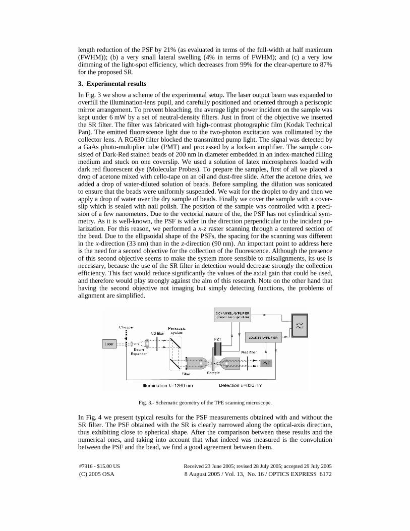
In Fig. 3 we show a scheme of the experimental setup. The laser output beam was expanded to overfill the illumination-lens pupil, and carefully positioned and oriented through a periscopic mirror arrangement. To prevent bleaching, the average light power incident on the sample was kept under 6 mW by a set of neutral-density filters. Just in front of the objective we inserted the SR filter. The filter was fabricated with high-contrast photographic film (Kodak Technical Pan). The emitted fluorescence light due to the two-photon excitation was collimated by the collector lens. A RG630 filter blocked the transmitted pump light. The signal was detected by a GaAs photo-multiplier tube (PMT) and processed by a lock-in amplifier. The sample con-sisted of Dark-Red stained beads of 200 nm in diameter embedded in an index-matched filling medium and stuck on one coverslip. We used a solution of latex microspheres loaded with dark red fluorescent dye (Molecular Probes). To prepare the samples, first of all we placed a drop of acetone mixed with cello-tape on an oil and dust-free slide. After the acetone dries, we added a drop of water-diluted solution of beads. Before sampling, the dilution was sonicated to ensure that the beads were uniformly suspended. We wait for the droplet to dry and then we apply a drop of water over the dry sample of beads. Finally we cover the sample with a cover-slip which is sealed with nail polish. The position of the sample was controlled with a preci-sion of a few nanometers. Due to the vectorial nature of the, the PSF has not cylindrical sym-metry. As it is well-known, the PSF is wider in the direction perpendicular to the incident po-larization. For this reason, we performed a x-z raster scanning through a centered section of the bead. Due to the ellipsoidal shape of the PSFs, the spacing for the scanning was different in the x-direction (33 nm) than in the z-direction (90 nm). An important point to address here is the need for a second objective for the collection of the fluorescence. Although the presence of this second objective seems to make the system more sensible to misalignments, its use is necessary, because the use of the SR filter in detection would decrease strongly the collection efficiency. This fact would reduce significantly the values of the axial gain that could be used, and therefore would play strongly against the aim of this research. Note on the other hand that having the second objective not imaging but simply detecting functions, the problems of alignment are simplified.
Fig. 3.- Schematic geometry of the TPE scanning microscope.
In Fig. 4 we present typical results for the PSF measurements obtained with and without the SR filter. The PSF obtained with the SR is clearly narrowed along the optical-axis direction, thus exhibiting close to spherical shape. After the comparison between these results and the numerical ones, and taking into account that what indeed was measured is the convolution between the PSF and the bead, we find a good agreement between them.
(C) 2005 OSA 8 August 2005 / Vol. 13, No. 16 / OPTICS EXPRESS 6172#7916 - $15.00 US Received 23 June 2005; revised 28 July 2005; accepted 29 July 2005

Fig. 4.- 2D gray-scale sections of the experimental 3-D PSFs as displayed by LabView. (a) PSF obtained with the standard 1.2 NA objective with clear pupil; and (b) PSF obtained with the same objective but with the SR filter inserted as the aperture stop.
To get a better insight into the experimental results, we have plotted, in Fig. 5, the axial and lateral profiles of the above measured PSFs. Note from this figure that no significant dila-tion is produced in the lateral direction. However, the axial FWHM is reduced from 1.43 μm to 1.13 μm. Since the lateral FWHM is, in both cases, about 0.99 μm, the eccentricity of the 3-D PSF is reduced by 33%.
Fig. 5.- Normalized axial and lateral PSF produced by the Olympus 1.2 NA objective (x). With empty dots (ο) we show the experimental results obtained when the SR filter is inserted in the illumination path.
4. Conclusions
In summary, we have demonstrated that quasi-isotropic 3-D resolution can be produced in TPE scanning microscopy by spatial modulation of the illumination beam, which involves a small modification of the microscope architecture. The proposed amplitude modulator, the SR filter, has a very simple structure: a shaded ring centered in the objective aperture stop. We have implemented the filter in a quite simple manner, and obtained the quasi-spherical spot using a conventional TPE scanning architecture. The outcome can be very useful not only in 3-D microscopy, but also in nano-fabrication and optical manipulation. The experimental re-sults are in quite good agreement with the theoretical predictions.
Acknowledgments
The authors wish to acknowledge stimulating discussions with Dr. P. Török (Imperial Col-lege, London). We also acknowledge Dr. K. Plamann for his help with the signal-processing software. We are also indebted to an anonymous, for his/her comments and suggestions,
(C) 2005 OSA 8 August 2005 / Vol. 13, No. 16 / OPTICS EXPRESS 6173#7916 - $15.00 US Received 23 June 2005; revised 28 July 2005; accepted 29 July 2005

which indeed help to improve the paper. This work was conducted in the frame of the Acción Integrada HF2003-0257, Ministerio de Ciencia y Tecnología, Spain and the Action Intégrée 07181VG Ministère des Affaires Etrangères, France, and mainly funded by the Plan Nacional I+D+I (grant DPI2003-4698), Ministerio de Ciencia y Tecnología and by the Generalitat Va-lenciana, Spain (Grant GV04B-186).
(C) 2005 OSA 8 August 2005 / Vol. 13, No. 16 / OPTICS EXPRESS 6174#7916 - $15.00 US Received 23 June 2005; revised 28 July 2005; accepted 29 July 2005