Embed Size (px)
Citation preview
538
Acta Cryst. (1987). B43, 538-543
Structural Studies of 2-Phenylthiazolidine
BY F. BAERT AND M. MULLER
Laboratoire de Dynamique des Cristaux Mol~culaires, UA 801 CNRS, Universitd des Sciences et Techniques de Lille 1, 59655 Villeneuve d'Ascq CEDEX, France
AND D. BARBRY AND D. COUTURIER
Laboratoire de SynthOse Organique, B~timent C 4 - UFR de Chimie, Universit~ des Sciences et Techniques de Lille 1, 59655 Villeneuve d'Ascq CEDEX, France
(Received 9 June 1986; accepted 12 March 1987)
Abstract
The crystal structure of 2-phenylthiazolidine (CgHI~NS, Mr=165.24) was solved by direct methods using three-dimensional X-ray diffraction data. The structure at 293 K is orthorhombic, space group P212~2~ with a = 18.148 (9), b = 8.397 (3), c= 5.627 (3) A, V=857.5 A3, Z = 4 , Dx = 1.27 Mgm -3, Mo K6, h = 0.7107 A, /z = 0.263 mm -~, F(000) = 352, R=0 .086 for 879 reflections with 1>3o-(I) . Only one H atom has been located from a difference synthesis. At 132 K, R =0.027 for 1881 reflections with I > 3 t r ( 1 ) recorded with the same crystal. No phase transition was observed, a = 18.013 (3), b=8.299(2) , c = 5 . 5 7 7 ( 1 ) A , V=833.7A3, Dx = 1 .31Mgm -3. All the H atoms were found on a difference map with this low-temperature data. A rigid-body thermal-motion analysis revealed a large libration of the molecule around the smallest axis of inertia at 293 K, which decreases strongly at low temperature. Structural results agree with 1H NMR studies in solution. Charge-density deformation maps exhibit unidentified peaks near the S atom.
Introduction
Thiazolidines are important heterocyclic systems in biology and medicine. They constitute the backbone of the penicillin structure and are formed in vivo from cysteine and endogenous aldehydes.
Very few papers report crystal structures of thiazolidines, and only the 4-carboxylate derivatives have received attention. These derivatives have been shown to exist in amino acid or zwitterionic forms according to the other substituents of the ring. (S)- Thiazolidine-4-carboxylic acid (Goodman, Chen, Benedetti, Pedone & Corradani, 1972; Loscalzo, Kal- len & Voet, 1973) adopts a zwitterionic form where the conformation of the thiazolidine ring is an envelope with S at the summit. An example of the amino acid form was found by Howard-Lock, Lock & Smalley (1985) in the structure of (S)-2,2,5,5- tetramethylthiazolidine-4-carboxylic acid.
0108-7681/87/060538-06501.50
We have investigated a great number ofthiazolidine derivatives by NMR spectroscopy; one of them, 2- phenylthiazolidine, known for its radioprotective properties, gave a suitable crystal for X-ray study.
Experimental
Synthesis
2-Phenylthiazolidine was obtained by condensa- tion of 2-aminoethanethiol hydrochloride and an excess of benzaldehyde with a suitable alkaline treat- ment, according to Schmolka & Spoerri (1957).
Data collection, structure solution and refinement
A plate-like crystal of dimensions 0 .15x0 .3x 0.5 mm was sealed in a Lindemann capillary for the X-ray study. Accurate cell dimensions from 25 reflec- tions with 20 <- 0 - 27 ° and intensities of Bragg reflec- tions were measured by an automatic four-circle diffractometer (Philips PW 1100) in the o9-20 mode using Mo Ka radiation. Two data sets at 293(2) and 132(0.5) K were recorded using similar o9-20 scans, where the scan width was fixed to 1.3 °, scan speed = 0.015° min -1"
2208 intensities up to (sin 0)/A <0 .807A -1 ( 0 - h_<28, 0_< k--- 13, 0 - < l - 9 ) were recorded at room temperature and 2114 intensities up to (sin 0)/A < 0.807,~ -~ (0--- h-<28, 0<-k<- 13, 0-< l-<9) were col- lected at low temperature. Three standard intensities monitored at intervals of 2 h showed no significant variation. Both data sets were corrected for Lorentz and polarization factors, but no absorption and extinction corrections were made. 879 reflections with 1> 3o-(1) in the high-temperature data set and 1881 reflections with 1 > 3o"(1) in the low-temperature data set.
The unit cell was found to be orthorhombic, space group P2~212~ with four molecules situated at general positions. The structure was solved by direct methods (MULTAN; Germain, Main & Woolfson, 1971) using 499 reflections of the high-temperature data set with normalized structure factors E -> 1.236. Only one
O 1987 International Union of Crystallography
BAERT, MULLER, BARBRY A N D COUTURIER 539
Table 1. Atomic coordinates (xl05, ×103 f o r H) and equivalent isotropic thermal parameters (A2 x 102) at
132 K
Ueq = 1~.. 2 U i .a*, a*. a, . aj. i j ] ]
x y z Ueq
S 71661 (2) 12470 (5) 12661 (8) 15 (1) N 76255 (7) 39080 (16) 35467 (28) 16 (1) C(2) 70691 (9) 26581 (18) 38885 (34) 15 (1) C(4) 83066 (9) 30865 (21) 27470 (35) 18 (1) C(I1) 58785 (9) 28969 (20) 61772 (36) 18 (1) C(10) 51655 (9) 35153 (23) 64837 (36) 21 (2) C(6) 62874 (8) 32865 (18) 41406 (30) 14 (1) C(7) 59782 (10) 42987 (23) 24204 (34) 24 (1) C(5) 81143 (10) 19219 (22) 6757 (37) 21 (2) C(9) 48629 (9) 45374 (22) 47874 (80) 22 (1) C(8) 52700 (10) 49325 (24) 27510 (37) 27 (2)
H(C2) 716 (1) 195 (2) 533 (4) 36 (5) H(N) 750 (1) 458 (2) 252 (3) 27 (6) H(CS) 817 (1) 250 (2) -86 (4) 24 (6) H'(C5) 851 (1) 92 (2) 61 (3) 38 (6) H(C4) 871 (1) 390 (2) 236 (3) 19 (5) H'(C4) 854 (1) 244(2) 418(3) 24(5) H(C7) 622 (I) 452 (2) 108 (3) 24 (5) H(C8) 508 (1) 564 (2) 156 (3) 27 (5) H(C9) 436 (l) 496 (2) 492 (3) 15 (4) H(CI0) 490 (1) 326 (2) 799 (4) 40 (7) H(CI 1) 608(1) 221 (2) 732(3) 29(6)
H atom was located from this set of data, and the final wR -- 0.087% for the 879 reflections above 3o'(I) (Sheldrick, 1976). All the H atoms were found for the low-temperature data set, using 1881 intensities above 3o-(1); wR--0 .023%. Positions and isotropic Debye-Waller factors of the H atoms were refined. Unit weights were used for the high-temperature data set while for the low-temperature data w is defined as 1 / 0"2(Fo) were 0"2(Fo) 2 m. 0.counting- {- (0"02 F2o) 2.
Scattering factors for S, C and N were taken from International Tables for X-ray Crystallography (1974) while the factors for H were those of Stewart, David- son & Simpson (1965). The absolute values of peaks and troughs in the final difference Fourier maps are respectively 0.3, 0.2 e,~-3 for the low-temperature data set and 0.4 and 0 - S e ~ -3 for the high- temperature data. (A/0")max are respectively 0.2 and 0.005 for the high- and low-temperature structures; the atom positions corresponding to the best R values of the low-temperature structure are listed in Table 1.* Table 2 gives bond lengths and angles.
Thermal vibration analysis
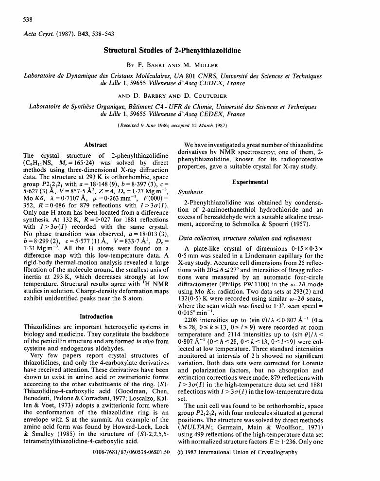
Thermal motions of S, C, N were described by anisotropic parameters while isotropic ones were assigned to the H atoms. Fig. 1 shows the thermal
* Lists of structure factors, anisotropic thermal parameters, mean planes and intermolecular distances have been deposited with the British Library Document Supply Centre as Supplementary Publi- cation No. SUP 43804 (23 pp.). Copies may be obtained through The Executive Secretary, International Union of Crystallography, 5 Abbey Square, Chester CH1 2HU, England.
Table 2. Bond lengths (,~ ) and bond angles (o) with e.s.d.'s in parentheses
S-C(2) 1.882 (2) N-H(N) 0.830 (18) S-C(5) 1-827 (2) C(2)-H(C2) 1.011 (18) N-C(2) 1.454 (2) C(4)-H(C4) 1.032 (15) N-C(4) 1.472 (2) C(4)-H'(C4) 1.048 (18) C(2)-C(6) 1-508 (2) C(11)-H(C11) 0.926 (18) C(4)-C(5) 1-545 (3) C(10)-H(C10) 0.986 (20) C(11)-C(10) 1-393 (2) C(7)-H(C7) 0.886 (18) C(11)-C(6) 1.392 (2) C(5)-H(C5) 0.987 (20) C(10)-C(9) 1-382 (4) C(5)-H'(C5) 1.101 (15) C(6)-C(7) 1.391 (2) C(9)-H(C9) 0.972 (14) C(7)-C(8) 1.392 (3) C(8)-H(C8) 0.949 (18) C(9)-C(8) 1.391 (4)
C(2)-S-C(5) 92-1 (1) C(ll)-C(10)-H(C10) 118.] (]1) C(2)-N-C(4) ]06.5 (1) C(9)-C(10)-H(CI0) ]2].4 (] l ) C(2)-N-H(N) 112-6 (12) C(2)-C(6)-C(11) ]19.3 (1) C(4)-N-H(N) ]09.0 (]2) C(2)-C(6)-C(7) 121.2 (]) S-C(2)-N 106.1 (1) C(11)-C(6)-C(7) 119.4(2) S-C(2)-C(6) 112.0 (1) C(6)-C(7)-C(8) 120.2 (2) S-C(2)-H(C2) 103.8 (10) C(6)-C(7)-H(C7) 120.7 (11) N-C(2)-C(6) 114.1 (1) C(8)-C(7)-H(C7) 119.1 (11) N-C(Z)-H(C2) 114.0(10) S-C(5)-C(4) 105.4(1) C(6)-C(2)-H(C2) 106.4 (10) S-C(5)-H(C5) 113.4 (10) N-C(4)-C(5) 109.2 (I) S-C(5)-H'(C5) 112"5 (9) N-C(4)-H(C4) 111-3 (9) C(4)-C(5)-H(C5) 109.1 (10) N-C(4)-H'(C4) 109.9 (9) C(4)-C(5)-H'(C5) 110.8 (9) C(5)-C(4)-H(C4) 114.6 (9) H(C5)-C(5)-H'(C5) 105-7 (14) C(5)-C(4)-H'(C4) 109.9 (9) C(10)-C(9)-C(8) 119.7 (2) H(C4)-C(4)-H'(C4) 101.7 (13) C(10)-C(9)-H(C9) 122-4 (9) C(10)-C(l 1)-C(6) 120.1 (2) C(8)-C(9)-H(C9) 117.8 (9) C(10)-C(11)-H(C11) 120.3 (11) C(7)-C(8)-C(9) 120.1 (2) C(6)-C(11)-H(C11) 119.6(11) C(7)-C(8)-H(C8) 118-4(10) C(I 1)-C(10)-C(9) 120.3 (2) C(9)-C(8)-H(C8) 121.4 (10)
ellipsoids of the atoms of 2-phenylthiazolidine at low and high temperatures. T, L and S tensors describing the thermal motion of the molecule were fitted to the Uu's of the S, C and N atoms using the least-squares procedure proposed by Schomaker & Trueblood (1968). The elements of these tensors are given in Table 3. The r.m.s, differences between observed U 0 and those calculated from TLS tensors do not exceed 0.0016A 2 (e.s.d.=0.0019) in the low-temperature state. The estimated precision of the ~ , from the least-squares refinement is about 0.0008/~2, so there are still significant non-rigid-body motions in the molecule even at 132 K.
The eigenvalues of T represent an isotropic transla- tion of the whole molecule, the motion being halved
H(CS)
C(SJ H'(C5) H(C8) ;(C7)
"~1C11)
H(C10)I
Fig. 1. Perspective view and numbering of the molecule: low- temperature state.
540 2 - P H E N Y L T H I A Z O L I D I N E
Table 3. Rigid-body vibration parameters for all the heavy atoms of the molecule
Low temperature High temperature T (A2x 10 -4) 165 (6) 1(6) - 1 4 (6) 337 (20) - 8 (21)
152 (12) 22 (8) 273 (33) 136 (10)
L (rad2x 10 -4) 30 (7) 1 (3) -5 (3) 105 (24) -18 (9) 13 (2) 7 (8) 32 (6)
10 (2) S (Aradx 10 -5) 76 (55) 52 (35) 73 (38) 167 (183) 52 (116)
-34(23) -46(31) 0(17) -166(76) -94(103) -42(25) -96(17) -30(189) -61(82) -207(56)
R.m.s. U(obs.)- U(calc.)(A 2) 0.0016 0.0052 E.s.d. Uj(obs.)(A 2) 0.0019 0.0063
- 2 3 (21) 27 (27)
327 (32) -26 (9) 24 (5) 35 (6)
205 (126) 118(57) -72(628)
Table 4. T and L referred to the inertial axes of the molecule
Low temperature
Principal axes Ine~ial Direct ion cosine (A 2 x 10 -") T (A 2 x 10 -4) (o) L (rad 2 x 10⁴) moment x y z
135 173 -5 -7 9.7 30 3 -1 43 0"890 -0"315 -0-328 159 159 -6 4.2 19 - I 202 0.449 0.732 0-513 115 117 3'1 3 209 0'078 -0-604 0.792
High temperature 363 357 -3 -22 34-5 105 2 -4 43 0"890 -0"318 -0.324 308 299 22 10'6 42 4 201 0.449 0.724 0"522 257 272 11"3 10 208 0'069 -0"611 0.788
at 132 K; also, there is a threefold difference between the l ibration motions of the molecule at room tem- perature and at 132 K. In Table 4 the quantit ies T and L are referred to the inertial axes of the molecule. At both temperatures the off-diagonal elements of the l ibration tensor L are quite small and it can be assumed that the molecule librates around the inertial axes.
The largest ampl i tude of vibration (5.8 ° ) at high temperature is around the axis with the smallest moment of inertia, the electronic density near the H atoms being strongly smeared by these l ibration motions and only the HC(9) atom close to the axis with the smallest moment of inertia was localized on a difference map.
Discuss ion
Molecular geometry
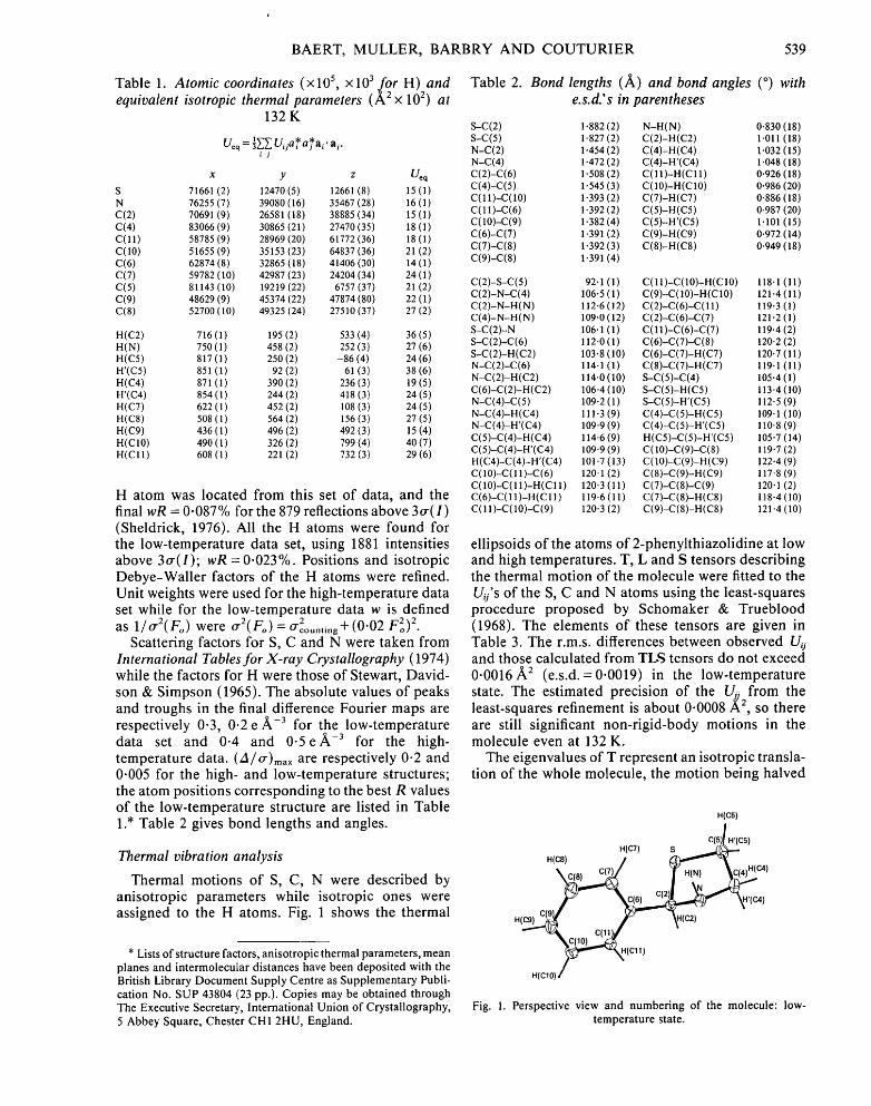
The bond lengths and bond angles of the molecules are reported in Table 2. Fig. 2(a) illustrates a com- parison of the bond lengths and angles of the thiazolidine ring found in our study with the values described by Howard-Lock et al. (1985) in 2,2,5,5- tetramethylthiazolidine-4-carboxylic acid. Bond angles and bond lengths are pretty close al though the average C-S distance is larger. The lengthening of the C-S bond claimed by Howard-Lock et al. with respect to the results found in thiazolidine-4- carboxylic acid (mean value 1.799 A) (Loscalzo et al., 1973) is probably not caused by the steric require- ments of the methyl groups. The compound described by Howard-Lock et al. can best be pictured by a plane through C(5)-S-C(2) , with N and C(4) atoms twisted out of the plane on opposite sides at 0.382 (4) and
-0 .269 (5) A (half-chair conformation). In contrast, 2-phenyl thiazol idine has an envelope conformat ion at 132 K with the N atom 0.596/~ out of the plane formed by C(2) -S-C(5) -C(4) . The phenyl substituent is equatorial and perpendicular to the mean plane of the heterocyclic ring.
1.54 7(8) C(5) 1.545{2) C(4)
/ !o2 .6 (4) lO9.s~5~ 1.839(7)/ 105.4(,1) !09.2(1) ~
1 . 8 2 7 ( ~ - - - - \ 1.469(7) / ~ 1.472(2) / 94.9 (3)
S < ~ 108.2(4) / 106.5 (1) 3 N
104.7(4) ~ 1.860(6) ~ 106.1(1) / ~ ~ / 1.473 (7)
C(2)
(a)
-:)5.1(5) C(5) -a1.1(1) C(4)
10.8 (4, / \49
C(2) -42.5(1)
(b)
Fig. 2. Comparison of the conformations of the thiazolidine ring observed in (S)-2,2,5,5-tetramethylthiazolidine-4-carboxylic acid and 2-phenylthiazolidine: (a) bond lengths (A), valence angles (o); (b) torsional angles (°). The underscored values refer to this study.
BAERT, MULLER, BARBRY AND COUTURIER 541
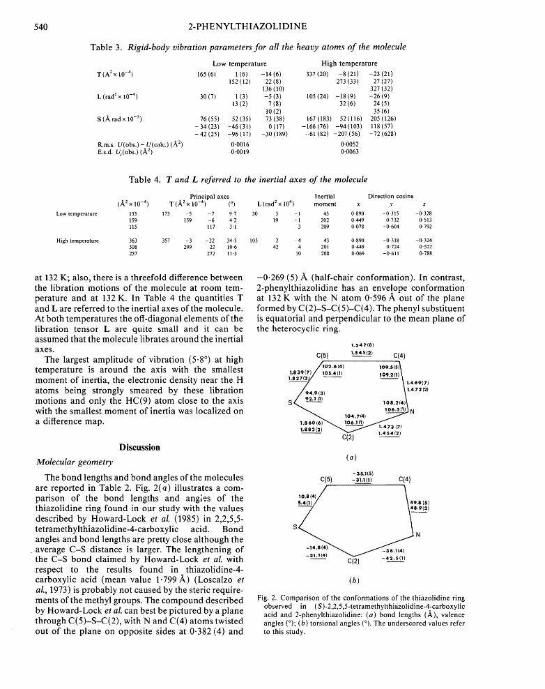
2-Phenylthiazolidine was also analyzed in solution by ~H NMR spectroscopy. The H(4)/H(5) part of the 400 MHz spectrum was first solved by hand and then by computer simulation. A structural analysis was attempted by Terol, Subra, Fernandez, Robbe, Chapat & Granger (1981) with LIS reagents but their shifts and couplings did not reproduce the experi- mental spectrum. Contrary to their conclusions, we think that the trans coupling constants are the largest (8.06 Hz) and smallest (3.57 Hz) ones, and that the mean values (6.56 and 6.79 Hz) are cis coupling constants; this is corroborated by full analysis of the spectra of 4-phenylthiazolidine {J[H'(C4), H(C5)] = 8.86 Hz and J[H'(C4), H'(C5)] =6.24 Hz} and of the more abundant isomer of 4-methyl-2- phenylthiazolidine {J[H'(C4) ,H(C5)]=9.62 and J[H'(C4), H'(C5)] = 5.86 Hz} (Barbry, 1986). In this last compound, the substituents prefer the equatorial position (like the 2-phenyl substituent in 2- phenylthiazolidine): the large couplings between H(C2) and H(N) (13.5 Hz) and between H(N) and H(C4) (12.5 Hz) are typical of antiperiplanar atoms. So the H atom linked to the N atom is axial as shown in Figs. 1 and 3. Treatment of our coupling constants for 2-phenylthiazolidine with the Altona equation (Haasnoot, De Leeuw & Altona, 1980) leads to the following dihedral angles:
H (C4)-C(4)-C(5)-H(C5) 40-0 °
H (C4)-C(4)-C(5)-H'(C5) 60.2 °
H'(C4)-C(4)-C(5)-H'(C5) 41.3 °
The values of cis dihedral angles of this compound in solution at 293 K are larger than those of the crystal (Fig. 3): 2-phenylthiazolidine appears to be confor- mationally less homogeneous in solution at room temperature than at 132 K in the solid state.
$ H(N) C( 7 ~ 54 ~ S
C(6) N ~ 113
(a) (b)
HtN) H ~ H(C4)
72 " ~ 111
H'(C4} S ' H'(C4) (c) (d)
H'(C5)
Molecular packing
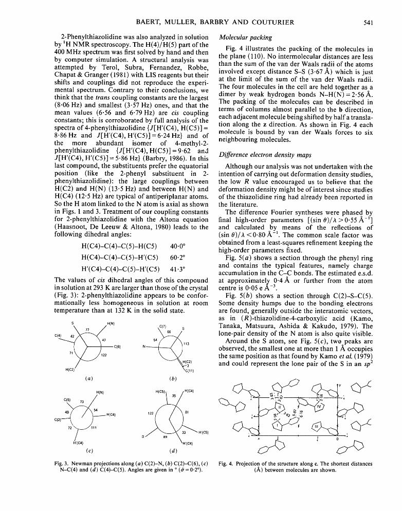
Fig. 4 illustrates the packing of the molecules in the plane (110). No intermolecular distances are less than the sum of the van der Waals radii of the atoms involved except distance S-S (3.67 A) which is just at the limit of the sum of the van der Waals radii. The four molecules in the cell are held together as a dimer by weak hydrogen bonds N - H ( N ) = 2.56 A. The packing of the molecules can be described in terms of columns almost parallel to the 5 direction, each adjacent molecule being shifted by half a transla- tion along the z direction. As shown in Fig. 4 each molecule is bound by van der Waals forces to six neighbouring molecules.
Difference electron density maps
Although our analysis was not undertaken with the intention of carrying out deformation density studies, the low R value encouraged us to believe that the deformation density might be of interest since studies of the thiazolidine ring had already been reported in the literature.
The difference Fourier syntheses were phased by final high-order parameters [(sin 0)/A > 0.55 ~ - ' ] and calculated by means of the reflections of (sin 0)/A < 0.80 A:-1. The common scale factor was obtained from a least-squares refinement keeping the high-order parameters fixed.
Fig. 5(a) shows a section through the phenyl ring and contains the typical features, namely charge accumulation in the C-C bonds. The estimated e.s.d. at approximately 0 .4A or further from the atom centre is 0.05 e A -3.
Fig. 5(b) shows a section through C(2)-S-C(5). Some density humps due to the bonding electrons are found, generally outside the interatomic vectors, as in (R)-thiazolidine-4-carboxylic acid (Kamo, Tanaka, Matsuura, Ashida & Kakudo, 1979). The lone-pair density of the N atom is also quite visible.
Around the S atom, see Fig. 5(c), tw( peaks are observed, the smallest one at more than 1Aoccupies the same position as that found by Kamo et al. (1979) and could represent the lone pair of the S in an sp 2
x J 0
Fig. 3. Newman projections along (a) C(2)-N, (b) C(2)-C(6), (c) Fig. 4. Projection of the structure along e. The shortest distances N-C(4) and (d) C(4)-C(5). Angles are given in o (~ = 0.20). (/~) between molecules are shown.
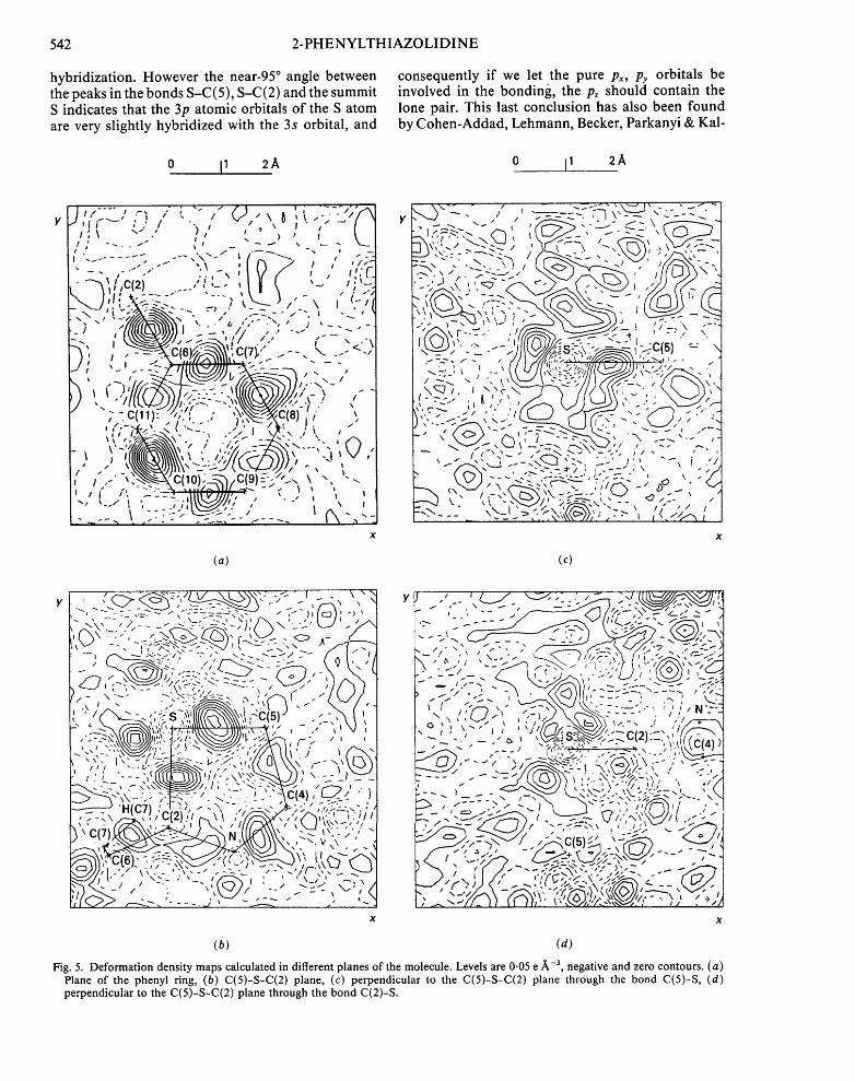
hybridization. However the near-95 ° angle between the peaks in the bonds S-C(5), S-C(2) and the summit S indicates that the 3p atomic orbitals of the S atom are very slightly hybridized with the 3s orbital, and
0 I1 2A
consequently if we let the pure Px, Py orbitals be involved in the bonding, the p. should contain the lone pair. This last conclusion has also been found by Cohen-Addad, Lehmann, Becker, Parkanyi & Kal-
0 11 2A
(@'; . X q 2 / { - -- . . - j
c ~
\ -
, ( <
\ 1 " '
\
' : S _ / -~ ," t "" , " ( . 2 - ,, ^ ' , ( . : - - - - ' ; ~ q ' i i I . ' / " ' , I / - \ " I " / / - ' I 1 7 ~,.~ v . , I ' L " > , ~ t I
' . ...... .--,'\ - t, ~ ' ,;';-'l ~ , , ' " ~ ' " ~ I I I :),-,,If>/ , , , : : : :d
" -"~ . / .C(21 _ , ' l , _ . ~1 i ( ' - , /;'-:.l
: , , ( - - 1 x \ , t _ . ~ ~ ' x ' / , ' - - ~ ' ' " / "' t -~_ , - ' 1
~ - , f ~ , ,... , , . , . , - .,,,.., ,-..-, - " ' ( \~ )1 . , ' , .= = - ' , , 7 . ' . " - ; - -- .. -.
] I I " ' " \ , , I I A ' t l I I I I ~ . , . " x _ - ..-/ t ~ - . . - "..~. I ~ " " " - . . . . ,
_ - ..,
~ f .). . - . - . : .~ ~ , . - \ , - _ . .
. ' , ' - ~ ~ - " I ' , - - " l ' . " ~ - / ~ , ' ' I ' . . ' ' . ; ; LK-;).~-,,<.' " - . . -', I ~ r ' , ; ~ ,
_ ,~,..z ~ ( . , ,.,,, : - " \ - - - , . , . ..,
I.-" "':~'-~-~'C(10)'~--,~----'1/C(9)~:.;//~" \ " , "~ ' I i ~ ' , ~ - ' ~ - - 2 " " ' V I I I I ' ~ " ~ • l " "' '--"~', , ; l l ~ / . / , " ,' " ', , ,
\ ":::-',- ~--~'>-" _/ _ ~ I X " "-- I ~ - , , " - % ~ (~ - ~ - ~-~--~ - ' ~ - ~ • I '~ -'~ -
X
542 2 -PHENYLTHIAZOLIDINE
(a) (c)
y Y
' ~ . "?-:.q \ ' . -" ",.~" I " -I
I ~ ' i x '~i I ~ ' . . . . i
~ ( ( ~ k'~ ,----,,\t631
~ ~ , ,;-q.,-T. TI ~ ~ . (4),~,.)i
~,~?! "F"~ "-'"i '-"" "" "--='" ; 1 __ H(C7)C~i)~,i~'~) ,f ......... ' / I ' , , ' I I ' I /
~ - _ _ , . ; \_,:) ~ ' ~ / r "-" . - . = - - ~ ' . - , (I
X X
(b) (d)
Fig. 5. Deformation density maps calculated in different planes of the molecule. Levels are 0.05 e/~-3, negative and zero contours. (a) Plane of the phenyl ring, (b) C(5)-S-C(2) plane, (c) perpendicular to the C(5)-S-C(2) plane through the bond C(5)-S, (d) perpendicular to the C(5)-S-C(2) plane through the bond C(2)-S.
BAERT, MULLER, BARBRY AND COUTURIER 543
man (1984) (notice that the S atom is not in the same environment) but they could not explain it b y modelling with an sp 2 deformation.
The difficulties of interpreting the results around the S show that more quantitative studies of this type are needed to elucidate the hybridization mode of the S atom where the d orbitals play an important role.
This work was supported in part by CNRS UA 801 (Dynamique des Cristaux Mol6culaires).
References
BARBRY, D. (1986). DSc Thesis, Univ. of Lille. COHEN-ADDAD, C., LEHMAN, M. S., BECKER, P., PARKANYI,
L. & KALMAN, A. (1984). J. Chem. Soc. Perkin Trans. 2, pp. 191,-196.
GERMAIN, G., MAIN, P. & WOOLFSON, M. M. (1971). Acta Cryst. A27, 368-376.
GOODMAN, M., CHEN, V., BENEDETTI, E., PEDONE, C. & COR- RADINI, P. (1972). Biopolymers, 11, 1779-1787.
HAASNOOT, C., DE LEEUW, F. & ALTONA, C. (1980). Tetrahe- dron, 36, 2783-2792.
HOWARD-LOCK, H., LOCK, L. & SMALLEY, P. S. (1985). Can. J. Chem. 63, 2411-2419.
International Tables for X-ray Crystallography (1974). Vol. IV. Birmingham: Kynoch Press. (Present distributor D. Reidel, Dordrecht.)
KAMO, J., TANAKA, N., MATSUURA, Y., ASHIDA, T. & KAKUDO, M. (1979). Bull. Chem. Soc. Jpn, 52, 706-710.
LOSCALZO, J., KALLEN, R. L. & VOET, D. (1973). Arch. Biochem. Biophys. 157, 426-430.
SCHMOLKA, I. & SPOERRI, P. (1957). J. Am. Chem. Soc. 79, 4716-4720.
SCHOMAKER, V. & TRUEBLOOD, K. H. (1968). Acta Cryst. B24, 63-76.
SHELDRICK, G. M. (1976). SHELX76. Program for crystal struc- ture determination. Univ. of Cambridge, England.
STEWART, R. F., DAVIDSON, E. R. & SIMPSON, W. T. (1965). J. Chem. Phys. 42, 3175-3187.
TEROL, A., SUBRA, G., FERNANDEZ, J. P., ROBBE, Y., CHAPAT, J. P. & GRANGER, R. (1981). Org. Magn. Reson. 17, 68-70.