Embed Size (px)
Citation preview
400 SAMUEL V. ABRAHAM
REFERENCES 1 Kipp, C. J. On keratitis from malarial fever. Trans. Amer. Ophth. Soc, 1880, v. 3, p. 91. 2 Hansen Grut, E. Sur deux formes typiques de kératite. Congrès périodique international des
sciences médicales, 8°™ Session, Copenhague, 1884. Compt. rend. 1886, v. 3, Sec. d'Opht., p. 34. "Emmert, E. Keratitis dendritica exulcerans mycotica; eine noch nicht beschriebene Form
ulcerierender Hornhautentzündung. Centralb. f. prakt. Augenh., 1885, v. 9, p. 302. * Ellett, E. C. A series of cases of malarial keratitis. Ophth. Ree, 1899, v. 8, p. 115. 1 . Dendritic keratitis of malarial origin. Journ. Amer. Med. Assoc, 1906, v. 46, p. 1984. 6 Charles, J. W. Dendritic keratitis. Amer. Jour. Ophth., 1904, v. 21, p. 97. 7 . Dendritic keratitis. Ophthalmology, 1906-7, v. 3, p. 604. 8 Grüter, W. Experimentale und klinische Untersuchungen über den sogenannten Herpes
corneae. Bericht, ü. d. deut. ophth. Gesell., 1920, v. 43, p. 162. "Shapiro, E., and Coles, H. D., Dendritic keratitis caused by abscessed teeth. Jour. Amer.
Dental Assoc, 1940, v. 27, August, p. 205.
THE CONVERGENCE FUNCTION IN RELATION TO THE BASAL METABOLISM*
S A M U E L V. A B R A H A M , M.D. Los Angeles, California
This is a continuation of a similar study1 and includes previously reported as well as new cases. It, as well as the previous report, published in 1938, is based on a classification of near horizontal phoria and duction findings at 13 inches. This classification is supported by the data found in at least 5,000 cases.
A report on this classification has recently been made.2 In brief, it stated that near horizontal phoria and duction findings could be divided into three groups : group I, the normals ; group II, the subnormals ; group III, the abnormals. Group I contained only those individuals who were without ocular complaints and who used their eyes sufficiently in daily activities to permit assumption of average demands upon the visual apparatus. Group II contained those whose outstanding symptom was one of tiring, both ocular and general. Group III contained the remaining cases. These cases required more detailed study. The chief symptom in this group was one of increased irritability or lowered threshold of tolerance.
♦Work aided by a grant from the Kramer Research Fund, Chicago, E. V. L. Brown, M.D., Director. Presented before the Los Angeles Ophthalmological Society in 1939.
In the presentation of the data forming the basis of the classification, it was stated that the base-in readings in the normal and subnormal groups were similar, that both groups (I and I I ) showed the ability to relax all the convergence used to bring the eyes from parallelism to near fixation (and even a little more than this). It was also pointed out that the abnormals (group II I ) differed definitely in this respect, usually not having this ability. The tendency in this group to fail to give up the convergence in use at the moment suggests an overactive convergence. It was noted, also, that the base-out readings in these cases may not be considered reliable indicators of the convergence reserve, as the basic findings are masked by this overactive convergence. This view was supported by further data obtained under cycloplegia. Under cyclo-plegia it was frequently found that group-Ill cases resembled group-II cases. This was also true for group-Ill cases after the problems involved were fully or partially solved.
The idea of a convergence spasm superimposed on or simultaneously present with a convergence weakness should not be considered difficult to accept. Such a
CONVERGENCE FUNCTION AND METABOLISM 401
state was considered by Bielschowsky3 as "by no means unusual. . . ." Whether or not the picture of an underactive convergence alone (group II) or combined with an increased irritability of convergence (group III) is a reflection of the general condition is a moot question, upon which there are many views but almost no data.
Bothman4 mentions convergence weakness in cases of exophthalmos following thyroidectomy. In these cases due to hyperthyroidism, movements were limited and the muscles, when exposed by surgery, showed the muscle changes described by Naffziger.5 Fridenberg6 mentions convergence insufficiency in the hypothyroid type but gives no data. Mayer7 considers examples of convergence insufficiency as usual in exophthalmic goiter. He does not feel able to explain the convergence weakness he found in three cases of myx-edema in which there was an average basal metabolic rate of —13.3 percent. He found, in cases of obesity and hypo-thyroidism, that ability to converge was "satisfactory" (by the approach-the-nose method). Zentmayer8 considered fatigue following overstimulation of the body as a cause of convergence insufficiency. Duane9 advised care of general health factors after correction of the errors of refraction and before giving prism exercises. Bielschowsky,10 Howe,11 and others have called attention to the relationship between the general health and convergence insufficiency. Davis12 admits that in some cases convergence insufficiency is due to some general health factor.
Heath13 says that muscular insufficiencies may be due to the general condition, such as heart trouble or nervous prostration. Fox14 says, ". . . the very complexity of the condition speaks for its close relationship to the general health." Maddox15 advises constitutional measures. Oppenheimer,16 in 1895, mentioned the use of strychnine for converg
ence weakness. Major Field17 pointed out that too many heterophorias disappear or decrease in amount upon improvement in the general physical condition to ignore the claim that the extraocular muscles are affected by the general condition. Howard18 said that in cases of hypothy-roidism there may occasionally be shown a weakened convergence (by the "approach" method). Bryant,19 emphasizing the importance of general health studies in the specialties, says that muscular imbalances of the eyes are not uncommon in the subthyroid individuals, the convergence being especially affected.
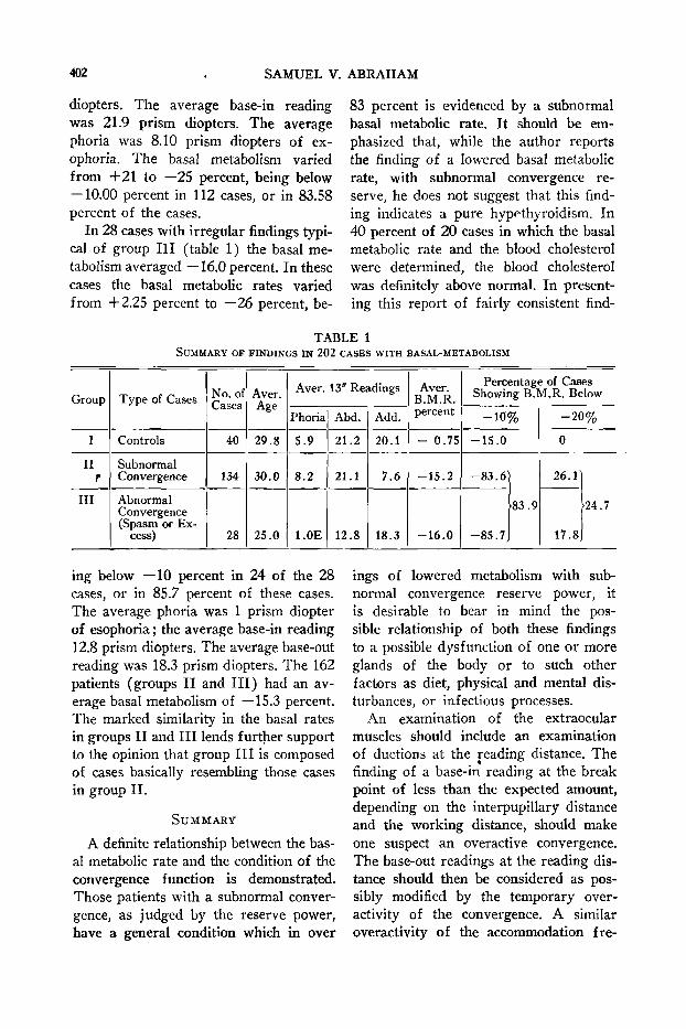
The author feels sure that further emphasis on the relation of the condition of the extraocular muscles to the general health should be made. It is thought that if laboratory proof of such a relationship could be presented, a definite step forward would be made both in our understanding of the extraocular muscle mechanism and in our therapy for heterophorias. In the previous study,1 for the first time, the relationship between convergence and basal metabolism was reported. In table 1 are summarized the findings in 202 patients in whom the basal metabolic rates had been obtained.
In 40 control cases (group I ) the basal metabolism averaged —0.75 percent. The average base-out reading at 13 inches was 20.0 prism diopters. The average base-in reading was 21.0 prism diopters. The average phoria was 5.6 prism diopters of exophoria. The basal metabolism varied from +35.00 to —19 percent, being below — 10.0 in 15 percent of the cases. In 134 cases with persistent ocular complaints after correction of the error of refraction, the tests showed subnormal convergence reserve (less than 15 prism diopters, base out, for 13 inches). In this group of subnormals (group II) the basal metabolism averaged —15.2 percent. The average base-out reading was 7.6 prism
402 SAMUEL V. ABRAHAM
diopters. The average base-in reading was 21.9 prism diopters. The average phoria was 8.10 prism diopters of ex-ophoria. The basal metabolism varied from +21 to —25 percent, being below — 10.00 percent in 112 cases, or in 83.58 percent of the cases.
In 28 cases with irregular findings typical of group III (table 1) the basal metabolism averaged —16.0 percent. In these cases the basal metabolic rates varied from +2.25 percent to —26 percent, be
ing below —10 percent in 24 of the 28 cases, or in 85.7 percent of these cases. The average phoria was 1 prism diopter of esophoria ; the average base-in reading 12.8 prism diopters. The average base-out reading was 18.3 prism diopters. The 162 patients (groups II and III) had an average basal metabolism of —15.3 percent. The marked similarity in the basal rates in groups II and III lends further support to the opinion that group III is composed of cases basically resembling those cases in group II .
SUMMARY
A definite relationship between the basal metabolic rate and the condition of the convergence function is demonstrated. Those patients with a subnormal convergence, as judged by the reserve power, have a general condition which in over
83 percent is evidenced by a subnormal basal metabolic rate. It should be emphasized that, while the author reports the finding of a lowered basal metabolic rate, with subnormal convergence reserve, he does not suggest that this finding indicates a pure hypcthyroidism. In 40 percent of 20 cases in which the basal metabolic rate and the blood cholesterol were determined, the blood cholesterol was definitely above normal. In presenting this report of fairly consistent find
ings of lowered metabolism with subnormal convergence reserve power, it is desirable to bear in mind the possible relationship of both these findings to a possible dysfunction of one or more glands of the body or to such other factors as diet, physical and mental disturbances, or infectious processes.
An examination of the extraocular muscles should include an examination of ductions at the reading distance. The finding of a base-in reading at the break point of less than the expected amount, depending on the interpupillary distance and the working distance, should make one suspect an overactive convergence. The base-out readings at the reading distance should then be considered as possibly modified by the temporary over-activity of the convergence. A similar overactivity of the accommodation fre-
TABLE 1 SUMMARY OF FINDINGS IN 202 CASES WITH BASAL-METABOLISM
Group
I
II r
III
Type of Cases
Controls
Subnormal Convergence
Abnormal Convergence (Spasm or Ex
cess)
No. of Cases
40
134
28
Aver. Age
29.8
30.0
25.0
Aver. 13" Readings
Phoria
5.9
8.2
1.0E
Abd.
21.2
21.1
12.8
Add.
20.1
7.6
18.3
Aver. B.M.R. percent
- 0.75
- 1 5 . 2
- 1 6 . 0
Percentage of Cases Showing B.M.R. Below
- 1 0 %
- 1 5 . 0
- 8 3 . 6 1
- 8 5 . 7
83 .9
- 2 0 %
0
26.1
17.8
24.7
CONVERGENCE FUNCTION AND METABOLISM 403
quently, although not always, will be found in these cases. Cycloplegia is, therefore, particularly desirable in these cases both for its effect on the accommodation and for the possible effect on the convergence. Under cycloplegia the previously overactive convergence may appear as a definitely subnormal convergence (as in group I I ) .
A complete discussion of the subject of heterophorias, their nature, their diagnosis, and their treatment would require a very extensive monograph. A proper discussion of this subject would require a consideration of the phorias and the ductions at varying fixation points including the entire field of fixation. It would also require a consideration of the subjects' "primary position of rest," "divergence center," "convergence center," "accommodation and convergence relationship," "convergence excess," "divergence insufficiency," "retinal correspondence," and many other related subjects. Such a discussion is obviously beyond the scope of this article. Some of these subjects are touched upon in another article, in preparation, on the "Nature of the heteorophorias."
In an individual case the cause or causes of discomfort must be determined by all the findings. However, such a high degree of correlation between the tests as used
and the general metabolic processes, suggests that one is justified in emphasizing the probability that correction of minor errors of refraction and periodic orthoptic training can be only of palliative value. Those who follow these methods to the exclusion of general care and therapy are subject to the criticism made of some nonmedicai practitioners, in that they delay proper medical care. The usual effort to use the convergence function, as well as the need therefore, throughout the waking hours should be expected to be more effective exercise than the short periods of special exercises, even if given daily. That neither the normally great demands on this function, nor the especially induced demands over a short period, produce lasting results, suggests that local exercises are of little therapeutic value. Therapeutic results have been good in a considerable number of cases, but the therapy has been so varied that a discussion thereof is a subject in itself.
CONCLUSION
The attempt has been made to show the correlation existing between the convergence function (as studied by use of the duction method at near) and the general health (using the basal-metabolism test as a laboratory indicator thereof).
REFERENCES
'Abraham, S. V. Convergence vs. basal metabolism. Trans. Intern. Ophth. Congress of 1937, 1938, v. 4, p. 54.
' . Near horizontal phoria tests. Amer. Jour. Ophth., 1943, v. 26, Feb., pp. 271-279. 8 Bielschowsky, A. Lectures on motor anomalies. Dartmouth College Publication, Hanover,
New Hampshire, 1940, p. 105. * Bothman, L. Endocrines in ophthalmology. Illinois Med. Jour., 1934, v. 65, May, p. 226. 5 Naffziger, H. C. Pathogenic changes in the orbit, etc. Arch, of Ophth., 1933, v. 9, Jan., p. 1. "Fridenberg, P. Some ophthalmologic implications, etc. New York Med. Jour., 1921, v. 114,
July, p. 38. 'Mayer, L. L. 'Zentmayer, W.
7, p. 11. "Duane, A. Quoted by Dunnington in Jour. Med. Soc. New York, 1931, v. 12, Dec, p. 919. 10 Bielschowsky, A. Motor anomalies of the eyes. Arch, of Ophth., 1936, v. 13, April, p. 369. " Howe, L. Muscles of the eye. New York, G. P. Putnam's Sons, 1908, v. 2, p. 90. "Davis, W. T. Diagnosis and treatment of phorias. Amer. Jour. Ophth., 1938, v. 21, Feb.,
p. 14S.
Ocular convergence, etc. Arch, of Ophth., 1931, v. 6, Sept., p. 389. The eye and the endocrine organs. Jour. Amer. Med. Assoc, 1917, v. 69, July
404 OTIS WOLFE, I, AND RUSSELL M. WOLFE 18 Heath, F. C. The conservative treatment of muscular insufficiencies. Amer. Jour. Ophth.
and Otol., 1894, v. 3, p. 149. "Fox, L. W. Heterophoria. Amer. Jour. Ophth., 1923, v. 6, Feb., p. 110. 15 Maddox, E. J. Investigation on relation, etc. Ophth. Rev., 1886, v. 5, p. 341. 16 Oppenheimer, H. S. Strychnine for convergence weakness. Trans. Amer. Ophth. Soc, 1895,
v. 7, p. 361. " Field, P. C. Phorometry of normal eyes. Arch, of Ophth., 1911, v. 40, p. 526. 18 Howard, H. J. Relationship of hypothyroidism to ophthalmology. Southern Med. Jour., 1939,
v. 32, Nov., p. 1112. "Bryant, B. L. Thyroid derangements, etc. Jour. Med., 1937, v. 18, May, p. 128.
HEREDITARY CATARACTA CAERULEA Six RELATED CASES
OTIS WOLFE, I, M.D., AND RUSSELL M. WOLFE, M.D.* Marshalltown, Iowa
This paper is being written as a report supplementary to that of three cases previously reported with a description of the technique employed.1 This aspirating technique was further discussed in another publication with a description of a new aspirating needle for soft cataract.2
Briefly, these first 3 cases were observed in a mother, aged 50 years, and two children, aged 16 and 18 years. The cataracts of these three were nearly indistinguishable except that the mother also had posterior subcapsular opacities involving the central two thirds of the lens of the right eye and the central one third of the lens of the left eye. The unusual blue punctate opacification of the remainder of the cortex was identical in mother and children.
Intracapsular extractions were performed on the mother's eyes, and full normal corrected vision resulted. The two children's eyes were operated on by the authors' double-aspirating-needle technique, as described by us in a previous article.2 Both children obtained normal corrected vision.
Case 4. The next patient, seen on January 20, 1942—nearly two years after the first patient had been examined-—was a healthy and well-developed woman, 36
♦Now Lt. (jg) (Ophth.), M.C., U.S.N.R.
years of age. She had been wearing glasses since her fourteenth year, and her vision was last corrected five years prior to her examination here. The patient said that she had never enjoyed good vision, nor, on the other hand, had she ever noticed any decrease in vision, even during the last few years. Her condition so closely resembled what we had observed in our previous cases, that following the examination the patient was carefully questioned. She had nearly completely lost track of her family, but on questioning her further we were able to establish that this patient's mother and the previous patient's mother had been first cousins. A brother had cataracts and had had poor vision since birth, and we assumed that he was suffering from the same pathologic changes. However, the patient had five normal-sighted sisters.
The patient's vision in the right eye with a -4 .00D. sph. = 0 -3 .00D. cyl. ax. 180° was only 20/200. For the left eye a -3 .00D. sph. O -4 .00 D. cyl. ax. 180° gave her only 20/200 vision also. Tension in each eye was 18 mm. Hg (Schiòtz). There was no external evidence of disease of the eyes, and the adnexa appeared to be quite normal. The positions of the eyes were primary, and movements were full in all cardinal directions. The pupils were round and equal