Embed Size (px)
Citation preview
A. FAUCHER, P. MAZEROLLES, J. JAUD ET J. GALY
r6seau cristallin est assur6e par des forces de van der Waals.
L'&ude structurale de la dim&hyl-6,6 germa-6 cyclo- und6canone, r6alis6e ~ - 4 0 ° C , montre une confor- mation chaise-bateau-chaise (pour la chaine h 4 CH2) et chaise-bateau-chaise-bateau (pour la chaine ~t 5 CH2) a partir de l'atome de germanium.
Le groupement carbonyle est sensiblement perpen- diculaire au plan moyen passant par l'h&6roatome et le carbone sp 2. I1 faut noter que dans le cristal, la distance G e - O ne permet pas de rendre compte des effets trans- annulaires observ6s (Guimon et al., 1977; Duboudin et al., 1977). I1 est done probable que la mol6cule &udi6e a une conformation diff6rente en phase vapeur et ~. l'&at cristallin.
Le CNRS, la DGRST et la DESR ont apport6 leur aide mat6rielle ~ la r6alisation de ce travail.
Rff~rences
445
ANET, F. A. L., JACQUES, M. ST, HENRICHS, P. M., CHENG, A. K., KRANE, J. & WONG, L. (1974). Tetrahedron, 30, 1629-1637.
DUBOUDIN, F., BOURGEOIS, G., FAUCHER, A. & MAZEROLLES, P. (1977). J. Organomet. Chem. 133, 29- 36.
GUIMON, C., PEISTER-GUILLOUZO, G., FAUCHER, A., MAZEROLLES, P. • LIMOUZIN, Y. (1977). J. Organomet. Chem. 131, 365-370.
International Tables for X-ray Crystallography (1974). Vol. IV. Birmingham: Kynoch Press.
LEDAAL, T. (1967). Tetrahedron Lett. pp. 4397-4402. LEDAAL, T. (1968). Tetrahedron Lett. pp. 651-656. MAZEROLLES, P. & FAUCHER, A. (1973). J. Organomet.
Chem. 63, 195-203. MOSSET, A., BONNET, J. J. & GALY, J. (1977). Acta Cryst.
B33, 2639-2644.
Acta Cryst. (1978). B34, 445-450
The Crystal and Molecular Structure ofToxisterol2-D Epoxide, C2sH440 2
By PETER F. LINDLEY AND MUDHAFAR M. MAHMOUD
Department of Crystallography, Birkbeck College, University of London, Malet Street, London WC1E 7HX, England
(Received 21 June 1977; accepted 17 July 1977)
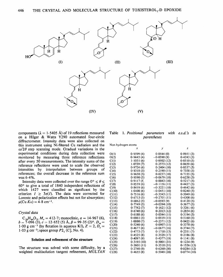
Crystals of toxisterol2-D epoxide are monoclinic, space group P2~, with a = 14.967 (6), b = 7.096 (3), c = 12.652 (5)/~,, fl = 99.35 (2) °. The structure has been refined to a residual of 0.067 for 1427 independent significant reflections measured on an automated four-circle diffractometer. In the compound, which is derived from ergosterol, ring A adopts a boat conformation in which the atoms defining the bow and stern, C(3) and C(10), are bridged by an O atom. Ring B is cleaved at the C(9)-C(10) bond and the second O atom forms an epoxide ring across the C(7) and C(8) positions. The geometry of the remainder of the molecule is similar to that found in other sterol structures.
Introduction Experimental
Photolysis of ergosterol (I) in ethanol and chromatography yield a number of products (Barton et al., 1975; Carlisle & Lindley, 1976) including two toxisterol 2 ethers, D and E (Barton, Barrett, Russell, Lindley & Mahmoud, 1976). On the basis of chemical and spectroscopic evidence structure (II) was proposed for the two ethers.
The present paper reports the complete details of the crystal structure analysis of toxisterol2-D epoxide (III). This structure enables configurations (IV) and (V) to be assigned to toxisterols2-D and -E respectively.
The compound was supplied by Professor Sir Derek Barton, FRS, and Dr A. G. M. Barrett of Imperial College, University of London, in the form of colourless prismatic crystals. A crystal fragment of dimensions 0.09 x 0.48 x 0.11 mm parallel to a, b and c respec- tively was mounted along the b axis and used for the X- ray measurements. Preliminary unit-cell dimensions and space-group information were obtained from precession photographs taken with Cu radiation. Accurate cell dimensions were obtained by least- squares refinement of the 0 values of the Cu Ka~
446 THE CRYSTAL AND MOLECULAR STRUCTURE OF TOXISTEROLz-D EPOXIDE
I
HO*" I~I~
(I)
1
(iv)
(II) (III)
I
(v)
components (2 = 1.5405 A) of 19 reflections measured on a Hilger & Watts Y290 automated four-circle diffractometer. Intensity data were also collected on this instrument using Ni-filtered Cu radiation and the 09/20 step scanning mode. Gradual variations in the experimental conditions during data collection were monitored by measuring three reference reflections after every 50 measurements. The intensity sums of the reference reflections were used to scale the observed intensities by interpolation between groups of references; the overall decrease in the reference sum was 6.4%.
Intensity data were collected over the range 0 ° < 0 < 60 ° to give a total of 1840 independent reflections of which 1427 were classified as significant by the criterion I > 3a(l) . The data were corrected for Lorentz and polarization effects but not for absorption; /t(Cu Ka) = 4.8 cm-k
Crystal data
C2sHa40 2, M r = 412.7; monoclinic, a = 14.967 (6), b = 7-096 (3), e = 12.652 (5) A, f l = 99.35 (2) °. D m = 1.00 g cm -3 (by flotation in aqueous KI), Z = 2, D c = 1.03 g cm-3; space group P2~ (C~, No. 4).
Solution and refinement of the structure
The structure was solved with some difficulty, by a weighted multisolution tangent refinement, M U L T A N
Table 1. Positional parameters with e.s.d.'s in parentheses
Non-hydrogen atoms x y z
O(1) 0.9599 (4) 0.0544 (0) 0.8951 (3) 0(2) 0.9445 (4) --0.0590 (9) 0.4243 (3) C(1) 1.1031 (6) 0.0502 (12) 0.8510 (5) C(2) 1.0739 (7) 0.2573 (12) 0.8659 (6) C(3) 0.9724 (6) 0.2404 (10) 0.8537 (5) C(4) 0.9318 (5) 0.2190 (11) 0.7358 (5) C(5) 0.9658 (5) 0.0237 (10) 0.7155 (5) C(6) 0.9590 (5) -0.0679 (10) 0.6238 (5) C(7) 0.9117 (5~ 0.0043 (10) 0.5217 (5) C(8) 0.8559 (6) -0.1156 (11) 0.4437 (5) C(9) 0.8419 (6) -0.3221 (10) 0.4642 (6) C(10) 1.0108 (6) -0.0451 (10) 0.8240 (5) C(l 1) 0.7516 (6) -0.3543 (11) 0.5049 (6) C(12) 0.6713 (5) -0.2701 (11) 0.4308 (6) C(13) 0.6862 (5) -0.0593 (9) 0.4120 (5) C(14) 0.7749 (5) -0.0394 (10) 0.3677 (5) C(15) 0.7742 (7) 0.1621 (12) 0.3261 (6) C(16) 0.6740 (6) 0.2013 (12) 0-2854 (6) C(17) 0.6188 (6) 0.0344 (11) 0.3194 (5) C(18) 0.6861 (5) 0.0519 (11) 0.5160 (5) C(19) 1.0080 (7) -0.2571 (12) 0.8425 (6) C(20) 0.5240 (6) 0.0907 (11) 0.3370 (6) C(21) 0.4677 (6) -0.0677 (16) 0.3744 (7) C(22) 0.4773 (7) 0.1718 (13) 0.2351 (7) C(23) 0.4525 (8) 0.3529 (17) 0.2186 (8) C(24) 0.4097 (9) 0.4377 (19) 0.1141 (8) C(25) 0.3195 (10) 0.5001 (31) 0.1234 (9) C(26) 0.2601 (I1) 0.3518 (31) 0.1556 (13) C(27) 0.2740 (9) 0.6260 (30) 0.0284 (12) C(28) 0.4655 (8) 0.5949 (20) 0.0774 (10)
P E T E R F. L I N D L E Y A N D M U D H A F A R M. M A H M O U D 447
Hydrogen atoms Table 1 (cont.)
x y z H(I 1) 1.1403 0.0312 0-7921 H(12) . 1.1405 -0.0060 0.9195 H(21) 1.0939 0.3164 0.9371 H(22) 1.0854 0.3479 0.8080 H(3) 0.9421 0.3368 0.8905 H(41) 0.8632 0-2216 0.7241 H(42) 0.9541 0.3141 0-6893 H(6) 0.9938 -0-1943 0.6265 H(7) 0.8937 0.1364 0.5225 H(91) * 0.8428 -0-3969 0.3968 H(92) 0.8955 -0-3679 0.5193 H(I 11) 0.7430 -0.4985 0.5149 H(112) 0.7560 -0-2994 0.5803 H(I 21) 0.6644 -0.3406 0.3595 H(122) 0.6142 -0.2884 0.4611 H (14) 0.7700 -0.1270 0- 3043 H ( 1 ~1) 0.8090 0.1686 0.2662 H(152) 0.7990 0.2471 0-3850 H(161) 0.6550 0.3204 0-3212 H(162) 0.6619 0.2185 0.2071 H(17) 0.6110 --0.0549 0.2570 H( 181 ) 0.6250 0.0307 0.5381 H(182) 0.6924 0.1882 0.4994 H(183) 0.7345 0.0098 0.5726 I-/~ 191) 1.0478 -0.3249 0.7960 H(192) 0.9473 -0.3070 0.8296 H(193) 1.0376 -0.2875 0.9208 H(20) 0-5318 0.1928 0.3948 H(211) 0.4069 -0.0136 0.3854 H(212) 0.4975 --0.1254 0.4422 H (213) 0.4560 --0.1654 0.3167 ~q(221) 0.4543 0.0767 0.1731 H(23) 0.4704 0.4384 0-2820 H(24) 0.4108 0-3281 0.0570 H(25) 0.3283 0.5889 0-1881 H(261) 0.1985 0.4058 0.1640 H(262) 0.2863 0.2924 0.2268 H(263) 0-2499 0.2501 0-1002 H(271) 0.3180 0.7160 0.0081 H(272) 0.2179 0.6807 0.0400 H(273) 0.2587 0-5322 -0.0385 H(281) 0.4343 0.6446 0.0080 H(282) 0.5290 0.5646 0.0754 H(283) 0.4643 0.7023 0.1337
74 (Germain, Main & Woolfson, 1971; Declercq, Germain, Main & Woolfson, 1973; Koch, 1974). In the computation of the normalized structure factors a molecular scattering factor was used for the 11-atom fragment involving the C and D rings with the addition of C(18) and C(20). After numerous attempts using up to 324 reflections with IEhl > 1-50 a tangent refine- ment using only 146 reflections (approximately five reflections per non-hydrogen atom in the asymmetric unit) with IEhl _ 1.67 yielded a phase set with values of 0.195 and 1.075 for RKarl e and the absolute figure of merit respectively; the corresponding combined figure of merit derived using unit weights for the components was 2.50. An E map computed with this phase set enabled 16 of the non-hydrogen atoms in the structure to be located. The remaining non-hydrogen atomic positions were determined by the iterative Fourier synthesis technique to give a residual of 0.33.
Refinement proceeded by the full-matrix least- squares method, using only the significant reflections and initially with all non-hydrogen atoms treated isotropically to give an R value of 0.174. A difference Fourier synthesis showed diffuse electron density maxima in the vicinity of the H atom positions but in subsequent calculations these atoms were placed in calculated positions assuming a C--H bond length of 1.0 A. No attempt was made to refine the positional or thermal parameters of the H atoms.
Further refinement, with non-hydrogen atoms treated anisotropically, proceeded by a partial full- matrix technique in which no more than 21 atoms and an overall scale factor were refined in any one cycle. The weighting scheme, w = [ 1 - exp( -a~ sin 2 0/22)]/(a2 + IFol + a31Fo 12) if IFol > 30.0, else w = a 4 with a 1 = 20.0, a 2 = 20.0, a 3 = 0.001 and a 4 --- 0-02, was used to make the average values of Z wA 2 uniform when analysed in terms of batches of increasing IFol and sin ~/2. The final values of R and R ' [= (E wA2/Z, WlFol2) 1/2] were 0.067 and 0.092 respec- tively; the residual for all the 1840 independent
Fig. 1. A stereodrawing of the molecule of toxisterol2-D epoxide viewed perpendicular to the plane defined by atoms C(4), C (5) and C (1).
448 THE CRYSTAL AND M O L E C U L A R STRUCTURE OF TOXISTEROL2-D EPOXIDE
reflections was 0.084. A final difference Fourier synthesis confirmed the correctness of the analysis.
Throughout the structure factor calculations, the atomic scattering factors listed by Hanson, Herman, Lea & Skillman (1964) were used and all computations were performed on the CDC6600 computer at the University of London Computer Centre. The final atomic coordinates are given in Table 1.* A satisfactory
* Lists of structure factors and anisotropic thermal parameters have been deposited with the British Library Lending Division as Supplementary Publication No. SUP 32947 (12 pp.). Copies may be obtained through The Executive Secretary, International Union of Crystallography, 13 White Friars, Chester CH 1 1NZ, England.
tensor analysis of the anisotropic thermal vibration parameters was obtained for all the non-hydrogen atoms.
Discussion
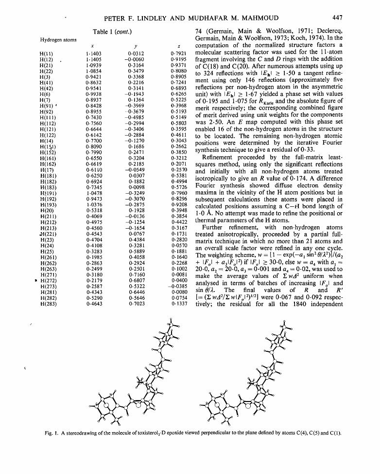
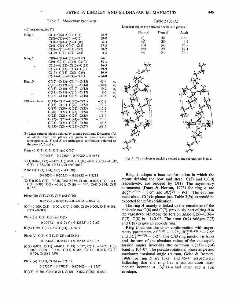
A stereodrawing of the molecule of toxisterolz-D epoxide is shown in Fig. 1. Fig. 2 is a schematic drawing showing the atomic labelling and the intra- molecular bond lengths and angles. Further details of the molecular geometry are recorded in Table 2.
C27 C28
1 • 4 4 3 (17) ~C25 ~2~ C22 ~%~ 1.344 (14)
C26 C23
~ ..~t;1 ~ ,.., O/o_,
g Y "/ .-.,,,, 1-,~.~97\ 1.~o ~,> /~ , , C19
C21 t,' l° / / C 1 3 C 1 % / / k ~ , _ _ , ~,. ":~:12 CSy,"~ <;~\.. ."~,.,~,, <1,> <.
"\c4 c3 ' I • ,b')_6 (9)
': 01 C10 C19 11.t-4 7 (11)
/ ' ~ ~ \ e 0 2 - - C7 - - C 6 / %, o~ ~< o 2 - c a - c 9 " _ \ C 1 - - C 1 0 - - C 5
~ " \ + ~ I - , , C 7 - - C 8 - - C 1 4 122.1 (6) ~ , ~ " ~ ~=,,~,.~c,~'~',~ C 12--C13 -C17 116" 0 (6)
riO 6 iT) Z ,~-),'% ,~+~,o~.,(~, ,o~.~7 C14- -C13- -C18 112"8 (6) / ~ + ° \ ,~-~ / C10- -01 - -C3 96-2 (4) / ~ '°o~c~,\.oO:~' %~/ C 2 - - C 3 - -C4 110.4 (6)
( ' " ' ~ '°'<"" <">vT '~N ,,~.,<,,~" \ - . / % ,,+\ ...... A ,~ ~ \
~ ~ ¢ ~ : = = = = = ~ I04 • 8, ~5) ~ 102.2(6)]
\ \,o,o,4
110.7 (,5) 117.1 (6) 117.2 (6) 108.3 (5)
Fig. 2. A schematic drawing of the molecule showing the intramolecular bond lengths (A) and angles (°). Estimated standard deviations are given in parentheses.
PETER F. L I N D L E Y A N D M U D H A F A R M. M A H M O U D 449
Table 2. Molecular geometry
(a) Torsion angles
Ring A
Ring C
-t
Ring D
r
17fl side chain
(°)
C(I)-C(2)-C(3)-C (4) -76.9 C(2)-C(3)-C(4)-C(5) 69.0 C (3)-C (4)-C (5)-C (10) 6.3 C(4)-C(5)-C(10)-C(1) -77.3 C(5)-C(10)-C(1)-C(2) 68.2 C(10)--C(1)--C(2)-C(3) 4.3
C (8)--C (9)--C ( l 1)-C (12) 54.1 C(9)-C(11)-C(12)-C(13) -55.3 C(11)-C(l 2)-C(I 3)-C(14) 56.5 C(12)-C(13)-C(14)-C(8) -59.9 C(13)-C(14)-C(8)-C(9) 59.9 C(14)-C(8)-C(9)-C(11) -54.8
C(I 7)-C(I 3)-C(14)-C(15) 45.1 tp o C(16)-C(17)-C(13)-C(14) -38-6 tp 1 C(15)-C(16)-C(17)-C(13) 19.2 tp 2 C (14)-C(15)-C(16)-C (17) 8.2 tp3 C(13)-C(14)-C(15)-C(16) -33.5 ~4
C(I 3)-C(17)-C(20)-C (21) -55.8 C(I 3)-C(17)-C (20)-C (22) -179.2 C(17)-C(20)-C(22)-C(23) -112.3 C (20)-C (22)-C (23)-C (24) 176.5 C(22)-C(23)-C(24)-C(25) 115.4 C (22)--C(23)-C (24)-C (28) -120.0 C (23)-C (24)-C (25)-C (26) -56.6 C (23)-C (24)-C (25)-C (27) 168.7
(b) Least-squares planes defined by atomic positions. Distances (/~) of atoms from the planes are given in parentheses where appropriate. X, Y and Z are orthogonal coordinates referred to the axes a*, b and c.
" r
Plane (i): C(1), C(2), C(3) and C(10)
0.0456X- 0.1948Y + 0.9798Z = 8.5683
[C(1) 0.026, C(2) -0.027, C(3) 0.019, C(10) --0.019, C(4) -1.342, C(5) -1.383, O(1) 0.811, C(19) 0.509]
Plane (ii): C(3), C(4), C(5) and C(10)
0.8603X + 0.3525 Y - 0.3683Z = 9.8233
1C(3) 0.027, C(4) -0.038, C(5) 0.039, C(10) -0.028, C(1) 1.341, C(2) 1.393, O(1) -0-801, C(19) -0.682, C(6) 0.144, C(7) 0.1581
Plane (iii): C(4), C(5), C(6) and C(10)
0.8675X + 0.3953 Y-0.3021Z = 10.4170
[C(4) 0.000, C(5) -0.001, C(6) 0.000, C(10) 0.000, C(3) 0. 168, C(7) -0.047]
Plane (iv): C(7), C(8) and 0(2)
0.3997X- 0.8121Y + 0.4252Z = 7.2169
[C(6) 1.196, C(9) 1.232, C(14) -1.243]
Plane (v): C(9), C(I 1), C(13)and C(14)
0.3544X + 0.5515 Y + 0.7551Z = 6.0176
[C(9) 0.022, C(ll) -0.022, C(13) 0.022, C(14) -0.022, C(8) 0.682, C(12) -0.674, C(15) 0.366, C(16) -0.211, C(17) -0.726, C(18) 1.450]
Plane (vi): C(15), C(16) and C(17)
0.0735X - 0.4705 Y - 0.8794Z = -1.6757
[C(13) -0.501, C(14)0.211, C(18) -2.029, C(20) -0-6891
Table 2 (cont.)
Dihedral angles (o) between normals to planes
Plane A Plane B Angle
(i) (ii) 113.0 (ii) (iii) 4.5 (iii) (iv) 95.9 (iv) (v) 89.1 (v) (vi) 153- 8
Fig. 3. The molecular packing viewed along the unit-cell b axis.
Ring A adopts a boat conformation in which the atoms defining the bow and stern, C(3) and C(10) respectively, are bridged by O(1). The asymmetry parameters (Duax & Norton, 1975) for ring ,4 are AC c"~-c~2) = 8.5 ° and AC c~3~ = 9.3 °. The environ- ment about C(4) is planar [see Table 2(b)] as would be expected for sp2-hybridization.
The ring A moiety is linked to the remainder of the molecule via C(6) and C(7), previously part of ring B in the ergosterol skeleton; the torsion angle C ( 5 ) - C ( 6 ) - C ( 7 ) - C ( 8 ) is - 1 4 0 . 0 °. The atom 0 ( 2 ) bridges C(7) and C(8) to give an epoxide ring.
Ring C adopts the chair conformation with asym- metry parameters AC c~1~) = 1.2 °, AC C(9)-C(11) = 2.5 ° and AC c(8)-c(9) -- 5.2 °. The C/D ring junction is trans and the sum of the absolute values of the endocyclic torsion angles involving the common C ( 1 3 ) - C ( 1 4 ) bond is 105.0 ° . The pseudo-rotational phase angle and maximum torsional angle (Altona, Geise & Romers, 1968) for ring D are 13.2 ° and 45.4 ° respectively, indicating that the ring has a conformation inter- mediate between a 13fl,14 a half chair and a 13fl envelope.
450 THE CRYSTAL AND MOLECULAR STRUCTURE OF TOXISTEROL2-D EPOXID'E
In the 17fl side chain, which lies approximately in the plane of the C and D rings, the H substituents at C(17) and C(20) are in an antiperiplanar conformation and C(22) is antiperiplanar with respect to the C(13)-C(17) bond; the torsion angle C(13) -C(17) - C(20)-C(22) is -179.2 °. C(21) is synclinal with respect to the C(13)-C(17) bond; the torsion angle C(13)-C(17)-C(20)-C(21) is -55 .8 °. The confor- mation of this portion of the molecule is closely similar to that found in the structures of other sterols in which the D ring is fully saturated (Duax & Norton, 1975, and references cited therein).
The molecular packing is shown by a stereodrawing along the b axis in Fig. 3. The molecules pack in layers approximately parallel to (101). There are no inter- molecular contacts that are significantly shorter than the sum of the corresponding van der Waals radii.
We thank the Ministry of Higher Education and Scientific Research of the Iraqi Government for a research scholarship (to MMM).
References
ALTONA, C., GEISE, H. J. & ROMERS, C. (1968). Tetrahedron, 24, 13-32.
BARTON, D. H. R., BARRETT, A. G. M., PENDLEt~URY, M. H., PHILLIPS, L., RUSSELL, R. A., WIDDOWSON, D. A., CARLISLE, C. H. & LINDLEY, P. F. (1975). Chem. Commun. pp. 101-102.
BARTON, D. H. R., BARRETT, A. G. M., RUSSELL, R. A., LINDLEY, P. F. & MAHMOUD, M. M. (1976). Chem. Commun. pp. 659-660.
CARLISLE, C. H. • LINDLEY, P. F. (1976). Acta Cry.~t. B32, 2653-2659.
DECLERCQ, J. P., GERMAIN, G., MAIN, P. & WOOLFSON, M. M. (1973). Acta Cryst. A29, 231-234.
DUAX, W. L. & NORTON, D. A. (1975). Atlas of Steroid Structure. New York, Washington and London: Plenum.
GERMAIN, G., MAIN, P. & WOOLFSON, M. M. (1971). Acta Cryst. A27, 368-376.
HANSON, H. P., HERMAN, F., LEA, J. D. & SKILLMAN, S. (1964). Acta Cryst. 17, 1040-1044.
KOCH, M. J. H. (1974). Acta Cryst. A30, 67-70.
Acta Cryst. (1978). B34, 450-453
Crystal and Molecular Structure of 1,3,7-Trimethyl-2,6-purinedione Hydrochloride Dihydrate (Caffeine Hydrochloride Dihydrate)
BY ANTHONY MERCER AND JAMES TROTTER
Department of Chem•try, University o f British Columbia, Vancouver, Brit&h Columbia V6 T 1 W5, Canada
(Received 11 February 1977; accepted 18 July 1977)
Crystals of the compound CsH~CIN402.2H20 are monoclinic, a = 12.391 (4), b = 6.524(1), c = 17.167 (6) A, fl = 118.82 (3) °, Z = 4, space group P2~/c. The structure was determined by direct methods and refined by full-matrix least-squares procedures to a final R of 0.064 for 1752 reflections with I _> 3o(1). The fused-ring system is essentially planar and protonated at the 9-position. The crystal contains two major types of hydrogen bonding involving N--H... O and O-H. . . CI interactions.
Introduction
The crystal structure determination of caffeine (I) hydrochloride dihydrate was undertaken primarily to provide the necessary information to complement an electron nuclear double resonance (ENDOR) study on this compound. The renewed interest in this area is due
O
CHs (I)
to the discovery that caffeine will inhibit the post- radiation repair of chromosomal aberrations in ir- radiated DNA (e.g. Konoplyannikov, 1975).
Experimental
The colourless, prismatic crystals were grown by evaporation from a saturated solution of caffeine in hydrochloric acid. The crystal chosen for study (ca 0.4 x 0.3 x 0.2 ram) was mounted with b parallel to the goniostat axis. Unit-cell and space-group data were obtained from film and diffractometer measurements. The unit-cell parameters were refined by a least-squares treatment of sin 2 0 values for 22 reflections measured
![CRYSTAL GARANTIES 2011 BAT10ï Mise en page 1 - Net … · 2011-05-05 · [CRYSTAL STUDIES] Crystal Studies, l’assurance complète de vos études à l’étranger ! Crystal Studies](https://img.pdfslide.fr/doc/110x75/5ebc95e11463d476e401c447/crystal-garanties-2011-bat10-mise-en-page-1-net-2011-05-05-crystal-studies.jpg)