Embed Size (px)
Citation preview
The triiodothyronine nuclear receptor c-ErbAK1 inhibitsavian MyoD transcriptional activity in myoblasts
Laetitia Daury, Muriel Busson, Franc°ois Casas, Isabelle Cassar-Malek, Chantal Wrutniak-Cabello, Gerard Cabello*
Unite d'Endocrinologie Cellulaire, UMR Di¡erenciation Cellulaire et Croissance (INRA, Universite Montpellier II, ENSAM),Institut National de la Recherche Agronomique (INRA), 2 place Viala, 34060 Montpellier Cedex 1, France
Received 16 August 2001; revised 16 October 2001; accepted 17 October 2001
First published online 30 October 2001
Edited by Giulio Superti-Furga
Abstract Thyroid hormone stimulates myoblast differentiation,through an inhibition of AP-1 activity occurring at the onset ofdifferentiation. In this study we found that the T3 nuclearreceptor c-ErbAKK1 (T3RKK1) is involved in a mechanismpreserving the duration of myoblast proliferation. Independentlyof the hormone presence, T3RKK1 represses avian MyoDtranscriptional activity. Using several mutants of T3RKK1, wefound that the hinge region plays a crucial role in the inhibition ofMyoD activity. In particular, mutations of two small basicsequences included in KK helices abrogate the T3RKK1/MyoDfunctional interaction. Similarly, the T3 receptor also repressesmyogenin transcriptional activity. Therefore, despite stimulatingavian myoblast differentiation by a T3-dependent pathway notinvolving myogenic factors, T3RKK1 contributes to maintain anoptimal myoblast proliferation period by inhibiting MyoD andmyogenin activity. ß 2001 Federation of European Biochemi-cal Societies. Published by Elsevier Science B.V. All rightsreserved.
Key words: T3 nuclear receptor; MyoD; Myogenin;Myoblast; Di¡erentiation
1. Introduction
Thyroid hormone is a major regulator of muscle develop-ment. In vivo, this hormone not only increases the number [1]and the diameter of myo¢bers [2], but also in£uences theirmetabolic and contractile features [3,4]. In addition, triiodo-thyronine (T3) promotes fetal to neonatal myosin isoformtransition [5]. In vitro studies of our team have provided ¢rstevidence that the T3 myogenic in£uence includes an increasedmyoblast withdrawal rate from the cell cycle leading to astimulation of terminal di¡erentiation [6,7]. Moreover, over-expression experiments established that the T3 nuclear recep-tor c-ErbAK1 (T3RK1) is involved in the myogenic activity ofthe hormone [8]. However, these data raised a contradictionbetween in vivo and in vitro experiments, by the observationthat T3 stimulates muscle development despite it reduces theduration of myoblast proliferation.
To conciliate these data, we searched for other mechanismsleading to a preservation of the proliferation period. Interest-ingly, a crucial mechanism involved in the T3 myogenic in£u-
ence, repression of AP-1 activity (Jun/Fos transcriptional ac-tivity) by liganded T3RK1, is only functional at a particularstage of myoblast progression in the myogenic program char-acterized by RXR expression [9,10]. In the same line, asMyoD is involved in the induction of myoblast withdrawalfrom the cell cycle [11,12], we have studied the possibility thata T3RK1-dependent mechanism could in£uence the activity ofthis myogenic factor. In the present work, we bring evidencethat independently of the T3 presence, T3RK1 inhibits CMD1(avian MyoD) transcriptional activity, through a functionalinteraction involving the hinge domain of the receptor.
2. Materials and methods
2.1. Cell cultures
Quail myoblasts of the QM7 cell line [13] were seeded at a platingdensity of 7000 cells/cm2. They were grown in Earle 199 mediumsupplemented with tryptose phosphate broth (0.2%), L-glutamine(2 mM), gentamicin (50 Wg/ml), and fetal calf serum (10%). Serumwas T3 depleted according to [14]. After hormonal depletion, T3 andT4 levels measured by radioimmunoassay were always lower than thedetection limit of the assay.
2.2. Plasmids and reporter genes
The myogenin-CAT reporter plasmid contains the 3131/+40 frag-ment of the chicken myogenin promoter upstream the chlorampheni-col acetyltransferase (CAT) coding sequence [15]. The expression vec-tors for chicken c-ErbAK1, c-ErbAK1 v1^36 and MyoD (pRSVc-erbAK1, pSG5-v1 and pRSV CMD1) have previously been described[16^18]. The expression vector encoding murine c-ErbAK1 (pSG5c-erbAK1) was constructed by insertion of a 1.2 kb fragment encodingmurine c-ErbAK1, cloned by PCR, in pSG5 vector. The expressionvector encoding murine c-ErbAK1 v1^256 (pSG5 K1t) has been de-scribed elsewhere [19]. The expression vector for rat Gal4/c-ErbAK1(pSVGal4K1) has been provided by Dr F. Flamant (ENS Lyon,France). The expression vectors for rat c-ErbAK1-D, c-ErbAK1-1,c-ErbAK1-2, and c-ErbAK1-3 mutants (pMT2 c-ErbAK1-D, pMT2c-ErbAK1-1, pMT2 c-ErbAK1-2, and pMT2 c-ErbAK1-3) have beenconstructed by Lee and Mahdavi [20]. Mutant and wild-type c-Er-bAK1 proteins used in this study are presented in Fig. 1. The expres-sion vector encoding N-CoR (pCEP4 N-CoR) has previously beendescribed [21].
2.3. Transient transfections and CAT assays
Transient transfections were performed using the calcium phos-phate co-precipitation procedure [9]. 1 Wg of pCMV L-galactosidaseexpression vector was cotransfected to provide an internal control oftransfection e¤ciency. After cell exposure to precipitates for 24 h, theDNA-containing medium was replaced with fresh medium containingT3 (1038 M) when indicated, and the cells were grown for a further 24h. L-Galactosidase activity was measured as previously described [22].CAT enzymatic activity was measured by following the kinetics of
0014-5793 / 01 / $20.00 ß 2001 Federation of European Biochemical Societies. Published by Elsevier Science B.V. All rights reserved.PII: S 0 0 1 4 - 5 7 9 3 ( 0 1 ) 0 3 0 6 3 - 0
*Corresponding author. Fax: (33)-4-67 54 56 94.E-mail address: [email protected] (G. Cabello).
FEBS 25454 12-11-01
FEBS 25454FEBS Letters 508 (2001) 236^240
chloramphenicol acetylation [9]. Results are expressed as percentageof control values after L-galactosidase normalization.
In parallel experiments, c-ErbAK wild-type and mutant expression,as well as localization of the proteins, was assessed in cytoimmuno-£uorescence experiments using RHTII antibody raised against theCOOH-terminus of the protein [23], according to the procedure de-scribed by Wrutniak et al. [23].
2.4. Statistical analysisStatistical analyses were performed using the paired t-test [24].
3. Results
3.1. c-ErbAK1 expression inhibits CMD1 transcriptionalactivity in myoblasts
The in£uence of avian c-ErbAK1 expression on CMD1transcriptional activity was studied in transient transfectionexperiments using the QM7 avian myoblast line, with a re-porter gene driven by a minimal myogenin promoter respond-ing to CMD1 [15]. Whereas CMD1 induced an 11-fold stim-ulation of myogenin promoter basal activity (P6 0.001), thisin£uence was fully abrogated by coexpression of avian c-Er-bAK1 (11-fold inhibition relative to CMD1 alone; P6 0.001,Fig. 2A). Moreover, addition of 1038 M T3 in the culturemedium did not signi¢cantly in£uence CMD1 transcriptionalactivity, and did not a¡ect the inhibitory activity of T3RK1.Similar data were obtained using another promoter regulatedby MyoD (L-tropomyosin promoter, data not shown).
3.2. The hinge domain of c-ErbAK1 is involved in the inhibitionof CMD1 transcriptional activity
To identify the T3RK1 functional domains involved in theinhibition of CMD1 transcriptional activity, we assessed theactivity of a truncated form of the receptor [25] harboring adeletion of the NH2-terminus, c-ErbAK1 v1^36. This T3RK1mutant displayed a signi¢cant inhibitory in£uence on CMD1transcriptional activity (2-fold inhibition of CAT activity rel-atively to CMD1 alone, P6 0.025, Fig. 2B). When correctedfor the frequency of cells displaying a c-ErbA staining in thenucleus assessed in cytoimmuno£uorescence experiments, therecorded inhibition was of the same magnitude than observedfor the integral receptor (Table 1). These data suggest that theamino-terminal part of c-ErbAK1 including amino acids 1^36is not essential for the functional interaction with CMD1.
In additional experiments, we used several mutants of mur-ine or rat c-ErbAK1. This approach was justi¢ed by the veryhigh conservation degree of the amino acid sequence of the T3
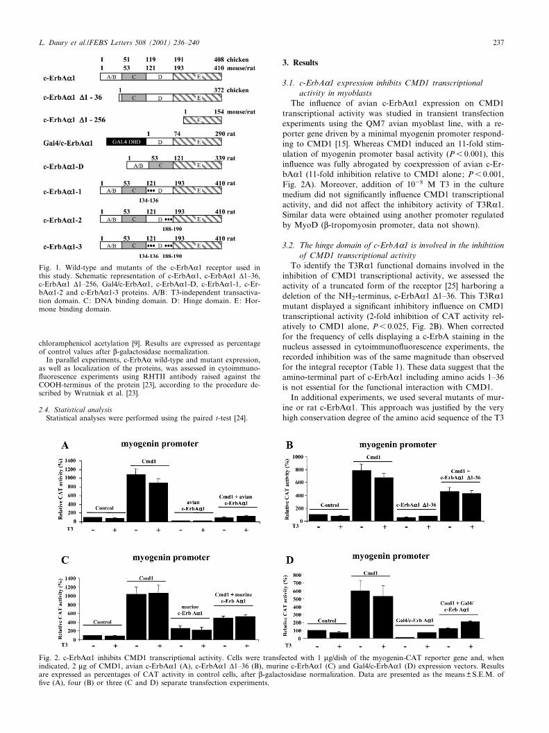
Fig. 1. Wild-type and mutants of the c-ErbAK1 receptor used inthis study. Schematic representation of c-ErbAK1, c-ErbAK1 v1^36,c-ErbAK1 v1^256, Gal4/c-ErbAK1, c-ErbAK1-D, c-ErbAK1-1, c-Er-bAK1-2 and c-ErbAK1-3 proteins. A/B: T3-independent transactiva-tion domain. C: DNA binding domain. D: Hinge domain. E: Hor-mone binding domain.
Fig. 2. c-ErbAK1 inhibits CMD1 transcriptional activity. Cells were transfected with 1 Wg/dish of the myogenin-CAT reporter gene and, whenindicated, 2 Wg of CMD1, avian c-ErbAK1 (A), c-ErbAK1 v1^36 (B), murine c-ErbAK1 (C) and Gal4/c-ErbAK1 (D) expression vectors. Resultsare expressed as percentages of CAT activity in control cells, after L-galactosidase normalization. Data are presented as the means þ S.E.M. of¢ve (A), four (B) or three (C and D) separate transfection experiments.
FEBS 25454 12-11-01
L. Daury et al./FEBS Letters 508 (2001) 236^240 237
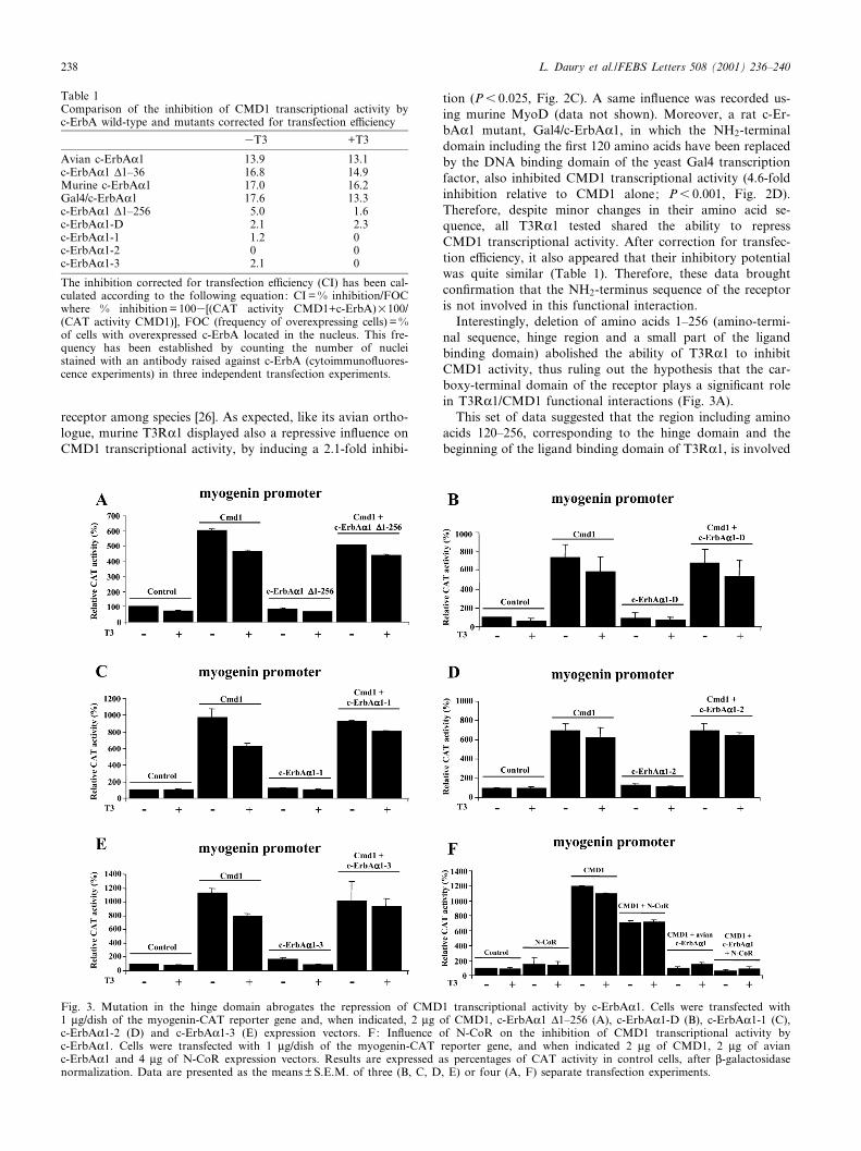
receptor among species [26]. As expected, like its avian ortho-logue, murine T3RK1 displayed also a repressive in£uence onCMD1 transcriptional activity, by inducing a 2.1-fold inhibi-
tion (P6 0.025, Fig. 2C). A same in£uence was recorded us-ing murine MyoD (data not shown). Moreover, a rat c-Er-bAK1 mutant, Gal4/c-ErbAK1, in which the NH2-terminaldomain including the ¢rst 120 amino acids have been replacedby the DNA binding domain of the yeast Gal4 transcriptionfactor, also inhibited CMD1 transcriptional activity (4.6-foldinhibition relative to CMD1 alone; P6 0.001, Fig. 2D).Therefore, despite minor changes in their amino acid se-quence, all T3RK1 tested shared the ability to repressCMD1 transcriptional activity. After correction for transfec-tion e¤ciency, it also appeared that their inhibitory potentialwas quite similar (Table 1). Therefore, these data broughtcon¢rmation that the NH2-terminus sequence of the receptoris not involved in this functional interaction.
Interestingly, deletion of amino acids 1^256 (amino-termi-nal sequence, hinge region and a small part of the ligandbinding domain) abolished the ability of T3RK1 to inhibitCMD1 activity, thus ruling out the hypothesis that the car-boxy-terminal domain of the receptor plays a signi¢cant rolein T3RK1/CMD1 functional interactions (Fig. 3A).
This set of data suggested that the region including aminoacids 120^256, corresponding to the hinge domain and thebeginning of the ligand binding domain of T3RK1, is involved
Table 1Comparison of the inhibition of CMD1 transcriptional activity byc-ErbA wild-type and mutants corrected for transfection e¤ciency
3T3 +T3
Avian c-ErbAK1 13.9 13.1c-ErbAK1 v1^36 16.8 14.9Murine c-ErbAK1 17.0 16.2Gal4/c-ErbAK1 17.6 13.3c-ErbAK1 v1^256 5.0 1.6c-ErbAK1-D 2.1 2.3c-ErbAK1-1 1.2 0c-ErbAK1-2 0 0c-ErbAK1-3 2.1 0
The inhibition corrected for transfection e¤ciency (CI) has been cal-culated according to the following equation: CI = % inhibition/FOCwhere % inhibition = 1003[(CAT activity CMD1+c-ErbA)U100/(CAT activity CMD1)], FOC (frequency of overexpressing cells) = %of cells with overexpressed c-ErbA located in the nucleus. This fre-quency has been established by counting the number of nucleistained with an antibody raised against c-ErbA (cytoimmuno£uores-cence experiments) in three independent transfection experiments.
Fig. 3. Mutation in the hinge domain abrogates the repression of CMD1 transcriptional activity by c-ErbAK1. Cells were transfected with1 Wg/dish of the myogenin-CAT reporter gene and, when indicated, 2 Wg of CMD1, c-ErbAK1 v1^256 (A), c-ErbAK1-D (B), c-ErbAK1-1 (C),c-ErbAK1-2 (D) and c-ErbAK1-3 (E) expression vectors. F: In£uence of N-CoR on the inhibition of CMD1 transcriptional activity byc-ErbAK1. Cells were transfected with 1 Wg/dish of the myogenin-CAT reporter gene, and when indicated 2 Wg of CMD1, 2 Wg of avianc-ErbAK1 and 4 Wg of N-CoR expression vectors. Results are expressed as percentages of CAT activity in control cells, after L-galactosidasenormalization. Data are presented as the means þ S.E.M. of three (B, C, D, E) or four (A, F) separate transfection experiments.
FEBS 25454 12-11-01
L. Daury et al./FEBS Letters 508 (2001) 236^240238
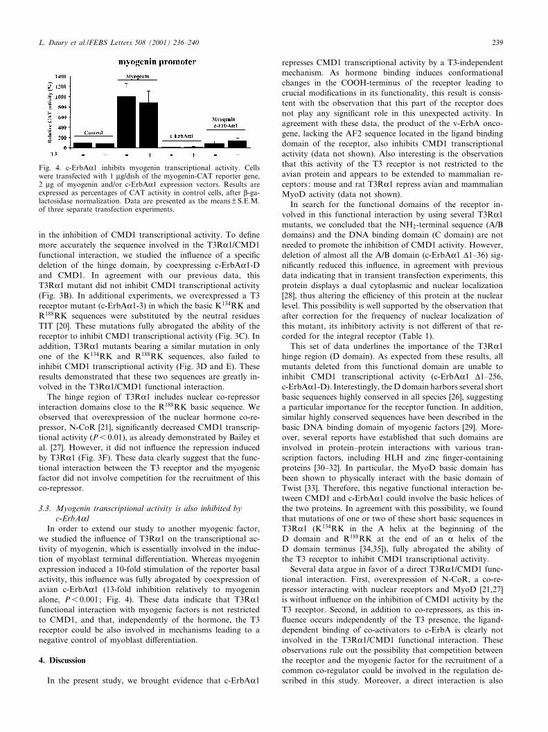
in the inhibition of CMD1 transcriptional activity. To de¢nemore accurately the sequence involved in the T3RK1/CMD1functional interaction, we studied the in£uence of a speci¢cdeletion of the hinge domain, by coexpressing c-ErbAK1-Dand CMD1. In agreement with our previous data, thisT3RK1 mutant did not inhibit CMD1 transcriptional activity(Fig. 3B). In additional experiments, we overexpressed a T3receptor mutant (c-ErbAK1-3) in which the basic K134RK andR188RK sequences were substituted by the neutral residuesTIT [20]. These mutations fully abrogated the ability of thereceptor to inhibit CMD1 transcriptional activity (Fig. 3C). Inaddition, T3RK1 mutants bearing a similar mutation in onlyone of the K134RK and R188RK sequences, also failed toinhibit CMD1 transcriptional activity (Fig. 3D and E). Theseresults demonstrated that these two sequences are greatly in-volved in the T3RK1/CMD1 functional interaction.
The hinge region of T3RK1 includes nuclear co-repressorinteraction domains close to the R188RK basic sequence. Weobserved that overexpression of the nuclear hormone co-re-pressor, N-CoR [21], signi¢cantly decreased CMD1 transcrip-tional activity (P6 0.01), as already demonstrated by Bailey etal. [27]. However, it did not in£uence the repression inducedby T3RK1 (Fig. 3F). These data clearly suggest that the func-tional interaction between the T3 receptor and the myogenicfactor did not involve competition for the recruitment of thisco-repressor.
3.3. Myogenin transcriptional activity is also inhibited byc-ErbAK1
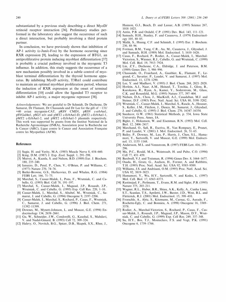
In order to extend our study to another myogenic factor,we studied the in£uence of T3RK1 on the transcriptional ac-tivity of myogenin, which is essentially involved in the induc-tion of myoblast terminal di¡erentiation. Whereas myogeninexpression induced a 10-fold stimulation of the reporter basalactivity, this in£uence was fully abrogated by coexpression ofavian c-ErbAK1 (13-fold inhibition relatively to myogeninalone, P6 0.001; Fig. 4). These data indicate that T3RK1functional interaction with myogenic factors is not restrictedto CMD1, and that, independently of the hormone, the T3receptor could be also involved in mechanisms leading to anegative control of myoblast di¡erentiation.
4. Discussion
In the present study, we brought evidence that c-ErbAK1
represses CMD1 transcriptional activity by a T3-independentmechanism. As hormone binding induces conformationalchanges in the COOH-terminus of the receptor leading tocrucial modi¢cations in its functionality, this result is consis-tent with the observation that this part of the receptor doesnot play any signi¢cant role in this unexpected activity. Inagreement with these data, the product of the v-ErbA onco-gene, lacking the AF2 sequence located in the ligand bindingdomain of the receptor, also inhibits CMD1 transcriptionalactivity (data not shown). Also interesting is the observationthat this activity of the T3 receptor is not restricted to theavian protein and appears to be extended to mammalian re-ceptors: mouse and rat T3RK1 repress avian and mammalianMyoD activity (data not shown).
In search for the functional domains of the receptor in-volved in this functional interaction by using several T3RK1mutants, we concluded that the NH2-terminal sequence (A/Bdomains) and the DNA binding domain (C domain) are notneeded to promote the inhibition of CMD1 activity. However,deletion of almost all the A/B domain (c-ErbAK1 v1^36) sig-ni¢cantly reduced this in£uence, in agreement with previousdata indicating that in transient transfection experiments, thisprotein displays a dual cytoplasmic and nuclear localization[28], thus altering the e¤ciency of this protein at the nuclearlevel. This possibility is well supported by the observation thatafter correction for the frequency of nuclear localization ofthis mutant, its inhibitory activity is not di¡erent of that re-corded for the integral receptor (Table 1).
This set of data underlines the importance of the T3RK1hinge region (D domain). As expected from these results, allmutants deleted from this functional domain are unable toinhibit CMD1 transcriptional activity (c-ErbAK1 v1^256,c-ErbAK1-D). Interestingly, the D domain harbors several shortbasic sequences highly conserved in all species [26], suggestinga particular importance for the receptor function. In addition,similar highly conserved sequences have been described in thebasic DNA binding domain of myogenic factors [29]. More-over, several reports have established that such domains areinvolved in protein^protein interactions with various tran-scription factors, including HLH and zinc ¢nger-containingproteins [30^32]. In particular, the MyoD basic domain hasbeen shown to physically interact with the basic domain ofTwist [33]. Therefore, this negative functional interaction be-tween CMD1 and c-ErbAK1 could involve the basic helices ofthe two proteins. In agreement with this possibility, we foundthat mutations of one or two of these short basic sequences inT3RK1 (K134RK in the A helix at the beginning of theD domain and R188RK at the end of an K helix of theD domain terminus [34,35]), fully abrogated the ability ofthe T3 receptor to inhibit CMD1 transcriptional activity.
Several data argue in favor of a direct T3RK1/CMD1 func-tional interaction. First, overexpression of N-CoR, a co-re-pressor interacting with nuclear receptors and MyoD [21,27]is without in£uence on the inhibition of CMD1 activity by theT3 receptor. Second, in addition to co-repressors, as this in-£uence occurs independently of the T3 presence, the ligand-dependent binding of co-activators to c-ErbA is clearly notinvolved in the T3RK1/CMD1 functional interaction. Theseobservations rule out the possibility that competition betweenthe receptor and the myogenic factor for the recruitment of acommon co-regulator could be involved in the regulation de-scribed in this study. Moreover, a direct interaction is also
Fig. 4. c-ErbAK1 inhibits myogenin transcriptional activity. Cellswere transfected with 1 Wg/dish of the myogenin-CAT reporter gene,2 Wg of myogenin and/or c-ErbAK1 expression vectors. Results areexpressed as percentages of CAT activity in control cells, after L-ga-lactosidase normalization. Data are presented as the means þ S.E.M.of three separate transfection experiments.
FEBS 25454 12-11-01
L. Daury et al./FEBS Letters 508 (2001) 236^240 239
substantiated by a previous study describing a direct MyoD/retinoid receptor interaction [36]. Preliminary studies per-formed in the laboratory also suggest the occurrence of sucha direct interaction, but probably involving a third proteinpartner.
In conclusion, we have previously shown that inhibition ofAP-1 activity (c-Jun/c-Fos) by the hormone occurring sinceRXR expression [9], leading to the expression of BTG1, anantiproliferative protein inducing myoblast di¡erentiation [37]is probably a crucial pathway involved in the myogenic T3in£uence. In addition, this study suggests the occurrence of anew mechanism possibly involved in a subtle control of myo-blast terminal di¡erentiation by the thyroid hormone appa-ratus. By inhibiting MyoD activity, T3RK1 could contributeto maintain an optimal myoblast proliferation period, whereasthe induction of RXR expression at the onset of terminaldi¡erentiation [10] could allow the liganded T3 receptor toinhibit AP-1 activity, a major myogenic repressor [38].
Acknowledgements: We are grateful to Dr Schmidt, Dr Dechesne, DrSamarut, Dr Flamant, Dr Chassande and Dr Lee for the gift of 3131/+40 avian myogenin-CAT, pRSV CMD1, pRSV c-erbAK1,pSVGal4K1, pSG5 K1t and pMT2 c-ErbAK1-D, pMT2 c-ErbAK1-1,pMT2 c-ErbAK1-2, and pMT2 c-ErbAK1-3 plasmids respectively.This work was supported by grants from the Institut National de laRecherche Agronomique (INRA), Association pour la Recherche surle Cancer (ARC), Ligue contre le Cancer and Association Franc°aisecontre les Myopathies (AFM).
References
[1] Sugie, H. and Verity, M.A. (1985) Muscle Nerve 8, 654^660.[2] King, D.M. (1987) J. Exp. Zool. Suppl. 1, 291^298.[3] Mutvei, A., Kuzela, S. and Nelson, B.D. (1989) Eur. J. Biochem.
180, 235^240.[4] Ianuzzo, D., Patel, P., Chen, V., O'Brien, P. and Williams, C.
(1977) Nature 270, 74^76.[5] Butler-Browne, G.S., Herlicoviez, D. and Whalen, R.G. (1984)
FEBS Lett. 166, 71^75.[6] Marchal, S., Cassar-Malek, I., Pons, F., Wrutniak, C. and Ca-
bello, G. (1993) Biol. Cell 78, 191^197.[7] Marchal, S., Cassar-Malek, I., Magaud, J.P., Rouault, J.P.,
Wrutniak, C. and Cabello, G. (1995) Exp. Cell Res. 220, 1^10.[8] Cassar-Malek, I., Marchal, S., Altabef, M., Wrutniak, C., Sa-
marut, J. and Cabello, G. (1994) Oncogene 9, 2197^2206.[9] Cassar-Malek, I., Marchal, S., Rochard, P., Casas, F., Wrutniak,
C., Samarut, J. and Cabello, G. (1996) J. Biol. Chem. 271,11392^11399.
[10] Downes, M., Mynett-Johnson, L. and Muscat, G.E. (1994) En-docrinology 134, 2658^2661.
[11] Gu, W., Schneider, J.W., Condorelli, G., Kaushal, S., Mahdavi,V. and Nadal-Ginard, B. (1993) Cell 72, 309^324.
[12] Halevy, O., Novitch, B.G., Spicer, D.B., Skapek, S.X., Rhee, J.,
Hannon, G.J., Beach, D. and Lassar, A.B. (1995) Science 267,1018^1021.
[13] Antin, P.B. and Ordahl, C.P. (1991) Dev. Biol. 143, 111^121.[14] Samuels, H.H., Stanley, F. and Casanova, J. (1979) Endocrinol-
ogy 105, 80^85.[15] Malik, S., Huang, C.F. and Schmidt, J. (1995) Eur. J. Biochem.
230, 88^96.[16] Forman, B.M., Yang, C.R., Au, M., Casanova, J., Ghysdael, J.
and Samuels, H.H. (1989) Mol. Endocrinol. 3, 1610^1626.[17] Casas, F., Rochard, P., Rodier, A., Cassar-Malek, I., Marchal-
Victorion, S., Wiesner, R.J., Cabello, G. and Wrutniak, C. (1999)Mol. Cell. Biol. 19, 7913^7924.
[18] Lin, Z.Y., Dechesne, C.A., Eldridge, J. and Paterson, B.M.(1989) Genes Dev. 3, 986^996.
[19] Chassande, O., Fraichard, A., Gauthier, K., Flamant, F., Le-grand, C., Savatier, P., Laudet, V. and Samarut, J. (1997) Mol.Endocrinol. 11, 1278^1290.
[20] Lee, Y. and Madhavi, V. (1993) J. Biol. Chem. 268, 2021^2028.[21] Horlein, A.J., Naar, A.M., Heinzel, T., Torchia, J., Gloss, B.,
Kurokawa, R., Ryan, A., Kamei, Y., Soderstrom, M., Glass,C.K. and Rosenfeld, M.G. (1995) Nature 377, 397^404.
[22] Nielsen, D.A., Chou, J., MacKrell, A.J., Casadaban, M.J. andSteiner, D.F. (1983) Proc. Natl. Acad. Sci. USA 80, 5198^5202.
[23] Wrutniak, C., Cassar-Malek, I., Marchal, S., Rascle, A., Heusser,S., Keller, J.M., Flechon, J., Dauca, M., Samarut, J., Ghysdael,J. and Cabello, G. (1995) J. Biol. Chem. 270, 16347^16354.
[24] Snedecor, G.W. (1961) Statistical Methods, p. 534, Iowa StateUniversity Press, Ames, IA.
[25] Bigler, J., Hokanson, W. and Eisenman, R.N. (1992) Mol. Cell.Biol. 12, 2406^2417.
[26] Marchand, O., Sa¢, R., Escriva, H., Van Rompaey, E., Prunet,P. and Laudet, V. (2001) J. Mol. Endocrinol. 26, 51^65.
[27] Bailey, P., Downes, M., Lau, P., Harris, J., Chen, S.L., Hama-mori, Y., Sartorelli, V. and Muscat, G.E. (1999) Mol. Endocri-nol. 13, 1155^1168.
[28] Andersson, M.L. and Vennstrom, B. (1997) FEBS Lett. 416, 291^296.
[29] Ma, P.C., Rould, M.A., Weintraub, H. and Pabo, C.O. (1994)Cell 77, 451^459.
[30] Bardwell, V.J. and Treisman, R. (1994) Genes Dev. 8, 1664^1677.[31] Osada, H., Grutz, G., Axelson, H., Forster, A. and Rabbitts,
T.H. (1995) Proc. Natl. Acad. Sci. USA 92, 9585^9589.[32] Williams, J.S. and Andrisani, O.M. (1995) Proc. Natl. Acad. Sci.
USA 92, 3819^3823.[33] Hamamori, Y., Wu, H.Y., Sartorelli, V. and Kedes, L. (1997)
Mol. Cell. Biol. 17, 6563^6573.[34] Rastinejad, F., Perlmann, T., Evans, R.M. and Sigler, P.B. (1995)
Nature 375, 203^211.[35] Wagner, R.L., Huber, B.R., Shiau, A.K., Kelly, A., Cunha Lima,
S.T., Scanlan, T.S., Apriletti, J.W., Baxter, J.D., West, B.L. andFletterick, R.J. (2001) Mol. Endocrinol. 15, 398^410.
[36] Froeschle, A., Alric, S., Kitzmann, M., Carnac, G., Aurade, F.,Rochette-Egly, C. and Bonnieu, A. (1998) Oncogene 16, 3369^3378.
[37] Rodier, A., Marchal-Victorion, S., Rochard, P., Casas, F., Cas-sar-Malek, I., Rouault, J.P., Magaud, J.P., Mason, D.Y., Wrut-niak, C. and Cabello, G. (1999) Exp. Cell Res. 249, 337^348.
[38] Su, H.Y., Bos, T.J., Monteclaro, F.S. and Vogt, P.K. (1991)Oncogene 6, 1759^1766.
FEBS 25454 12-11-01
L. Daury et al./FEBS Letters 508 (2001) 236^240240