Embed Size (px)
Citation preview
Recueil des Travaux Chimiques des Pays-Bas, 10913, March 1990 133
Recueil Review
Red. Trav. Chim. Pays-Bas 109, 133-146 (1990) 0165-05 l3/90/03 l33-14$4.00
X-Ray absorption spectroscopic studies on iron in soybean lipoxygenase: a model for mammalian lipoxygenases#
M. C. Feiters* ’, H. helens*, C. A. Veldink*, J. F. C. Wegenthart*, S. Navaratnam**, J. C. Allen**, H.-F. NoltingY and C. HermesY
*Department of Bio-organic Chemistry, Bijvoet Centre for Biomolecular Research, Utrecht University, P .O. Box 80.07s. NL-3508 TB Utrecht. The Netherlands ** North-East Wales Institute of Higher Education, Connah ’s Quay, Deeside, Clwyd CHS 4BR, United Kingdom
NotkestraJe 85. 0-2000 Hamburg 52. Federal Republic of Germany ‘Present address: Department of Organic Chemistry, Catholic University of Nijmegen, Toernooiveld, NL-65-25 ED Nijmegen. The Netherlands (Received July 5th, 1989)
European Molecular Biology Laboratory Outstation at Deutsches Elektronen Synchrotron,
Abstract. X-ray absorption spectroscopy studies are contributing both to the refinement of the geometry of metal sites already known from crystallographic studies and the elucidation of the coordination sphere in metal sites in metalloproteins where only limited information was available. Specific problems in the analysis of coordinating imidazoles can be overcome by applying multiple- scattering simulations. The model for the lipoxygenase iron site derived from the EXAFS study, a coordination sphere predominated by imidazoles, may require some adjustment in view of the possi- bility of coordination by water, and has received independent confirmation from studies of amino acid sequence homologies and the interaction with catechols. The spectroscopy of lipoxygenase is not consistent with the presence of the organic cofactor, PQQ, nor with its coordination to iron, but would be consistent with the presence, but not the coordination to iron, of the carboxy-hydroxy-indoloquinone-glutamic acid cofactor, CHIQG. From the observed amino acid sequence homologies it is concluded that plant lipoxygenases can be good models for the studies of mammalian lipoxygenases.
Abbreviations
CHIQG
DESY EMBL EDTB
EDTP
EPR ETE
EXAFS
9-HPOD
4-( 2-carboxy-4,5-dihydro-6-hydroxy-4,5- -dioxo- 1 H-indol-7-yl)glutamic acid [“pro-PQQ”, 4-(2-carboxy-6-hydroxy- indolo-4,5-quinon-7-yl)glutamic acid] Deutsches Elektronen Synchrotron European Molecular Biology Laboratory N,N,N’,N’-tetrakis [( 1 H-benzimidazol-2- -yl)methyl]- 1,2-diamine N,N,N ’,N’-tetrakis[( 1 H-pyrazol- 1 -yl)- methyl]-1 ,2-diamine Electron Paramagnetic Resonance 5Z,8Z,11 Z,14Z-eicosatetraenoic acid (arachidonic acid) (1) Extended X-Ray Absorption Fine Structure 9-hydroperoxy- 1 OE, 1 2Z-octadecadienoic acid
13(S)-HPOD
NMR NWO
O D
PQQ
SERC
SRS TIEOH
XAES XANES XAS
13s-hydroperoxy-9 Z, 1 1 E-octadecadi- enoic acid (3) Nuclear Magnetic Resonance Netherlands Organization for Scientific Research 9Z,12Z-octadecadoienoic acid (linoleic acid) (2) 4,5-dihydro-4,5-dioxo- 1 H-pyrrolo[2,3-f]- quinoline-2,7,9-tricarboxylic acid (5) (methoxatin or Pyrrolo-Quinoline- Quinone) (British) Science and Engineering Re- search Council Synchrotron Radiation Source 1,1,2-tris( I-methyl- 1 H-imidazol-2-yl)- ethanol X-Ray Absorption Edge Spectroscopy X-Ray Absorption Near-Edge Structure X-Ray Absorption Spectroscopy
- # Dedicated to Prof. Dr. W. Drenfh on the occasion of his retire-
ment from the Chair of Physical Organic Chemistry at the University of Utrecht.
I34 M . C. Feiters et al. / X-Ray absorption spectroscopic studies on iron in soybean lipoxygenase
1. Introduction
Lipoxygenases (E.C. 1.13.1 1.12) catalyze the regio- and stereo-selective dioxygenation of polyunsaturated fatty acids containing one or more 1 ZPZ-pentadiene systems (Scheme l), like linoleic acid (OD, 2) or arachidonic acid (ETE, l), to form fatty-acid hydroperoxides. For example, the primary reaction product of dioxygenation of OD by soybean lipoxygenase-1 at pH 9.0 is 13s-hydroperoxy- -9Z,11 E-octadecadienoic acid [ I3(S)-HPOD, 31’.
COOH
(-
13(Sl HPOD ( 3 )
Scheme I . Substrates and products of lipoxygenase reactions. ETE, 5 Z,8Z,1 I Z,14 Z-eicosatetraenoic acid; OD, 9Z, l2 Z - -octadecadienoic acid; I3(S)-HPOD, 13 S-hydroperoxy- -9Z.11 E-octadecadienoic acid.
In mammalian tissues, lipoxygenases, being involved in the initial steps of the biosynthesis of the leukotrienes and lipoxins from ETE2, are of great physiological and pathologi- cal importance. Until recently, however, investigations have focussed on plant lipoxygenases (see Ref. 3 for a review), because of their larger abundance and relative ease of purifi- cation, in spite of the fact that less details are known of their physiological role. They have been suggested to play a role in germination4 and implicated in the development of flavours and off-flavours’. Although in recent years research on mammalian lipoxygenases has developed rapidly, many interesting results in work on soybean lipoxygenases have
0 8
06 a
i l n 0 0 4 n <
02
C O
been reported by various groups since 1984, including the amino acid sequences of soybean lipoxygenase-I6, -27 and -3’, magnetic susceptibility studies of various enzyme species including native, yellow and anaerobically substrate- reduced yellow soybean lipoxygenase-lY,’O, spectroscopic studies on lipo~ygenase-2”.’~, the substitution of Fe by the 57Fe isotope and subsequent Mossbauer studies on the iron in soybean lipoxygenase- 1 I3,l4, application of modified sub- strates and high oxygen pressures’’-‘*, detailed studies on the mode of action of various inhibitor^'^-^', an electron paramagnetic resonance (EPR) of the effect of sub- stitution of the water oxygen by 170, the discovery of an organic cofactor2’, and the elucidation of the iron environ- ment by the interpretation of the extended X-ray absorption fine structure (EXAFS)26. In this review, we will discuss the application of X-Ray Absorption Spectroscopy (XAS) to metalloproteins generally, and to lipoxygenase and models in particular, and put our recent EXAFS and other results in the context of other progress in lipoxygenase research.
I . 1. Iron in plant lipoxygenases
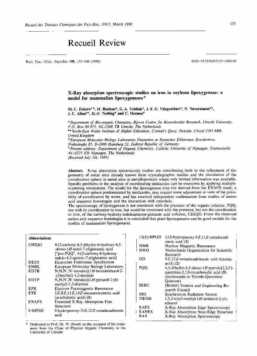
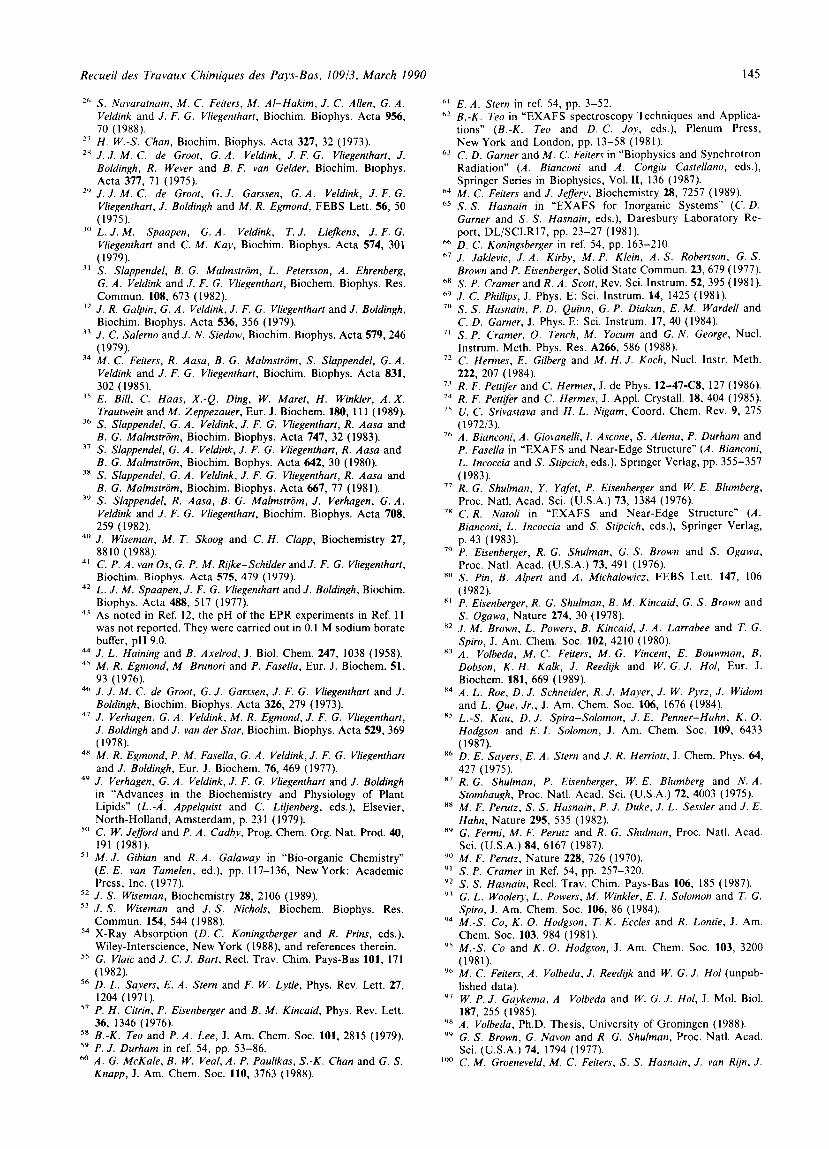
Native soybean lipoxygenase-l ( M , 94,03S6, 1 atom of iron per molecule of enzyme27) is virtually EPR-~ilent”.’~ and has no characteristic features in the ultraviolet- (UV) and visible-light absorption spectrum (Figure 1, trace a) apart from strong protein backbone absorption at approx. 200 nm and a band around 280nm ascribed to tyrosine and tryptophan absorption’*.”’. The iron in this enzyme species has been established to be high-spin Fe(I1) ( S 2 ) by ‘H NMR spectroscopy on the interaction with alcohols and magnetic susceptibility measurements3’. Addition of nitric oxide, NO, gives an electron paramagnetic resonance (EPR) signal at g 4’*, which is explained by the formation of an antiferromagnetically coupled Fe(I1)- NO complex with resulting S 3/2”. The effect of NO is not analogous to that of molecular oxygen. Magnetic susceptibility studies reveal no effect on the electronic state of the iron upon deoxy- genation’. The fact that anaerobic addition of 1 equivalent of fatty acid substrate after deoxygenation does not yield the EPR signal characteristic of enzyme-product inter- action proves that the deoxygenation procedure is effec- t i ~ e ~ ~ . The most plausible interpretation is that there is no coordination of dioxygen to the iron atom in native lipoxy-
20 7 5 6 0 4 3 9-value
3CJ0 40C 50C 600 c >‘? c 2> 2 32 W a v e l e n g t h ( n r n ) M a g n e t i c ‘ l u x d e n s i t ) ( T ’
Fig. I . Light absorption and EPR spectra of lipoxygenase. Left panel: Near-UV and visible absorption spectra of soybean lipoxy- genase-I, 0.38 mM, in air-saturated 0 .2 M Tris-HCI buffer, pH 9.0, 5 “c. Right panel: EPR spectra of soybean lipoxygenase-1. 0.30 mM, in 0.2 M Tris-HCI buffer, pH 9.0. Temperature, 15 K , modulation amplitude, I mT, microwave frequency. 9.307 GHz. Adapted from ReJ 29. (a) native enzyme, (b) yellow enzyme, (c)purple enzyme.
Recueil des Travaux Chimiques des Pays-Bas, 10913. March 1990 135
genase, which is perhaps surprising as it would seem to be the obvious place for dioxygen binding and stereoselective orientation with respect to the fatty-acid substrate. It is not excluded that conditional binding of dioxygen exists, i.e. dioxygen is only bound by the iron in the enzyme after or during interaction with the fatty-acid substrate. The possi- bility of a weak interaction of dioxygen with iron, without effect on the electronic state of the iron, has been left open', and it is interesting to note that recent Mossbauer studies of horse-liver alcohol dehydrogenase with Fe(I1) substituted in its catalytic site have revealed just such a new mode of oxygen binding, wiz. with weak spin coupling via a coordi- nated water molecule'5. The absence of a line broadening in the EPR signal of the complex with NO in H2"0 points to the absence of iron-coordinated water in this enzyme species, but it cannot be excluded that NO has displaced a water molecule coordinated to iron in the native enzyme24. Addition of 1 molar equivalent of 13(S)-HPOD to the native soybean lipoxygenase-1 at pH 9 leads to the formation of the so-called yellow enzyme species. This has a shoulder in the UV visible ~ p e c t r u m ~ ~ . ~ ' ( E ~ ~ , , 3000 M - ' . cm I)', the intensity of which is correlated3' with that of EPR signals around g 6, pointing to the presence of high-spin Fe(II1) (S 5 / 2 ) in a readily distorted ligand field slightly deviating from axial symmetry37, and typically representing 80% of the iron (Figure I , trace b)". Effects of pH on these signals have also been noted, and shown by simulations to be due to a large extent to line width changes". The g 6 EPR signal can be made to appear more homogeneous with respect to its ligand symmetry, viz. with more perfect axial ligand symmetry, by the addition of cyanide or ethanol (0.05%, v/v), neither of which inhibit the enzyme at the concentration used3'. The absence of an effect of "C substi- tution in cyanide on the EPR spectrum proves that it does not coordinate to iron3'. However, this does not imply that water does not coordinate to iron in this enzyme species, as it has recently been shown that substitution of the oxygen in water by I7O leads to a line broadening in the EPR spec- trum at pH 7, even in the presence of cyanide or ethanolz4. A marked reduction of the intensity of the EPR signal of yellow enzyme upon storage at 4 °C has been observed, but the reductant is unknown3'. The amount of EPR-visible iron is also lower when the yellow enzyme is prepared at concentrations exceeding 0.5 mM, as has been noted during sample preparation for EXAFS and magnetic susceptibility measurements", the non-EPR-visible raction still being high-spin Fe( 11) as in the native enzyme. On the other hand, the phenomenon that dilution of native enzyme to concen- trations below 1 pM "activates" it4(' could be interpreted as an oxidation to yellow enzyme, but without known oxidant. Addition of more than one molar equivalent of 13(S)- -HPOD to native or yellow soybean lipoxygenase-1 leads to the formation of a purple enzyme species". Its spec- troscopic properties are in fact dominated by those of the yellow enzyme, as the features characteristic of the purple enzyme, a 570 nm absorption (cS7" 10,000 M ~ ' . cm ~ ' ) cor- related with a g = 4.3 EPR signal, represent at most 10% of the iron (Figure 1, trace c)~'. This enzyme species is unstable with respect to reversion to yellow enzyme, a reaction which is slowed down in the presence of dioxy- gen3". These results can be compared with those of other plant lipoxygenases that have a preference for the formation of 9-HPODs from OD4'. Similar spectroscopic changes as for soybean lipoxygenase-1 are observed upon addition of equimolar and excess amounts of 13(S)-HPOD to pea lipoxygenase at pH 742, with larger deviations from perfect axial ligand symmetry as the only difference. These may be due to the different pH, which was in both cases chosen in correspondence with the optimum for enzymic activity,
rather than to the different source of the enzyme. With soybean lipoxygenase-2, the yellow and purple enzyme species are not formed consecutively but concomitantly, and more EPR-visible iron at g = 6 was found with I3(S)-HPOD than with 9(R)- or 9(S)-HPOD".43, and with 13(S)-HPOD at lower pH''. The effect of ethanol on soybean lipoxygenase-2 is identical to that on soybean lipoxygenase-1". These findings show that the iron sites n plant lipoxygenases are quite similar, in spite of differences in the regio- and stero-selectivity of the product formation.
I .2. Mechanism i f lipoxygenase catalysis
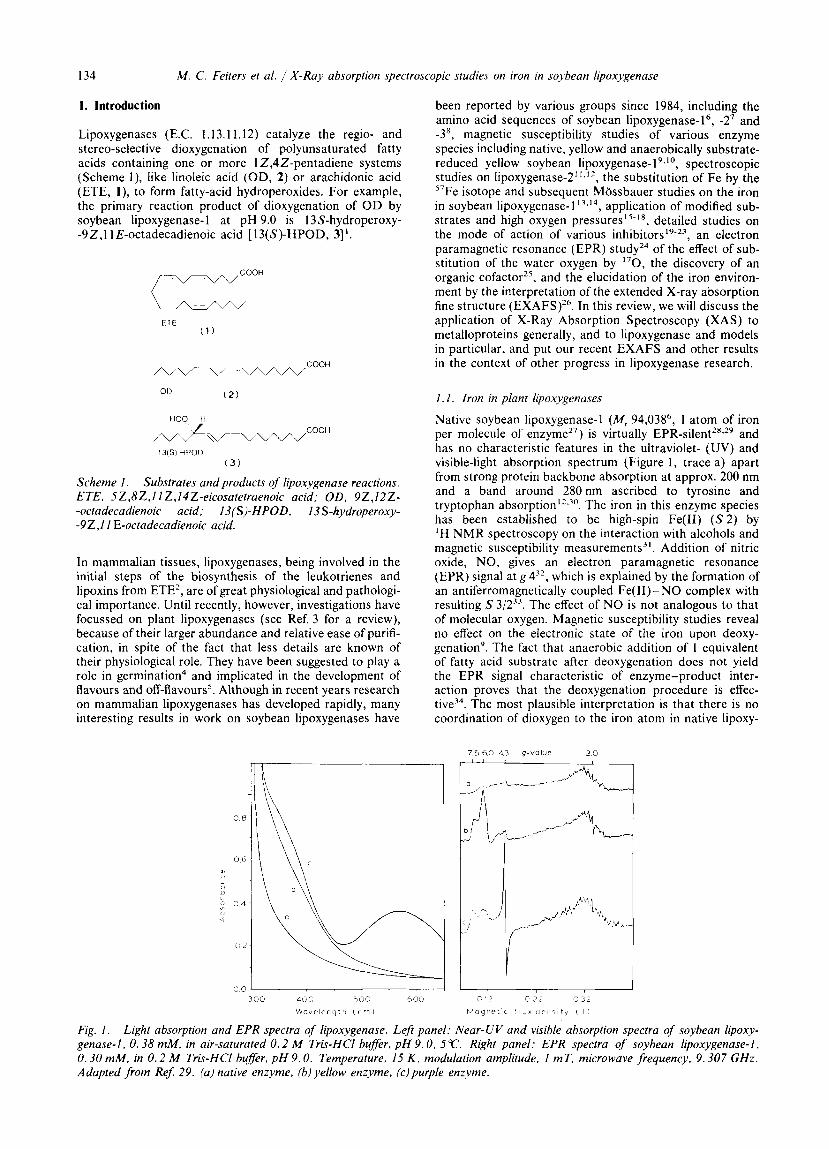
The yellow enzyme species is active in the dioxygenation (Scheme 2 , left half)2x.2y. The reaction of native enzyme with 13(S)-HPOD to yellow enzyme is then a necessary activation, which is related to the observed lag phase44. In order to account for the formation of the 3(S)-HPOD needed for activation, it has been suggested that the native enzyme should be active as ell^'^^^. Recent kinetic experi- ments with substrate concentrations below the enzyme concentration suggest that dilution to concentrations below 1 pM is sufficient for activation, and reaction with 13(S)-HPOD may not be compuIsory4".
E - Fell:) lnat lve e n z y m e )
,- 13s-HPOD
react ton p r o d u c t s \ o f 135-HPOD
. . 135 HPOD E FeI;:I , OH +RD , i' iyellov,;nzyrnrni\( \,\
E-Fel I I I l ROO- E-Fell11 HO OR '
E - F e l l l ) ROO t - F e 1 1 1 )
Scheme 2. Proposed mechanism for the aerobic and anaerobic reactions catalyzed by soybean lipoxygenase- I , adapted from Refs. 10 and 28, suggesting a key role for the reduction step of yellow enzyme with OD in the catalytic cycles. RH = OD, R' = 1inole.vl radical. RO' = 13(S)-HPOD alkoxy radical, ROO' = 13(S)-HPOD peroxy radical, R O O - = 13(S)-HPOD peroxy anion.
The yellow enzyme interacts with the fatty-acid substrate, yielding the fatty-acid radical, a proton, and an electron reducing the iron (Scheme 2, central part)2s. Evidence for the formation of the fatty-acid radical comes from EPR spin-trapping experiments46 in the so-called anaerobic reaction, where iron shuttles between the Fe(I1) and Fe(II1) states, being reduced by O D and oxidized by 13(S)-HPOD, yielding inoleyl and alkoxy radicals, respectively (Scheme 2, right half)47. Leakage of radicals from the enzyme is also proposed to account for the so-called co-oxidation, i.e. the bleaching of susceptible compounds, e.g. carotenes, during the lipoxygenase-catalyzed di~xygenation~. The valence change of iron upon interaction of yellow enzyme with O D
136 M . C. Feiters et al. / X-Ray absorption spectroscopic studies on iron in soybean lipoxygenase
was suggested by stopped-flow kinetics48 and EPR*’, indi- cating the disapearance of the spectroscopic features of yellow enzyme, and was definitely established by magnetic susceptibility measurementsko. The fatty-acid radical, with its double bonds conjugated, reacts with dioxygen to yield a peroxy radical which picks up a proton and electron to give the product hydroperoxide, and leaves the enzyme with the iron in the Fe(II1) state (Scheme 2)28. The base which interacts with the fatty acid substrate has so far not been identified. It has been noted that pH changes in the range 7-9 have an effect on the line shape of the EPR signal of the yellow enzyme39 and the native enzyme-NO complex22, which can be explained by the presence of an ionizable group with a pK, in that region near the iron. Changes in the UV spectrum upon reaction of native enzyme to yellow enzyme have been interpreted as due to tyrosine ionization12, but it should be noted that it is dangerous to draw conclusions from the UV spectra of yellow enzyme in view of the strong absorptions of products of the decomposition of 13(S)-HPOD, e.g. the 13-0x0- -9,l l - d i e n e ~ ~ ” . ~ ~ in the UV and near-UV. It could be argued that the water molecule coordinated to iron24 is the base, as the proximity of the Fe(II1) ion probably promotes its ioni- zation, and that the activation is only needed to get the iron in the right valence state to create the base. Although one might consider that the pK, of the proton to be abstracted from C-1 1 in OD could be relatively low because of the energy gained from the conjugation of the double bond, and that proton abstraction might be facilitated by the con- comitant reduction of Fe(II1) to Fe(II), or covalent binding of Fe at the terminal pentadienyl carbon15, it has been argued that there is no base strong enough in the enzyme for removal of the hydrogen as a proton, nor enough oxidizing power on the iron to remove it as a hydrogen radical, and that the initial step in catalysis should be one-electron oxidation of the C- 12- C- 13 double bond in OD by Fe(II1) to lower the pK, of the proton at C-1 150,5’. The proposal that the novel organic quinone cofactor is the base and also coordinates to iron so that proton abstraction and concomitant reduction should be facilitated2, is dis- cussed below. It has been suggested that the fatty-acid intermediate that reacts with dioxygen is a soft carbanion rather than a radi- ca150.52 , but the introduction of 0x0 groups which should stabilize such an intermediate does not make better sub- strates”. Others have presented evidence from studies with substrate modification and high oxygen pressures, that an organoiron intermediate, with oxygen insertion in the iron-carbon bond, is more likely”.
2. Introduction to X-ray absorption spectroscopy (XAS) of metalloproteins
2.1 . Theory of EXAFS
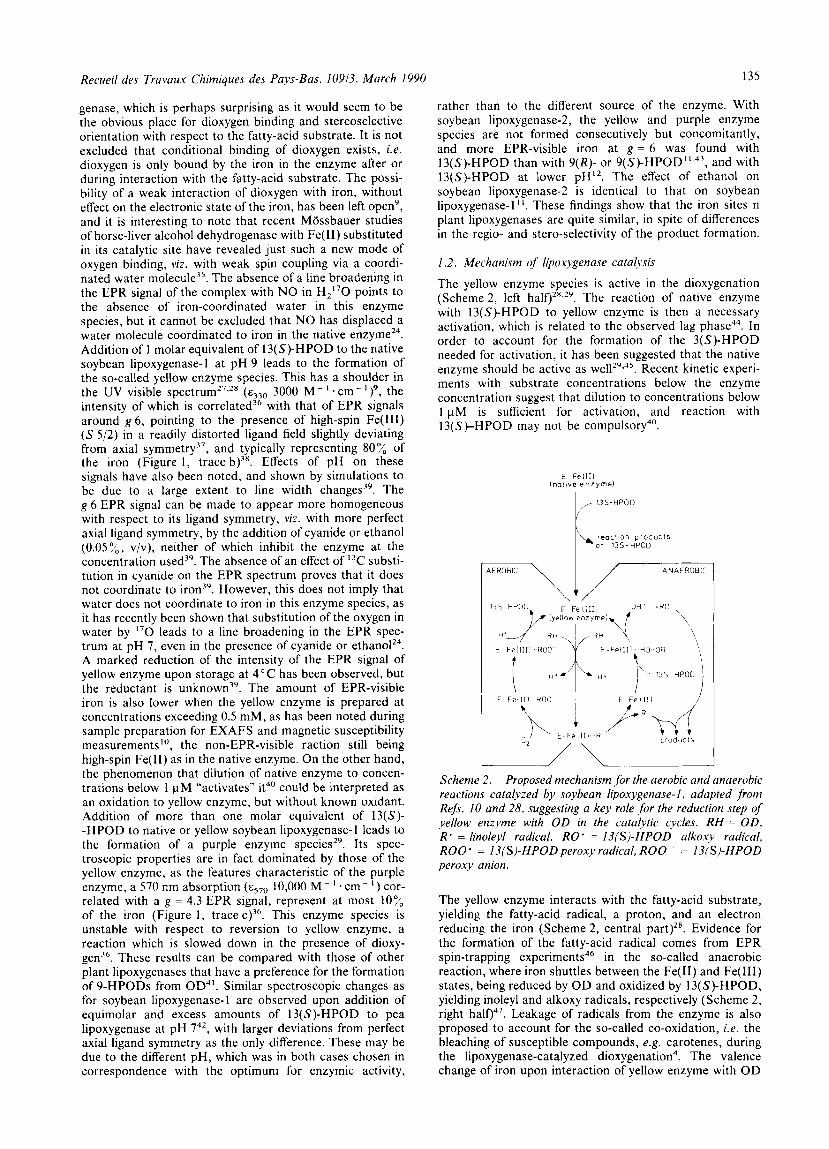
The acronym EXAFS (Extended X-Ray Absorption Fine Structure) refers to the post-edge oscillations which are typically observed in the X-ray absorption spectrum ex- tending in an energy range of some hundreds of eV starting 50 eV above an edge step. In Figure 2, the procedure of ex- traction of the EXAFS from the X-ray absorption spectrum of the model compound poly[ p-hexakis(2-methylimidazol- ato-N,N’)triiron(II)] (see below) is shown, the result being presented in k space, defined by k 2 = 2m. ( E - E,)/h, with E being the energy of the incident beam, and E, the threshold energy of the edge under consideration, in this case the iron edge. The measurement and analysis of EXAFS has developed
1 5
L 0 a n v1
7 I i
7- r ~ + r - 7 - 7 7 ,*-TT-l -150 100-50 0 5 0 100 150 200 250 300 350 LOO L50 500
energy lev1
k ( s - ’ I
Fig. 2. Extraction of EXAFS from the X-ray absorption spec- trum at the iron edge of poly[p-hexakis(2-methylimidazolato- -N,N’)triiron(II)] with the programme EXBACKI, now incorpo- rated in the programme EXBACK, available from the SERC Daresbury Laboratory, U.K. Top panel: a value for E , has been chosen (approx. 7120 eV), a second-order polynomial is extrapolated from the pre-edge to obtain the absorption coefficient in the absence of iron, p ’ , and a third-order poly- nomial is fitted through the post-edge region to mimick the smooth decay in the absence of backscatterers. p,. Bottom panel: the EXAFS, defined by (pA-po) /po , where pa = p-p’, has been extracted, and converted into k space, with k 2 = 2 m . ( E - Eo)/h , where E is the energy of the incident beam, and Eo the threshold energy of the edge under considera- tion. k’ weighting is applied.
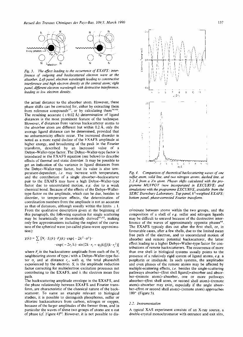
into a poweful structural too154.55 since it was shown that Fourier transformation of the EXAFS with respect to the photoelectron wave vector yields a radial distribution function in which the peaks represent shells of atoms, with the peak positions relating to the distances to the absorber atom56. Because of the photoelectric effect at the absorber atom due to irradiation with X-rays of energies above the edge energy, a photoelectron wave leaves the absorber atom and is backscattered by the surrounding atoms, leading to interference effects in the electron density at the absorber atom and hence corresponding fluctuations in its absorption coefficient (Cf. Figure 3). While travelling between the absorber and backscatterer, the electron undergoes phase shifts which cause the posi- tion of the peak in the Fourier transform to be shifted from
Recueil des Travaux Chimiques des Pays-Bas, 10913, March 1990 137
/ N N
Fig. 3. The effect leading to the occurrence of EXAFS: inter- ference of outgoing and backscattered electron wave at the absorber. Leji panel, electron wavelength leading to constructive interference and high electron density at the central atom; right panel, different electron wavelength with destructive interference. leading to low electron density.
*electron N
the actual distance to the absorber atom. However, these phase shifts can be corrected for, either by extracting them from reference compounds5', or by calculating them58-60. The resulting accurate ( f 0.02 A) determination of ligand distances is the most prominent feature of the technique. However, if distances from various backscatterer atoms to the absorber atom are different but within 0.2 A, only the average ligand distance can be determined, provided that no anharmonicity effects occur. The increased disorder is noted as a more rapid decline of the EXAFS amplitude at higher energy, and broadening of the peak in the Fourier transform, described by an increased value of a Debye-Waller-type factor. The Debye-Waller-type factor is introduced in the EXAFS equation (see below) to describe effects of thermal and static disorder. It may be possible to get an indication of the variance in ligand distances from the Debye-Waller-type factor, but its value is also tem- perature-dependent, i.e. may increase with temperature, and the contribution of a single absorber-backscatterer pair to the EXAFS may have a high Debye-Waller-type factor due to uncorrelated motion, e.g. due to a weak chemical bond. Because of the effects of the Debye-Waller- type factor on the amplitude, which can be due, besides to disorder, to temperature effects, the determination of coordination numbers from the amplitude is not as accurate as that of distances, although usually within the limits f 1. From the qualitative description given at the beginning of this paragraph, the following equation for single scattering may be heuristically or theoretically derived6',62, making only few approximations including the neglect of the curva- ture of the spherical wave (so-called plane-wave approxima- tion):
~ ( k ) = 1 [N;S,(k).F,(k).exp( - 2 k 2 . a 2 ) . I
.exp( - 2 r I / ? i ) . s i n [ 2 k . r , + cp,(k)]/(k.r;)]
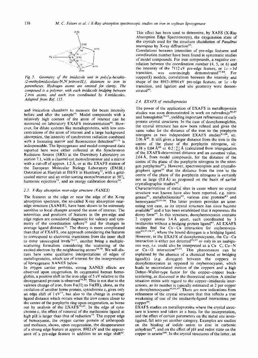
where F, is the backscatterer amplitude from each of the N, neighbouring atoms of type i with a Debye-Waller-type fac- tor a, and at distance r,, with q, the total phaseshift experienced by the electron. S, is the amplitude reduction factor correcting for multielectron excitation processes not contributing to the EXAFS, and ?i the electron mean free path. The backscattering amplitude envelope in the EXAFS, and the phase relationship between EXAFS and Fourier trans- form, are characteristic of the chemical nature of the back- scatterer. To name an example relevant to biological studies, it is possible to distinguish phosphorus, sulfur or chlorine backscatterers from carbon, nitrogen or oxygen, because of the larger amplitudes of the former three, and in particular the waves of these two groups of atoms are K out of phase (4 Figure 4)63. However, it is not possible to dis-
4 5 6 7 8 9 10 11
k I a-1 I
L! 00 '-. - t t
1 2 3 L 5 6 7 8 9 10 R I A 1
Fig. 4 . Comparison of theoretical backscattering waves of one sulfu: atom, solid line, and two nitrogen atoms, dashed line, at 2 . 2 A from a Z n atom. Phases shifis calculated with the pro- gramme MUFPOT (now incorporated in EXCURVE) and simulations with the programme EXCUR VE. available from the SERC Daresbury Laboratory. Top panel, k3-weighted EXAFS; bottom panel, phase-corrected Fourier transform.
criminate between atoms within the two groups, and the composition of a shell of e.g. sulfur and nitrogen ligands may be difficult to unravel because of the destructive inter- ference of the waves of approximately opposite phaseP4. The EXAFS typicaly dies out after the first shell, or, in favourable cases, after a few shells, due to the limited mean free path of the electron, and to uncorrelated motion of absorber and remote potential backscatterer, the latter effect leading to a higher Debye-Waller-type factor for con- tributions of remote backscatterers. The occurrence of more than one shell in biological systems usually points to the presence of a relatively rigid system of ligand atoms, e.g. a porphyrin or imidazole. In such systems, the amplitudes and even phases of the remote atoms may be affected by multiple-scattering effects, i.e. besides the single-scattering pathways absorber-(first shell 1igand)-absorber and absor- ber-(remote atom)-absorber, one or more pathways absorber-(first shell atom, or second shell atom)-(remote atom)-absorber may exist, especially if the angle absor- ber-(first or second shell atom)-(remote atom) approaches 180" (Figure 5) .
2.2. Instrumentation
A typical XAS experiment consists of an X-ray source, a double-crystal monochromator with entrance and exit slits,
138 M . C. Feiters et al. / X-Ray absorption spectroscopic studies on iron in soybean lipoxygenase
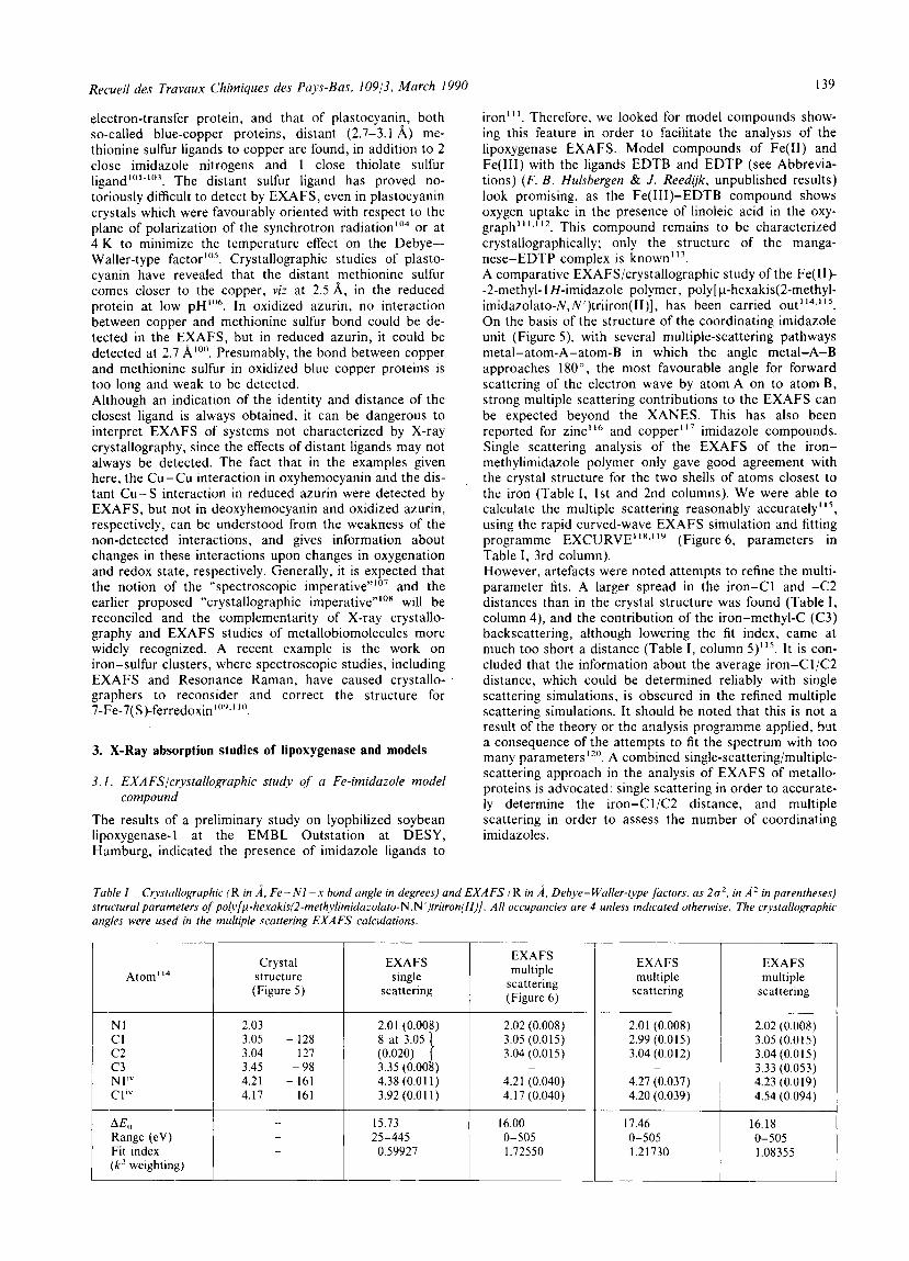
Fig. 5 . Geometry of the imidazole unit in polyfp-hexakis- (2-methylimidazolato-N,N’)triiron(II)], distances to iron in parentheses. Hydrogen atoms are omitted for clarity. The compound is a polymer. with each imidazole bridging between 2 iron atoms, and each iron coordinated by 4 imidazoles. Adapted from Ref: 11.5.
and ionization chambers to measure the beam intensity before and after the sample65. Model compounds with a relatively high content of the atom of interest can be measured on laboratory EXAFS instrumentation66. How- ever, for dilute systems like metalloproteins, with low con- centrations of the atom of interest and a large background absorption, the intensity of synchrotron radiation combined with a focussing mirror and fluorescence d e t e ~ t i o n ~ ~ - ~ ’ is indispensable. The lipoxygenase and model compound data reported here were either collected at the Synchrotron Radiation Source (SRS) at the Daresbury Laboratory on station 7.1, with a channel-cut monochromator and a mirror with a cut-off of approx. 1.2 A, or at the EXAFS station of the European Molecular Biology Laboratory (EMBL) Outstation at Hasylab at DESY in Hamburg72, with a gold- coated mirror and an order-sorting monochromator at 50 % harmonic rejection73, and an energy calibration device74.
2.3. X-Ray absorption near-edge structure (XANES)
The features at the edge or near the edge of the X-ray absorption spectrum, the so-called X-ray absorption near- edge structure (XANES), have been shown to be extremely sensitive to local changes around the atom of interest. The intensities and positions of features in the pre-edge and edge region are considered diagnostic for valency and sym- metry of the coordination sphere75 and for changes in average ligand distance76. The theory is more complicated than that of EXAFS, one approach considering the features to correspond to electronic transitions from the inner shell to outer unoccupied level^^^.^^, another being a multiple- scattering formalism considering the scattering of the excited electron by the neighbouring atom^^^.^'. We will dis- cuss here some qualitative interpretations of edges of metalloproteins, which are of interest for the interpretation of lipoxygenase XANES below. In oxygen carrier proteins, strong XANES effects are observed upon oxygenation. In oxygenated human hemo- globin, a positive shift in the iron edge of 5 eV relative to the deoxygenated protein is ~ b s e r v e d ~ ~ . ~ ~ . This is not due to the valence change of iron, from Fe(I1) to Fe(III), alone, as the oxidation of another heme protein, cytochrome c, gives only an edge shift of 1 eV77, but also to the change in average ligand distance which occurs when the iron comes closer to the centre of the porphyrin ring upon oxygenation, as borne out by analysis of the EXAFS79.8’. In the edge of cyto- chrome c, the effect of removal of the methionine ligand at high pH is larger than that of reduction77. The copper edge of hemocyanin, the oxygen carrier protein of arthropods and molluscs, shows, upon oxygenation, the disappearance of a strong edge feature at approx. 8982 eV and the appear- ance of a pre-edge feature in addition to an edge shift82.
This effect has been used to determine, by XAES (X-Ray Absorption Edge Spectroscopy), the oxygenation state of the crystals used for the structure elucidation of Panulirus interruptus by X-ray diffractionx3. Correlations between intensities of pre-edge features and coordination number have been found in systematic studies of model compounds. For iron compounds, a negative cor- relation between the coordination number (4, 5 , or 6) and the intensity of the 71 12 eV pre-edge feature, or Is-+ 3d transition, was convincingly d e m ~ n s t r a t e d ~ ~ . ’ ~ . For copper(1) models, correlations between the intensity and shape of the 8983-8984 eV pre-edge feature, or Is+ 4p transition, and ligation and site geometry were demon- stratedn5.
2.4. EXAFS of metalloproteins
The power of the application of EXAFS in metalloprotein studies was soon demonstrated in work on r u b r e d o ~ i n ~ ~ . ~ ~ and hemoglobin7’~*’, yielding important refinements of early protein crystal structures. In the case of deoxyhemoglobin, the crystal structure has now been refined and gives the same value for the distance of the iron to the porphyrin nitro ens as two independent EXAFS studies8’*88, viz.
centre of the plane of the porphyrin nitrogens, viz. 0.38 f 0.04 vs. 0.2 ::; A (calculated from triangulation of the EXAFS-determined distance and an assumed value, 2.04 A, from model compounds, for the distance of the centre of the plane of the porphyrin nitrogens to the nitro- gen porphyrins” ). However, spectroscopists and crystallo- graphers agree” that the distance from the iron to the centre of the plane of the porphyrin nitrogens is certainly not as large (0.8A;) as proposed on the basis of earlier crystallographic studies”. Characterizations of metal sites in cases where no crystal structure was known have also been reported, e.g. nitro- genase”, metallothioneinsY2, various zinc proteins63 and h e m o ~ y a n i n ~ ~ , ~ ~ - ’ ~ . The latter protein provides an inter- esting test case, as its crystal structure has since become available97 and it has been established that it represents the deoxy formx3. In this structure, deoxyhemocyanin contains 2 copper atoms 3.4A apart, each coordinated by 3 imidazoles without a bridging protein ligand97. All EXAFS studies find the Cu- Cu interaction for oxyhemocya- ninx2.Y3.Y4.Y7 , where the bound dioxygen is a bridging ligand. However, in the EXAFS of deoxyhemocyanin, the Cu-Cu interaction is either not d e t e ~ t e d ~ ~ , ~ ~ or only in an ambigu- ous way, i.e. could also be interpreted as a Cu-C, Cu-N or Cu- 0 interaction’2*’3. This phenomenon can be explained by the absence of a chemical bond or bridging Iigand(s) (e.g. dioxygen) between the coppers in deoxyhemocyanin as opposed to oxyhemocyanin, which leads to uncorrelated motion of the coppers and a high Debye-Waller-type factor for the copper-copper back- scattering, as discussed in the theoretical section. A similar situation exists with regard to the copper-imidazole inter- actions, as its number is typically estimated at 2 per copper in deoxyhem~cyanin~~*’~~~~. There are now indications from refinement of the crystal structure that this reflects a true weakening of one of the irnidazole-ligand interactions per copper9’. EXAFS studies on metalloproteins where the crystal struc- ture is known and taken as a basis for the interpretation, and the effect of certain parameters on the metal site inves- tigated, fall into yet another category. Examples are studies on the binding of iodide anion to zinc in carbonic anhydrase”, and on the effect of pH and redox state on the copper in azurin”’. In the crystal structures of the latter, an
2.06 i ”. It still gives a larger distance from the iron to the
Recueil des Travaux Chimiques des Pays-Bas, 10913, March 1990
Atom114
139
Crystal structure (Figure 5)
electron-transfer protein, and that of plastocyanin, both so-called blue-copper proteins, distant (2.7-3.1 A) me- thionine sulfur ligands to copper are found, in addition to 2 close imidazole nitrogens and 1 close thiolate sulfur 1igand 1 0 I - 103 . The distant sulfur ligand has proved no- toriously difficult to detect by EXAFS, even in plastocyanin crystals which were favourably oriented with respect to the plane of polarization of the synchrotron radiationlo4 or at 4 K to minimize the temperature effect on the Debye-- Waller-type factor“”. Crystallographic studies of plasto- cyanin have revealed that the distant methionine sulfur comes closer to the copper, viz at 2.5 A, in the reduced protein at low pH“’’. In oxidized azurin, no interaction between copper and methionine sulfur bond could be de- tected in the EXAFS, but in reduced azurin, it could be detected at 2.7 Presumably, the bond between copper and methionine sulfur in oxidized blue copper proteins is too long and weak to be detected. Although an indication of the identity and distance of the closest ligand is always obtained, it can be dangerous to interpret EXAFS of systems not characterized by X-ray crystallography, since the effects of distant ligands may not always be detected. The fact that in the examples given here, the Cu-Cu interaction in oxyhemocyanin and the dis- tant Cu-S interaction in reduced azurin were detected by EXAFS, but not in deoxyhemocyanin and oxidized azurin, respectively, can be understood from the weakness of the non-detected interactions, and gives information about changes in these interactions upon changes in oxygenation and redox state, respectively. Generally, it is expected that the notion of the “spectroscopic imperative”lo7 and the earlier proposed “crystallographic imperative”’0x will be reconciled and the complementarity of X-ray crystallo- graphy and EXAFS studies of metallobiomolecules more widely recognized. A recent example is the work on iron-sulfur clusters, where spectroscopic studies, including EXAFS and Resonance Raman, have caused crystallo- graphers to reconsider and correct the structure for 7-Fe-7( S)-ferredoxin 10’.I ’(’.
3. X-Ray absorption studies of lipoxygenase and models
3. I . EXAFS/crystallographic study of a Fe-irnidazole model
The results of a preliminary study on lyophilized soybean lipoxygenase-1 at the EMBL Outstation at DESY, Hamburg, indicated the presence of imidazole ligands to
compound
iron”’. Therefore, we looked for model compounds show- ing this feature in order to facilitate the analysis of the lipoxygenase EXAFS. Model compounds of Fe(I1) and Fe(II1) with the ligands EDTB and EDTP (see Abbrevia- tions) (F. B . Hulsbergen & J . Reedijk, unpublished results) look promising, as the Fe(II1)-EDTB compound shows oxygen uptake in the presence of linoleic acid in the oxy- graph” I . ’ 1 2 . This compound remains to be characterized crystallographically; only the structure of the manga- nese-EDTP complex is knownl13. A comparative EXAFS/crystallographic study of the Fe( 11)- -2-methyl-1 H-imidazole polymer, poly[ p-hexakis(2-methyl- imidazolato-N,N’)triiron( II ) ] , has been carried out’ 14.’ Is.
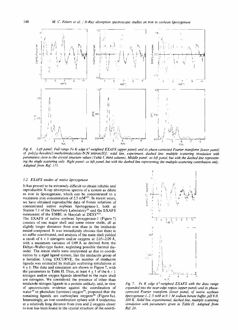
On the basis of the structure of the coordinating imidazole unit (Figure 5 ) , with several multiple-scattering pathways metal-atom-A-atom-B in which the angle metal-A-B approaches ISO”, the most favourable angle for forward scattering of the electron wave by atom A on to atom B, strong multiple scattering contributions to the EXAFS can be expected beyond the XANES. This has also been reported for zinc’16 and copper1I7 imidazole compounds. Single scattering analysis of the EXAFS of the iron- methylimidazole polymer only gave good agreement with the crystal structure for the two shells of atoms closest to the iron (Table I , 1st and 2nd columns). We were able to calculate the multiple scattering reasonably accurately’ I s ,
using the rapid curved-wave EXAFS simulation and fitting programme EXCURVE’1X.’’9 (Figure 6, parameters in Table I, 3rd column). However, artefacts were noted attempts to refine the multi- parameter fits. A larger spread in the iron-C1 and -C2 distances than in the crystal structure was found (Table I , column 4), and the contribution of the iron-methy1-C (C3) backscattering, although lowering the fit index, came at much too short a distance (Table I , column 5)I15. It is con- cluded that the information about the average iron-CliC2 distance, which could be determined reliably with single scattering simulations, is obscured in the refined multiple scattering simulations. It should be noted that this is not a result of the theory or the analysis programme applied, but a consequence of the attempts to fit the spectrum with too many parameters 12”. A combined single-scattering/muItiple- scattering approach in the analysis of EXAFS of metallo- proteins is advocated: single scattering in order to accurate- ly determine the iron-C1/C2 distance, and multiple scattering in order to assess the number of coordinating imidazoles.
Tahle I Crystallographic (R in A , Fe-NI --x bond angle in degrees) and EXAFS (R in A. Debjle-Waller-type factors, as 202 , in A’ in parentheses) structural parameters of poly~~-hexakis(2-methylimidazolalo-N,N~)triiron(ll)]. All occupancies are 4 unless indicated otherwise. The crystallographic angles were used in the multiple scattering EXAFS calculations.
N I CI c 2 c 3 N I ” CI‘”
A& Range (eV) Fit index ( k ’ weighting)
2.03
3.04 127 3.05 - 128
3.45 -98 4.21 - 161 4.17 161
EXAFS single
scattering
2.01 (0.008) 8 at 3.05 a (0.020) 3.35 (0.00 ) 4.38 (0.01 1 ) 3.92 (0.01 1)
15.73 25-445 0.59927
EXAFS multiple
scattering (Figure 6 )
2.02 (0.008) 3.05 (0.015) 3.04 (0.015)
4.21 (0.040) 4.17 (0.040)
-
16.00 0-505 1.72550
EXAFS multiple
scattering
2.01 (0.008) 2.99 (0.015) 3.04 (0.012)
4.27 (0.037) 4.20 (0.039)
-
17.46 0-505 1.21730
EXAFS multiple
scattering
2.02 (0.008) 3.05 (0.015) 3.04 (0,015) 3.33 (0.053) 4.23 (0.019) 4.54 (0.094)
16.18 0-505 1.08355
140 M. C. Feiters et al. / X-Ray absorption spectroscopic studies on iron in soybean lipoxygenase
~~ ~
3 5 7 9 1 1 3 5 7 9 11 3 5 7 9 11 klA 1) k ( A ~ l ) k iA '!
~~ _____
Fig. 6. Left panel: Full range Fe K edge k3-weighted EXAFS (upper panel) and its phase-corrected Fourier trans$orm (lower panel) of poly[~-hexakis(2-methylimidazolato-N,N')triiron(II)]: solid line, experiment, dashed line, multiple scattering simulation with parameters close to the crystal structure values (Table I , third column). Middle panel: as left panel, but with the dashed line represent- ing the single scattering only. Right panel: as left panel, but with the dashed line representing the multiple-scattering contribution only. Adapted from Ref: 115.
3.2. EXAFS studies of native lipoxygenase
It has proved to be extremely difficult to obtain reliable and reproducible X-ray absorption spectra of a system as dilute as iron in lipoxygenase, which can be concentrated to a maximum iron concentration of 2.5 mMI2'. In recent years, we have obtained reproducible data of frozen solutions of concentrated native soybean lipoxygenase- 1, both at Station 7.1 of the Daresbury Laboratory26 and the EXAFS instrument of the EMBL in Hasylab at DESY"'. The EXAFS of native soybean lipoxygenase-1 (Figure 7) consists of one major shell and some minor shells, all at slightly longer distances from iron than in the imidazole model compound. It was immediately obvious that there is no sulfur coordinated, and analysis of the main shell yielded a result of 6 f- 1 nitrogens and/or oxygens at 2.05-2.09 A, with a maximum variance of 0.09A as derived from the Debye-Waller-type factor, neglecting possible thermal dis- order. The minor shells were interpreted as due to coordi- nation by a rigid ligand system, like the imidazole group of a histidine. Using EXCURVE, the number of imidazole ligands was estimated by multiple scattering simulations at 4 f- 1. The data and simulation are shown in Figure 7, with the parameters in Table 11. Thus, at least 4 f- 1 of the 6 t 1 nitrogen and/or oxygen ligands identified in the main shell are nitrogens. We considered the presence of other than imidazole nitrogen ligands in a protein unlikely, and, in view of spectroscopic evidence against the coordination of water3' or phenolate (tyrosine) oxygen2', proposed that the remaining ligands are carboxylate oxygens" (Figure Sa). Interestingly, an iron coordination sphere with 4 imidazoles at a relatively long distance from iron and 2 oxygens closer to iron has been found in the crystal structure of the coordi-
1 I
d 4 I
R I A I
Fig. 7 . Fe K edge k'-weighted EXAFS with the data range expanded into the near-edge region (upper panel) and its phase- corrected Fourier transform (lower panel) of native soybean lipoxygenase-1,2.5 mM in 0.1 M sodium borate bufler,pH 9.0, 200 K . Solid line, experimental, dashed line, multiple scattering simulation with parameters given in Table I I . Adapted from ReJ 26.
Recueil des Travaux Chimiques des Pays-Bas. 10913. March 1990 141
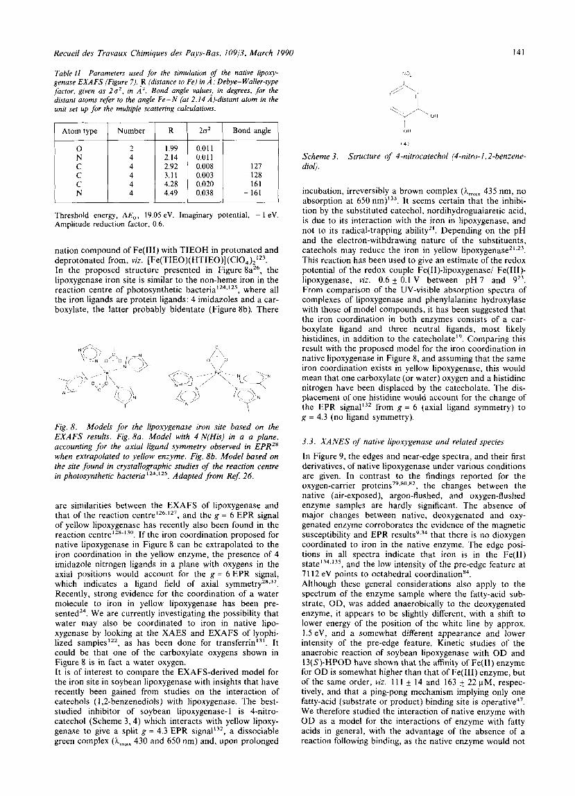
Table I 1 Parameters used for the simulation ,of the native lipoxy- genase EXA FS (Figure 7) . ,R (distance to Fe) in A ; Debye- Waller-type factor, given as 2a2, in A'. Bond angle values, in degrees. for the distant atoms refer to the angle Fe-N (at 2.14 A)-dictant atom in the unit set up for the multiple scattering calculations.
Atom type Number
C N 4
R 1 21s' 1 Bond angle
0.008 0.003
- 161
Threshold energy, AE", 19.05 eV. Imaginary potential, - 1 eV. Amplitude reduction factor, 0.6.
nation compound of Fe(1II) with TIEOH in protonated and deprotonated from, viz. [Fe(TIEO)(HTIEO)] (C104)2'23. In the proposed structure presented in Figure 8a26, the lipoxygenase iron site is similar to the non-heme iron in the reaction centre of photosynthetic b a ~ t e r i a l ~ ~ . ' ~ ' , where all the iron ligands are protein ligands: 4 imidazoles and a car- boxylate, the latter probably bidentate (Figure 8b). There
Fig. 8. Models for the lipoxygenase iron site based on the EXAFS results. Fig. 8a. Model with 4 N(His) in a a plane, accounting for the axial ligand symmetry observed in EPR2' when extrapolated to yellow enzyme. Fig. 86. Model based on the site found in crystallographic studies of the reaction centre in photosynthetic bacterial 24*125. Adapted from Re$ 26.
are similarities between the EXAFS of lipoxygenase and that of the reaction ~ e n t r e ~ ~ ~ . ' ~ ' , and the g = 6 EPR signal of yellow lipoxygenase has recently also been found in the reaction ~ e n t r e ' ~ " ' ~ " . If the iron coordination proposed for native lipoxygenase in Figure 8 can be extrapolated to the iron coordination in the yellow enzyme, the presence of 4 imidazole nitrogen ligands in a plane with oxygens in the axial positions would account for the g = 6 EPR signal, which indicates a ligand field of axial ~ y m m e t r y ~ " ~ ~ . Recently, strong evidence for the coordination of a water molecule to iron in yellow lipoxygenase has been pre- ~ e n t e d ~ ~ . We are currently investigating the possibility that water may also be coordinated to iron in native lipo- xygenase by looking at the XAES and EXAFS of lyophi- lized samples122, as has been done for t ran~fer r in '~ ' . It could be that one of the carboxylate oxygens shown in Figure 8 is in fact a water oxygen. It is of interest to compare the EXAFS-derived model for the iron site in soybean lipoxygenase with insights that have recently been gained from studies on the interaction of catechols ( 1,2-benzenediols) with lipoxygenase. The best- studied inhibitor of soybean lipoxygenase-1 is 4-nitro- catechol (Scheme 3,4) which interacts with yellow lipoxy- genase to give a split g = 4.3 EPR signal'32, a dissociable green complex (A,,, 430 and 650 nm) and, upon prolonged
ri 0,
Scheme 3. diol).
Structure of 4-nitrocatechol (4-nitro- I ~ 2-benzene-
incubation, irreversibly a brown complex (h,,, 435 nm, no absorption at 650 r ~ m ) ' ~ ~ . It seems certain that the inhibi- tion by the substituted catechol, nordihydroguaiaretic acid, is due to its interaction with the iron in lipoxygenase, and not to its radical-trapping ability". Depending on the pH and the electron-withdrawing nature of the substituents, catechols may reduce the iron in yellow l i p o ~ y g e n a s e ~ ~ . ~ ~ . This reaction has been used to give an estimate of the redox potential of the redox couple Fe(I1)-lipoxygenase/ Fe(II1)- lipoxygenase, viz. 0.6 f 0.1 V between pH 7 and 923. From comparison of the UV-visible absorption spectra of complexes of lipoxygenase and phenylalanine hydroxylase with those of model compounds, it has been suggested that the iron coordination in both enzymes consists of a car- boxylate ligand and three neutral ligands, most likely histidines, in addition to the catecholate". Comparing this result with the proposed model for the iron coordination in native lipoxygenase in Figure 8, and assuming that the same iron coordination exists in yellow lipoxygenase, this would mean that one carboxylate (or water) oxygen and a histidine nitrogen have been displaced by the catecholate. The dis- placement of one histidine would account for the change of the EPR signal'32 from g = 6 (axial ligand symmetry) to g = 4.3 (no ligand symmetry).
3.3. XANES of native lipoxygenase and related species
In Figure 9, the edges and near-edge spectra, and their first derivatives, of native lipoxygenase under various conditions are given. In contrast to the findings reported for the oxygen-carrier protein^^'.'^,^^ , the changes between the native (air-exposed), argon-flushed, and oxygen-flushed enzyme samples are hardly significant. The absence of major changes between native, deoxygenated and oxy- genated enzyme corroborates the evidence of the magnetic susceptibility and EPR r e s ~ l t s ~ . ~ ~ that there is no dioxygen coordinated to iron in the native enzyme. The edge posi- tions in all spectra indicate that iron is in the Fe(I1) state 134,135 , and the low intensity of the pre-edge feature at 71 12 eV points to octahedral coordination". Although these general considerations also apply to the spectrum of the enzyme sample where the fatty-acid sub- strate, OD, was added anaerobically to the deoxygenated enzyme, it appears to be slightly different, with a shift to lower energy of the position of the white line by approx. 1.5 eV, and a somewhat different appearance and lower intensity of the pre-edge feature. Kinetic studies of the anaerobic reaction of soybean lipoxygenase with O D and 13(S)-HPOD have shown that the affinity of Fe(I1) enzyme for OD is somewhat higher than that of Fe(II1) enzyme, but of the same order, viz. 11 1 f 14 and 163 5 22 pM, respec- tively, and that a ping-pong mechanism implying only one fatty-acid (substrate or product) binding site is operative4'. We therefore studied the interaction of native enzyme with O D as a model for the interactions of enzyme with fatty acids in general, with the advantage of the absence of a reaction following binding, as the native enzyme would not
142 M. C. Feiters er al. / X-Ray absorption spectroscopic studies on iron in soybean lipoxygenase
I 1 6
1 4
1 2
1 0
0 ; 0 8 0
06 0
0 0 4 3 -
O 0 f O 0 1
I
0 0 1. I 20 10 0 10 20 30 40 50
0 2
I
--I -1
I _-
t 0 2 ' 1
20 10 0 10 20 30 40 50
E n e r g y ( e V 1
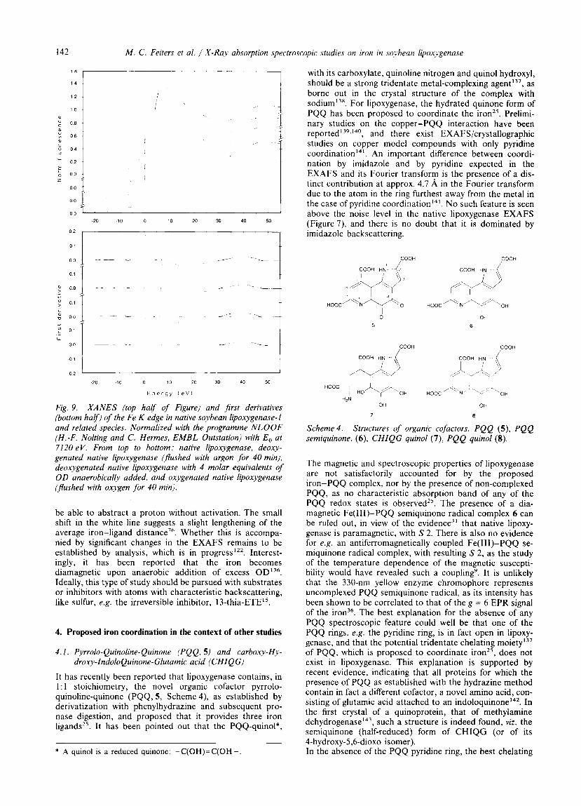
Fig. 9. XANES (top harf of Figure) and first derivatives (bottom harf) of the Fe K edge in native soybean lipoxygenase-1 and related species. Normalized with the programme NLOOF (H.-F. Nolting and C. Hermes, EMBL Outstation) with E, at 7120 eV. From top to bottom: native lipoxygenase. deoxy- genated native lipoxygenase (pushed with argon for 40 min). deoxygenated native lipoxygenase with 4 molar equivalents of OD anaerobically added, and oxygenated native lipoxygenase (Jlushed with oxygen for 40 min).
be able to abstract a proton without activation. The small shift in the white line suggests a slight lengthening of the average iron-ligand distance7'. Whether this is accompa- nied by significant changes in the EXAFS remains to be established by analysis, which is in progress122. Interest- ingly, it has been reported that the iron becomes diamagnetic upon anaerobic addition of excess OD'36. Ideally, this type of study should be pursued with substrates or inhibitors with atoms with characteristic backscattering, like sulfur, e.g. the irreversible inhibitor, 13-thia-ETE".
4. Proposed iron coordination in the context of other studies
4. I . P-vrrolo-Quinoline-Quinone (PQQ, 5) and carboxy-Hy- droxy-IndoioQuinone-Giutarnic acid (CHIQG)
It has recently been reported that lipoxygenase contains, in 1 : 1 stoichiometry, the novel organic cofactor pyrrolo- quinoline-quinone (PQQ, 5, Scheme 4), as established by derivatization with phenylhydrazine and subsequent pro- nase digestion, and proposed that it provides three iron l i g a n d ~ ~ ~ . It has been pointed out that the PQQ-quinol*,
~
* A quinol is a reduced quinone: -C(OH)=C(OH -
with its carboxylate, quinoline nitrogen and quinol hydroxyl, should be a strong tridentate metal-complexing agent'37, as borne out in the crystal structure of the complex with sodium'3X. For lipoxygenase, the hydrated quinone form of PQQ has been proposed to coordinate the iron25. Prelimi- nary studies on the copper-PQQ interaction have been r e p ~ r t e d ' ~ ' , ' ~ ~ , and there exist EXAFS/crystallographic studies on copper model compounds with only pyridine c~ord ina t ion '~ ' . An important difference between coordi- nation by imidazole and by pyridine expected in the EXAFS and its Fourier transform is the presence of a dis- tinct contribution at approx. 4.7 A in the Fourier transform due to the atom in the ring furthest away from the metal in the case of pyridine c~ord ina t ion '~ ' . No such feature is seen above the noise level in the native lipoxygenase EXAFS (Figure 7), and there is no doubt that it is dominated by imidazole backscattering.
COOH COOH * /
3
COOH HN
COOH COOH
COOH HN -
WJ '00' 1 Ho+\oH HOOC c ' ,OH
H2N OH OH
7 8
Scheme4. semiquinone, (6) , CHIQG quinol (7), PQQ quinol (8).
Structures of organic cofactors. PQQ (S), PQQ
The magnetic and spectroscopic properties of lipoxygenase are not satisfactorily accounted for by the proposed iron-PQQ complex, nor by the presence of non-complexed PQQ, as no characteristic absorption band of any of the PQQ redox states is observedz5. The presence of a dia- magnetic Fe(II1)-PQQ semiquinone radical complex 6 can be ruled out, in view of the evidence3' that native lipoxy- genase is paramagnetic, with S 2. There is also no evidence for e.g. an antiferromagnetically coupled Fe(II1)-PQQ se- miquinone radical complex, with resulting S 2, as the study of the temperature dependence of the magnetic suscepti- bility would have revealed such a coupling'. It is unlikely that the 330-nm yellow enzyme chromophore represents uncomplexed PQQ semiquinone radical, as its intensity has been shown to be correlated to that of the g = 6 EPR signal of the iron3'. The best explanation for the absence of any PQQ spectroscopic feature could well be that one of the PQQ rings, e.g. the pyridine ring, is in fact open in lipoxy- genase, and that the potential tridentate chelating moiety'37 of PQQ, which is proposed to coordinate iron25, does not exist in lipoxygenase. This explanation is supported by recent evidence, indicating that all proteins for which the presence of PQQ as established with the hydrazine method contain in fact a different cofactor, a novel amino acid, con- sisting of glutamic acid attached to an i n d o l o q ~ i n o n e ' ~ ~ . In the first crystal of a quinoprotein, that of methylamine dehydr~genase'~' , such a structure is indeed found, viz. the semiquinone (haif-reduced) form of CHIQG (or of its 4-hydroxy-5,6-dioxo isomer). In the absence of the PQQ pyridine ring, the best chelating
Recueil des Truvaux Chimiques des Puys-Bas. 10913, March 1990 I43
group in CHIQG would be the catechol or semiquinone moiety present in its reduced and half-reduced forms, respectively. As discussed above, catechols are known to inhibit lipoxygenase, and their addition has pronounced effects on the UV-visible and EPR . If one considers that the catechols may have displaced a pos- sible endogenous CHIQG catecholate ligand, one would expect the yellow enzyme to have the spectroscopic features of such a lipoxygenase-catechol complex also in the absence of exogenous catechol. In the spectra of lipoxy- genase without exogenous catechol (Figure 1 ), only the 570-nm chromophore of the purple enzyme, correlated with the g = 4.3 EPR signal representing at most 10% of the iron, would be consistent with such a coordination. Reduc- tion of CHIQG does not interfere with the ability of the cofactor to act as the proton-abstracting base2', as both the ionized hydrated quinone as well as the ionized quinol could perform this task. However, after reduction, addition of e.g. ethanol at the C-5 carbonyl, which gives a good explanation for its effect on the pH dependence of the EPR spectra2'.2s.3Y , would be impossible. Complexation of the cofactor to iron would block the redox chemistry of the cofactor, as well as its abilities to add ethanol at the C-5 carbonyl, or to act as the proton-abstracting base. It should be noted that although PQQ, or CHIQG, and other quinones have been known to be present in a variety of metalloproteins, e.g. amine ~ x i d a s e ' ~ ~ . ' ~ ' and the reaction centre of photosynthetic b a c t e ~ i a ' ~ ~ , ' ~ ' , the spec- t r o s ~ o p i c ' ~ ~ - ' ~ ' and crystallographic e ~ i d e n c e ' ~ ~ . ' ~ ' availa- ble suggests that they are at best close to the metal, but not coordinating to it. The proximity of the quinone moiety of
CHIQG to Fe, without coordination, but sufficiently close for electron exchange, as in the reaction centre of photo- synthetic bacteria, would make it a suitable candidate to be the proton-abstracting base, either as the ionized quinol or as the ionized hydrated quinone, and could account for a number of phenomena. The presence of a reduced cofactor could account for the phenomenon of the gradual dis- appearance of the EPR signal of the yellow enzyme'0,3'. The observed binding of ethanol in the proximity of iron could be explained by its addition at the C-5 carbonyl in the oxidized cofactor2'. As CHIQG resembles the indole- containing amino acid tryptophan, the features ascribed to tryptophan in the UV-visibleI2 and f l u o r e ~ c e n c e ' ~ ~ ~ ' ~ ' spec- tra could be in part due to the organic cofactor. As far as electron exchange between the iron and the cofactor or the substrates is concerned, it has been reported that the one- electron oxidation of Fe(I1) by Br:a is approx. 20 times faster upon complexation with imidazole"".
4.2. Amino acid sequences of lipoxygenase
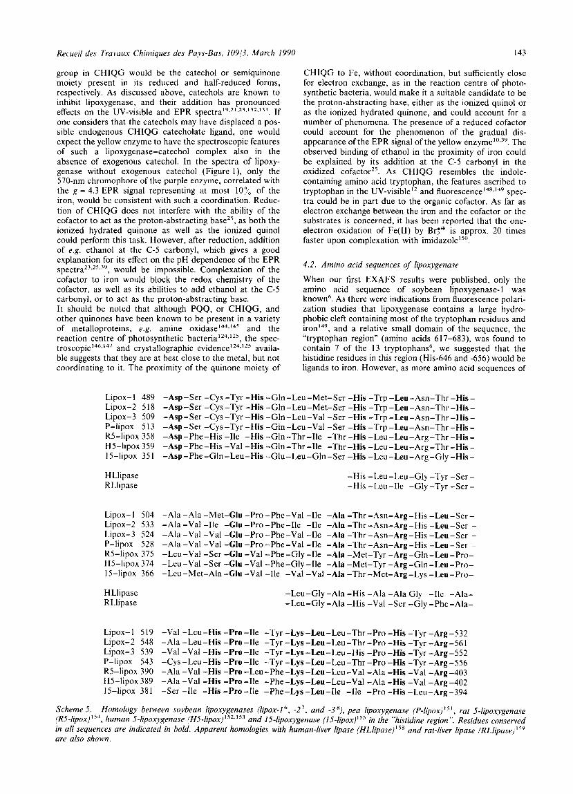
When our first EXAFS results were published, only the amino acid sequence of soybean lipoxygenase- 1 was known6. As there were indications from fluorescence polari- zation studies that lipoxygenase contains a large hydro- phobic cleft containing most of the tryptophan residues and iron'4y, and a relative small domain of the sequence, the "tryptophan region" (amino acids 6 17-683), was found to contain 7 of the 13 tryptophans', we suggested that the histidine residues in this region (His-646 and -656) would be ligands to iron. However, as more amino acid sequences of
Lipox- 1 489 -Asp-Ser -Cys -Tyr -His -Gln -Leu-Met-Ser -His -Trp -Leu-Asn-Thr -His - Lipox-2 5 18 -Asp-Ser -Cys -Tyr -His -Gln-Leu-Met-Ser -His -Trp -Leu-Asn-Thr -His - Lipox-3 509 -Asp-Ser -Cys -Tyr -His -Gln-Leu-Val -Ser -His -Trp -Leu-Asn-Thr -His - P-lipox 51 3 -Asp-Ser -Cys -Tyr -His -Gh-Leu-Val -Ser -His -Trp -Leu-Asn-Thr -His - R5-lipox 358 -Asp -Phe -His -1le -His -Gln -Thr -1le -Thr -His -Leu -Leu -Arg -Thr -His - H5-lipox 359 -Asp -Phe -His -Val -His -Gln -Thr -1le -Thr -His -Leu-Leu -Arg -Thr -His - 15-lipox 351 -Asp-Phe-Gln-Leu-His -Glu-Leu-Gln-Ser -His -Leu-Leu-Arg-Gly -His-
HLlipase RLlipase
-His -Leu-Leu-Gly -Tyr -Ser - -His -Leu-Ile -Gly -Tyr -Ser -
Lipox-l 504 -Ala -Ala -Met-Glu -Pro -Phe-Val -1le -Ala -Thr -Am-Arg-His -Leu-Ser - Lipox-2 533 -Ala -Val -1le -Glu -Pro -Phe-Ile -1le -Ala -Thr -Asn-Arg-His -Leu-Ser - Lipox-3 524 -Ala -Val -Val -Glu -Pro -Phe-Val -1le -Ala -Thr -Asn-Arg-His -Leu-Ser - P-lipox 528 -Ala -Val -Val -Glu -Pro -Phe-Val -He -Ala -Thr -Am-Arg-His -Leu-Ser -
R5-lipox 375 -Leu-Val -Ser -Clu -Val -Phe-Gly -1le -Ala -Met-Tyr -Arg-Gln -Leu-Pro- H5-lipox 374 -Leu-Val -Ser -Glu -Val -Phe-Gly -1le -Ala -Met-Tyr -Arg-Gln-Leu-Pro- 15-lipox 366 -Leu-Met-Ala -Glu -Val -1le -Val -Val -Ala -Thr -Met-Arg-Lys -Leu-Pro-
HLlipase RLlipase
-Leu-Gly -Ala -His -Ala -Ala Gly -1le -Ala- -Leu-Gly -Ala -His -Val -Ser -Gly -Phe-Ala-
Lipox- 1 5 19 -Val -Leu-His -Pro -1le -Tyr -Lys -Leu-Leu-Thr -Pro -His -Tyr -Arg-532 Lipox-2 548 -Ala -Leu-His -Pro -1le -Tyr -Lys -Leu-Leu-Thr -Pro -His -Tyr -Arg-561 Lipox-3 539 -Val -Val -His -Pro -1le -Tyr -Lys -Leu-Leu-His -Pro -His -Tyr -Arg-552 P-lipox 543 -Cys -Leu-His -Pro -1le -Tyr -Lys -Leu-Leu-Thr -Pro -His -Tyr -Arg -556 R5-lipox 390 -Ala -Val -His -Pro -Leu -Phe -Lys -Leu -Leu -Val -Ala -His -Val -Arg -403 H5-lipox 389 -Ala -Val -His -Pro-Ile -Phe-Lys -Leu-Leu-Val -Ala -His -Val -Arg-402 15-lipox 381 -Ser -1le -His -Pro-Ile -Phe-Lys -Leu-Ile -1le -Pro -His -Leu-Arg-394
Scheme 5 . Homology between soybean lipoxygenases (lipox-1 ', -2', and -3 '), pea lipoxygenase ( P - l i p o ~ ) ~ ~ ' , rat 5-lipoxygenuse (R5-lipox) "", human 5-lipoxygenuse (H5-lipox) ' ' 2 , ' 5 3 and 15-lipoxygenase (15-lip0x)"~ in the "histidine region". Residues conserved in all sequences are indicated in bold. Apparent homologies with human-liver lipase (HLlipase) '" and rat-liver lipase (RLlipase) I")
are also shown.
144 M . C. Feiters et al. / X-Ray absorption spectroscopic studies on iron in soybean lipoxygenase
soybean Iipoxygenase'.' and the pea l ip~xygenase'~' became available, it was clear that although most of the tryptophans in this region and His-656 are conserved, His-646 is not. In view of the similarities in spectroscopic properties of the iron site in soybean lipoxygenase-1 and -2"*'* and pea l i p ~ x y g e n a s e ~ ~ , it is expected that the iron ligands are in homologous places in all plant lipoxygenases. Another potential iron-coordinating region in the sequence has been identified7, containing 6 conserved histidines in three soybean lipoxygena~es~.', and a pea lipoxygenaseI5' (cf: Scheme 5). Recently, two sequences of human 5-lipoxy- genase have been published indeper~dent ly '~** '~~, as well as those of a rat 5 - l i po~ygenase '~~ and a human 15-lipoxy- g e n a ~ e ' ~ ~ , and it is now possible to extrapolate the homol- ogy in the plant lipoxygenases to the mammalian lipoxy- genases 156.
Five of the six histidines in the "histidine region" are con- served (cf- Scheme 5) , and none outside it, making those histidines strong candidates for iron coordination. In the "histidine region", an aspartic acid, a glutamic acid, and a lysine residue are also conserved. All these residues could be involved in covalent binding of CHIQG, and the car- boxylic acids could be iron ligands in addition to the histidines. Thus, there appears to be one amino acid con- figuration for iron coordination in all lipoxygenases, which makes the plant lipoxygenases good models for studies on the mammalian lipoxygenases. However, it should be noted that it was recently reported that 2 of these histidines, viz. His-362 and -372 in human 5-lipoxygenase, could be changed into Ser by genetic engineering technology without impairing the enzymic Although iron has been implied to be important for the mammalian lipoxygenases, its presence, and that of possible cofactors, still remain to be demonstrated. A similarity between part of this region in the human lipoxy- genase and the interface-binding domain in human lipo- protein lipaseI5' and rat hepatic l i p a ~ e ' ~ ~ has been noticed'52. The exciting possibility exists that one region in the protein is involved in both iron coordination and micelle/membrane recognition. However, the identification of the interface-binding domain was done in experiments with modification of a serine'" which is not in a homol- ogous position in lipoxygenase (cf. Scheme 5) . This serine has now been established to be highly conserved in lipase, in the sequence -Gly-x-Ser-x-Gly-'6'. In view of the absence of this sequence in the lipoxygenase "histidine region", it is unlikely that it should also be involved in interface binding. We have not found homologies with the amino acid sequences of the reaction centre of photosynthetic bacteria (cf Ref. 162, and references therein). In the reaction centre, two histidine ligands are provided by the L and M protein each, separated by 39 and 46 residues, respectively, with a conserved but non-coordinating histidine at 2 1 residues from the first histidine in the L chain, and the coordinating glutamate at 15 residues from the first histidine in the M chain (4 14 residues in the lipoxygenase "histidine region", Scheme 5). Clearly, profound differences in the protein chemistry exist, which make one class of proteins photo- synthetic reaction centres, and the other lipoxygenases, in spite of the spectroscopic and structural similarities of the iron site discussed above. In the lipoxygenase sequences, no conserved histidines exist in pairs, as in the sequences of iron/manganese superoxide d i s r n u t a s e ~ ' ~ ~ , and the His-His- -x-x-Tyr-Val- sequence characteristic of that class of pro- teins is absent. However, it is interesting to note that the crystal structure of Escherichia coli superoxide dismutase contains an area of unconnected electron density close to the iron site, which may be an organic c ~ f a c t o r ' ~ ~ . The only other protein, besides lipoxygenase, known to
contain both PQQ (or a related cofactor) and non-heme iron, Rhodococcus nitrile hydratase, has recently been se- quencedIh5, and found to have its histidines further apart than lipoxygenase, without homology to lipoxygenase. Re- mote analogies to lipoxygenase exist in the sequences of pterin-dependent amino acid hydroxylases, where histidines have been implied as iron l i g a r ~ d s ' ~ . ' ~ ~ . In these proteins, there are two conserved histidines 5 residues apart, in the sequence -Cys-His-Glu-Leu-Leu-Gly-His-Val-Pro-167, with one of the leucines conserved between them in the same position as in the soybean lipoxygenase sequence-], amino acids 493-498 (-His-Glu-Leu-Met-Ser-His-, conserved in all plant lipoxygenases) and 498-503 (-His-Trp-Leu-Asn-- Thr-His-, conserved in all lipoxygenases).
Acknowledgements
The authors thank NWO, the Netherlands Organization for Scientific Research, the SERC, the British Science and Engineering Research Council, and the EMBL, the Euro- pean Molecular Biology Laboratory, for support.
References and notes
I M . Hamberg and B. Samuefson, J.Biol. Chern. 242, 5329 (1967). ' B. Samuelsson, S.-E. Dahlen, J . A . Lindgren, C . A . Rouzer and
C . N. Serhan, Science 237, 1171 (1987). G . A . Veldink and J. F. G . Vliegenihari in "Advances in Inorganic Biochemistry", Vol. VI ( G . L . Eichhorn and L . G . Murzili, eds.), Elsevier, p. 139 (1984). M . Vernooy-Gerriisen, J. L . M . Leunissen, G . A . Veldink and J. F. G. Vliegenthari, Plant Physiol. 76, 1070 (1984).
' G. A. Veldink, J. F. G . Vliegenihari and J. Boldingh, Prog. Chem. Fats other Lipids 15, 131 (1977). ' D. Shibutu, J . Steczko, J. E. Dixon, M . Hermodson, R.
Yazdanparast and B. Axelrod, J. Biol. Chem. 262, 10080 (1987). D. Shibaia, J. Steczko, J. E. Dixon, P. C. Andrews, M . Hermodson, R. Yazdanparasi and B. Axelrod, J. Biol. Chem. 263, 6216 (1988). R. Yenofsky, M . Fine and C . Liu, Mol. Gen. Genet. 211, 215 (1988).
' L . Petersson, S . Slappendel and J. F. G . Vliegenihari, Biochim. Biophys. Acta 828, 81 (1985).
I n L . Peternon, S . Slappendel, M . C . Feiters and J. F. G . Vliegenthart, Biochim. Biophys. Acta 913, 228 (1987).
I ' M . C . Feiters, R. Aasa, B. G . Malmsirom, G . A . Veldink and J . F. G. Vliegenihart, Biochim. Biophys. Acta 873, 182 (1986). J. E . Draheim, R. T. Carroll, T. B . McNemar, W . R. Dunham, R. H. Sands and M . 0. Funk, Jr., Arch. Biochem. Biophys. 269, 208 (1989).
l 3 M . 0. Funk, R. T. Carroll, J. F. Thompson and W . R. Dunham, Plant Physiol. 82, 1139 (1986).
l 4 M . 0. Funk, J. Am. Oil Chem. Soc. 64, 642 (1987). I s E . J. Corey, M . diilarcuo and S . P. T. Maisuda, Tetrahedron
I' E. J. Corey and M . diilurcao, Tetrahedron Lett. 27,3589 (1986). l 7 E. J . Corey and R. Nagaia, J. Am. Chem. Soc. 109, 8107 (1987). I' E. J. Corey and J. C . Walker, J. Am. Chem. SOC. 109, 8108
(1987). l 9 D. D . Cox, S . J. Benkovic, L . M . Bloom, F. C . Bradley, M . J.
Nelson, L . Que, Jr. and D . M . Wallick, J. Am. Chem. SOC. 110, 2026 (1988).
'O J.-B. Galey, S. Bombard, C . Chopard, J.-J. Girerd, F. Lederer, D. -C. Thang, N.-H. Nam, D . Mansuy and J . 4 . Choitard, Biochemistry 27, 1058 (1988).
" C . Kemal, P. Louis-Flamberg, R. Krupinski-Olsen and A . L . Shorter, Biochemistry 26, 7064 (1987).
'' M . J. Nelson, J. Biol. Chem. 262, 12137 (1987). 23 M . J . Nelson, Biochemistry 27, 4273 (1988). 24 M . J. Nelson, J. Am. Chem. Soc. 110, 2985 (1988). 25 R. A. van der Meer and J. A . Duine, FEBS Lett. 235, 194 (1988).
Lett. 27, 3585 (1986).
Recueil des Travaux Chimiques des Pays-Bas, 10913, March 1990 145
” S. Navaratnam, M . C. Feiters, M. Al-Halnm, J . C. Allen, G. A . Veldink and J . F. G. Vliegenthart, Biochim. Biophys. Acta 956, 70 (1988).
” H. W . 3 . Chan, Biochim. Biophys. Acta 327, 32 (1973). ” J . J . M . C. de Groot, G. A . Veldink, J. F. G. Vliegenthart, J.
Boldingh, R. Wever and B. F. van Gelder, Biochim. Biophys. Acta 377, 71 (1975).
2” J . J . M . C. de Groot, G. J . Garssen, G. A . Veldink, J . F. G. Vliegenthart, J . Boldingh and M . R. Egmond, FEBS Lett. 56, 50 (1975).
‘I) L. J . M. Spaapen, G. A . Veldink, T. J . Liefkens, J . F. G. Vliegenthart and C. M. Kay, Biochim. Biophys. Acta 574, 301 (1979).
‘ I S. Slappendel, B . G. Malmstrom, L . Petersson, A . Ehrenberg, G. A . Veldink and J . F. G. Vliegenthart, Biochem. Biophys. Res. Commun. 108, 673 (1982).
” J . R. Galpin, G. A . Veldink, J. F. G. Vliegenthart and J . Boldingh, Biochim. Biophys. Acta 536, 356 (1979).
’’ J . C. Salerno and J . N. Siedow, Biochim. Biophys. Acta 579,246 ( 1979).
’‘ M. C. Feiters, R. Aasa, B . G. Malmstrom, S . Slappendel, G. A . Veldink and J . F. G. Vliegenthart, Biochim. Biophys. Acta 831, 302 (1985).
35 E. Bill, C. Haas, X.-Q. Ding, W . Mare!, H . Winkler, A. X . Trautwein and M. Zeppezauer, Eur. J . Biochem. 180, 1 1 1 (1989).
” S. Slappendel, G. A . Veldink, J . F. G. Vliegenthart, R. Aasa and B. G. Malrnstrom, Biochim. Biophys. Acta 747, 32 (1983).
37 S. Slappendel, G. A . Veldink, J . F. G. Vliegenthart, R. Aasa and B. G. Malmstrom, Biochim. Bophys. Acta 642, 30 (1980).
’’ S. Slappendel, G. A . Veldink, J. F. G. Vliegenthart, R. Aasa and B. G. Malmstrom, Biochim. Biophys. Acta 667, 77 (1981).
’’ S. Slappendel, R. Aasa, B . G. Malrnstrom, J . Verhagen, G. A . Veldink and J . F. G. Vliegenthart, Biochim. Biophys. Acta 708, 259 (1982).
40 J . Wiseman, M. T. Skoog and C. H . Clapp, Biochemistry 27, 8810 (1988).
4 1 C. P. A . van Os, G. P. M. Rijke-Schilder and J . F. G. Vliegenthart, Biochim. Biophys. Acta 575, 479 (1979).
4 2 L . J . M. Spaapen, J . F. G. Vliegenthart and J . Boldingh, Biochim. Biophys. Acta 488, 517 (1977).
43 As noted in Ref. 12, the pH of the EPR experiments in Ref. 1 1 was not reported. They were carried out in 0.1 M sodium borate buffer, pH 9.0.
44 J . L . Haining and B. Axelrod, J . Biol. Chem. 247, 1038 (1958). 45 M. R. Egmond, M. Brunori and P. Fasella, Eur. J . Biochem. 51,
93 (1976). 4’ J . J. M. C. de Groot, G. J . Garssen, J . F. G. Vliegenthart and J .
Boldingh, Biochim. Biophys. Acta 326, 279 (1973). 47 J . Verhagen, G. A . Veldink, M. R. Egmond, J . F. G. Vliegenthart,
J . Boldingh and J . van der Star, Biochim. Biophys. Acta 529, 369 (1978).
4’ M. R. Egmond, P. M. Fasella, G. A . Veldink, J . F. G. Vliegenthart and J . Boldingh, Eur. J . Biochem. 76, 469 (1977).
4y J . Verhagen, G. A . Veldink, J. F. G. Vliegenthart and J. Boldingh in “Advances in the Biochemistry and Physiology of Plant Lipids” (L.-A. Appelquist and C. Liljenberg, eds.), Elsevier, North-Holland, Amsterdam, p. 231 (1979).
’” C. W . Jefford and P. A . Cadby, Prog. Chem. Org. Nat. Prod. 40, 191 (1981).
5 ’ M. J . Gibian and R. A . Galaway in “Bio-organic Chemistry” (E . E. van Tamelen, ed.), pp. 117-136, New York: Academic Press, Inc. (1977).
5 2 J. S . Wiseman, Biochemistry 28, 2106 (1989). ” J . S. Wiseman and J . S. Nichols, Biochem. Biophys. Res.
Commun. 154, 544 (1988). 54 X-Ray Absorption (D. C. Koningsberger and R. Prins, eds.),
Wiley-Interscience, New York (l988), and references therein. 55 G. VIaic and J . C. J . Bart, Recl. Trav. Chim. Pays-Bas 101, 171
(1982). ” D. L . Sayers, E. A . Stern and F. W . Lytle, Phys. Rev. Lett. 27,
1204 (1971). 57 P. H . Citrin, P. Eb-enberger and B. M . Kincaid, Phys. Rev. Lett. 36, 1346 (1976).
5R B.-K. Teo and P. A . Lee, J . Am. Chem. SOC. 101, 2815 (1979). 5y P. J . Durham in ref. 54, pp. 53-86. ’’ A. G. McKale, B . W . Veal, A . P. Paulikas, S.-K. Chan and G. S .
Knapp, J . Am. Chem. SOC. 110, 3763 (1988).
‘ I E. A . Stern in ref. 54, pp. 3-52. B.-K. Teo in “EXAFS spectroscopy Techniques and Applica- tions” (B.-K. Teo and D. C. Joy, eds.), Plenum Press, New York and London, pp. 13-58 (1981).
” C. D. Garner and M. C. Feiters in “Biophysics and Synchrotron Radiation” (A . Bianconi and A . Congiu Castellano, eds.), Springer Series in Biophysics, Vol. 11, 136 (1987).
h4 M. C. Feiters and J . Jeffery, Biochemistry 28, 7257 (1989). ” S. S. Hasnain in “EXAFS for Inorganic Systems” (C. D.
Garner and S. S. Hasnain, eds.), Daresbury Laboratory Re- port, DLISCI.RI7, pp. 23-27 (1981).
” D. C. Koningsberger in ref. 54, pp. 163-210. ” J . Jaklevic, J . A . Kirby, M. P. Klein, A . S . Robertson, G. S.
Brown and P. Eisenberger, Solid State Commun. 23,679 (1977). “ S. P. Cramer and R. A . Scott, Rev. Sci. Instrum. 52, 395 (1981). ’’) J . C. Phillips, J . Phys. E : Sci. Instrum. 14, 1425 (1981). ’I’ S. S. Hasnain, P. D. Quinn, G. P. Diakun, E. M . Wardell and
C. D. Garner, J . Phys. E: Sci. Instrum. 17, 40 (1984). 7 1 S . P. Cramer, 0. Tench, M. Yocum and G. N. George, Nucl.
Instrum. Meth. Phys. Res. A266, 586 (1988). 72 C. Hermes, E. Gilberg and M . H. J . Koch, Nucl. Instr. Meth.
222, 207 (1984). ” R. F. Pettifer and C. Hermes, J . de Phys. 12-47-C8, 127 (1986). 74 R. F. Pettifer and C. Hermes, J. Appl. Crystall. 18, 404 (1985). 75 U. C. Srivastava and H. L. Nigam, Coord. Chem. Rev. 9, 275
( 1972/3). ” A . Bianconi, A . Giovanelli, I . Ascone, S. Alema, P. Durham and
P. Fasella in “EXAFS and Near-Edge Structure” ( A . Bianconi, L . Incoccia and S. Stipcich, eds.), Springer Verlag, pp. 355-357 (1983). R. G. Shulman, Y. Yafet, P. Eisenberger and W . E. Blumberg, Proc. Natl. Acad. Sci. (U.S.A.) 73, 1384 (1976).
’’ C. R. Natoli in “EXAFS and Near-Edge Structure” ( A . Bianconi, L . Incoccia and S. Stipcich, eds.), Springer Verlag, p. 43 (1983).
”) P. Eisenberger, R. G. Shulman, G. S. Brown and S. Ogawa, Proc. Natl. Acad. (U.S.A.) 73, 491 (1976).
‘(I S. Pin, B. Alpert and A . Michalowicz, FEBS Lett. 147, 106 (1982).
’‘ P. Eisenberger, R. G. Shulman, B . M. Kincaid, G. S. Brown and S. Ogawa, Nature 274, 30 (1978).
” J. M. Brown, L . Powers, B. Kincaid, J . A . Larrabee and T. G. Spiro, J . Am. Chem. SOC. 102, 4210 (1980).
” A. Volbeda, M. C. Feiters, M . G. Vincent, E. Bouwman, B . Dobson, K . H . Kalk, J . Reedijk and W . G. J . Hol, Eur. J . Biochem. 181, 669 (1989).
x4 A . L. Roe, D. J. Schneider, R. J . Mayer, J . W . Pyrz, J . Widom and L. Que, Jr., J . Am. Chem. SOC. 106, 1676 (1984). L . 3 . Kau, D. J . Spira-Solomon, J . E . Penner-Hahn, K . 0. Hodgson and E. I . Solomon, J . Am. Chem. SOC. 109, 6433 (1987).
” D. E. Sayers, E. A . Stern and J . R. Herriott, J . Chem. Phys. 64, 427 (1975).
’’ R. G. Shulman, P. Eisenberger, W . E . Blumberg and N. A . Stombaugh, Proc. Natl. Acad. Sci. (U.S.A.) 72, 4003 (1975).
’’ M . F. Perutz, S . S . Hasnain, P. J . Duke, J . L . Sessler and J . E. Hahn, Nature 295, 535 (1982).
” G. Fermi, M. F. Perutz and R. G. Shulman, Proc. Natl. Acad. Sci. (U.S.A.) 84, 6167 (1987).
’)O M. F. Perutz, Nature 228, 726 (1970). ‘ ) I S. P. Cramer in Ref. 54, pp. 257-320. ’)* S. S. Hasnain, Recl. Trav. Chim. Pays-Bas 106, 185 (1987). ’)’ G. L . Woolery, L . Powers, M. Winkler, E. I . Solomon and T. G.
‘)4 M.-S. Co, K . 0. Hodgson, T. K . Eccles and R. Lontie, J. Am.
’’ M.-S. Co and K . 0. Hodgson, J . Am. Chem. SOC. 103, 3200
’)‘ M. C. Feiters, A . Volbeda, J . Reedijk and W . G. J . Hol (unpub-
‘)7 W . P. J . Gaykema, A . Volbeda and W . G. J . Hol, J . Mol. Biol.
” A . Volbeda, Ph.D. Thesis, University of Groningen (1988). ’)’ G. S. Brown, G. Navon and R. G. Shulman, Proc. Natl. Acad.
loo C. M. Groeneveld, M . C. Feiters, S . S. Hasnain, J . van Run, J .
77
Spiro, J . Am. Chem. SOC. 106, 86 (1984).
Chem. SOC. 103, 984 (1981).
(1981).
lished data).
187, 255 (1985).
Sci. (U.S.A.) 74, 1794 (1977).
146 M . C. Feiters et al. / X - R a y absorption spectroscopic studies on iron in soybean lipoxygenase
1 0 1
1 0 2
1113
104
105
Il l6
107
I I I X
I I)Y
I 1 0
I l l
I I2
I 1 1
I14
I 1 5
I I6
117
I 1 8
119
I20
121
I22
I 2 7
I24
12s
I26
I27
I2M
I29
I 7 0
1 3 1
1 7 2
I 7 3
I 3 4
ReediJk and G. W . Canters, Biochim. Biophys. Acta 873, 214 (1986). E. T. Adman and L . H. Jensen, Isr. J . Chem. 21, 8 (1981). G. E . Norris, B. F. Anderson and E. N. Baker, J . Am. Chem. SOC. 108, 2782 (1986). J. M. Guss and H. C. Freeman, J . Mol. Biol. 169, 521 (1983). R. A . Scott, J . E. Hahn, S . Doniach, H . C. Freeman and K . 0. Hodgson, J . Am. Chem. SOC. 104, 5364 (1982). J . E. Penner-Hahn, M. Murata, K . 0. Hodgson and H. C. Freeman, Inorg. Chem. 28, 1826 (1989). H. C. Freeman, “Coordination Chemistry” ( J . P. Laurent, ed.), Pergamon Press, Osford, U.K. 21, 29 (1981). G. N. George and S. J. George, Trends in Biochem. Sci. 13, 369 (1988). J. Galloway, Nature 318, 602 (1985). G. H . Stout, S . Turley, L . C. Sieker and L . H. Jensen, Proc. Natl. Acad. Sci. (U.S.A.) 85, 1020 (1988). C. D. Stout, J . Biol. Chem. 263, 9256 (1988). M. C. Feiters, J. F. G. Vliegenthart, J . Reedijk and B. G. Malmstrom, Inorg. Chim. Acta 79, 148 (1983). M . C. Feiters, Ph.D. Thesis, University of Utrecht (1984). F. B. Hulsbergen, W . L . Driessen, J . Reedijk and G. C. Verschoor, Inorg. Chem. 23, 3588 (1984). A . L . Spek, A . J . M . Duisenberg and M . C. Feiters, Acta Cryst. C 39, 1212 (1983). M. C. Feiters, S . Navaratnam, M. Al-Hakim, J . C. Allen, A . L . Spek, G. A. Veldink and J. F. G. Wegenthart, J . Am. Chem. SOC. 110, 7746 (1988). R. F. Pettifer, D. F. Foulis and C. Hermes, J. de Phys. 12-47-C8, 545 (1986). R. W. Strange, N. J. Blackburn, P. F. Knowles and S. S. Hasnain, J . Am. Chem. SOC. 109, 7157 (1987). S. J. Gurman, N . Binsted and I . Ross, J. Phys. C: Solid State Phys. 17, 143 (1984). S. J . Gurman, N. Binsted and I . Ross, J. Phys. C: Solid State Phys. 19, 1845 (1986). P. A . Lee, P. H. Citrin, P. Eisenberger and B. M . Kincaid, Rev. Mod. Phys. 53, 789 (1981). One of the results reported earlier (Ref. 112) has later been shown to be an artefact, due to misalignment of the sample cell in the EXAFS apparatus, so that a mixture of the EXAFS of an iron contamination in the aluminium sample cell and that of lipoxygenase was obtained. The iron-aluminium back- scattering was then mistaken for iron-sulfur backscattering. L . M. van der Heydt, M . C. Feiters, G. A . Veldink, J. F. G. Vliegenthart, S. Navaratnam, J . C. Allen, H.-F. Nolting and C. Hermes, unpublished data. S. M. Gorun, G. C. Papaejihvmiou, R. B . Frankel and S. J . Lippard, J . Am. Chem. SOC. 109, 4244 (1987). J . Deisenhofer, 0. Epp, K . Miki, R. Huber and H. Michel, Nature 318, 618 (1985). J . P. Allen, G. Feher, T. 0. Yeates, H . Komyia and D . C. Rees, Proc. Natl. Acad. Sci. (U.S.A.) 84, 5730 (1987). P. Eisenberger, M. Y. Okamura and G. Feher, Biophys. J . 37, 523 (1982). G. Bunker, E. A . Stern, R. E. Blankenship and W . W . Parson, Biophys. J . 37, 539 (1982). V. Petrouleas and B. A . Diner, Biochim. Biophys. Acta 849,264 (1986). S. Itoh, X.-S. Tang and K . Satoh, FEBS Lett. 205, 275 (1986). R. Aasa, L.-E. AndrPasson, S. Styring and T. Vanngird, FEBS Lett. 243, 156 (1989). S. S. Hasnain, R. W . Evans, R. C . Garratt and P. F. Lindley, Biochem. J . 247, 369 (1987). J . R. Galpin, L. G. M. Tielens, G. A . Veldink, J. F. G. Vliegenthart and J. Boldingh, FEBS Lett. 69, 179 (1976). L. J. M . Spaapen, J. Verhagen, G. A . Veldink and J. F. G. Vliegenthart, Biochim. Biophys. Acta 617, 132 (1980). H.-F. Nolting, M.D. Thesis, University of Miinster, F.R.G. (1985).
I H.-F. Nolting, C. Hermes, R. F. Pettifer, G. Henkel and B. Krebs (unpublished data).
I76
I27
I z x
I 7 9
140
141
142
147
I44
145
I 4 6
147
14X
14Y
I so
I S 1
I 5 2
I S 3
I 5 4
I S 5
I 5 6
157
I 5 8
I59
T. h. Cheesbrough and B. Axelrod, Biochemistry 22, 3837 (1983). J . B. Noar, E . J. Rodriguez and T. C. Bruice, J . Am. Chem. SOC. 107, 7198 (1985). T. Ishida, M. Doi, K . Tomita, H. Hayashi, M. Inoue and T. Urakami, J. Am. Chem. SOC. 111, 6822 (1989). S. Suzuki, M . Sakurai, S . Itoh and Y. Ohshiro, Chem. Lett. 777 (1988). S. Suzuki, M. Sakurai, S . ltoh and Y. Ohshiro, Inorg. Chem. 27, 591 (1988). N. J. Blackburn, R. W. Strange, A . Farooq, M. S . Haka and K . D. Karlin, J . Am. Chem. SOC. 110, 4263 (1988). R. A . van der Meer, B . W . Groen and J . A . Duine, FEBS Lett. 246, 109 (1989). F. M. D. Vellieux, F. Huitema, H . Groendijk, K . H. Kalk, J. Franz, Jzn., J . A . Jongejan, J. A . Duine, K . Petratos, J. Drenth and W. G. J. Hol, EMBO J . 8, 2171 (1989). M. Ameyama, M. Hayashi, K . Matsushita, E. Shinagawa and 0. Adachi, Agric. Biol. Chem. 48, 562 (1984). C. L . Lobenstein-Verbeek, J . A . Jongejan, J. A . Frank and J. A . Duine, FEBS Lett. 170, 305 (1984). T. J. Williams and M. C. Falk, J . Biol. Chem. 261, 15949 (1986). G. C. Dismukes, H. A . Frank, R. Friesner and K . Sauer, Biochim. Biophys. Acta 764, 253 (1984). A. Finazzi-Agro, L . Avigliano, G. A . Veldink, J. F. G. Vliegenthart and J. Boldingh, Biochim. Biophys. Acta 326, 462 (1973). A. Finazzi A g o , L . Avigliano, M . R. Egmond, G. A . Veldink and J . F. G. Vliegenrhart, FEBS Lett. 52, 73 (1975). B. J. Parsons, M. Al-Hakim, G. 0. Phillips and A . J. Swallow, J . Chem. SOC., Faraday Trans. 82, 1575 (1986). P. M. Ealing and R. Casey, Biochem. J . 253, 915 (1988). R. A . F. Dixon, R. E. Jones, R. E. Diehl, C. D . Bennett, S. Kargman and C. A . Rouzer, Proc. Natl. Acad. Sci. (U.S.A.) 85, 416 (1988). T. Matsumoto, C. D. Funk, 0. Rddmark, J . -0 . Hoog, H. Jornvall and B. Samuelsson, Proc. Natl. Acad. Sci. (U.S.A.) 85, 26 and correction, p. 3406 (1988). J . M . Balcarek, T. W . Theisen, M . N. Cook, A . Varrichio, S.-M. Hwang, M. W. Strohsacker and S. T. Crooke, J. Biol. Chem. 263, 13937 (1988). E. Sigal, D. Grunberger, C. S . Craik, G. H. Caughey and J. A . Nadel, J . Biol. Chem. 263, 5328 (1988). Our alignment is based on alignments given in Refs. 7, 8, 148 and 151. A partial alignment of 6 lipoxygenases was published while this manuscript was in preparation: C. D. Funk, S. Hoshito, T. Matsumoto, 0. Rddmark and B. Samuehon, Proc. Natl. Acad. Sci. (U.S.A.) 86, 2587 (1989). C. D. Funk, H. Gunne, H. Steiner, T. Izumi and B. Samuelsson, Proc. Natl. Acad. Sci. (U.S.A.) 86, 2592 (1989). K . L . Wion, T. G. Kirchgessner, A. J . Lusis, M . C. Schotz and R. M. Lawn, Science 235, 1638 (1987). M . C. Komaromy and M . C. Schotz, Proc. Natl. Acad. Sci. (U.S.A.) 84, 1526 (1987). A. Guidoni, F. Benkouka, J . De Caro and M . Rovery, Biochim. Biophys. Acta 660, 148 (1981). E. Antonian, Lipids 23, 1101 (1988).
I h 2 H. Komiya, T. 0. Yeates, D. C. Rees, J . P. Allen and G. Feher, Proc. Natl. Acad. Sci. (U.S.A.) 85, 9012 (1988).
l h 3 J . I . Harris, A . D. Aufiet , F. D. Northrop and J. E . Walker, Eur. J . Biochem. 106, 297 (1980).
l h 4 W. C. Stallings, T. B . Powers, K . A . Pattridge, J. A . Fee and M. L . Ludwig, Proc. Natl. Acad. Sci. (U.S.A.)80, 3884 (1983).
I h 5 0. Ikehata, M . Nishiyama, S. Horinouchi and T. Beppu, Eur. J . Biochem. 181, 563 (1989).
I h 6 K . K . Andersson, D. D. Cox, L . Que, Jr., T. Flatmark and J . Haavik, J. Biol. Chem. 263, 18621 (1988).
l h 7 F. D. Ledley, A . G. DiLella, S . C. M . Kwok and S. L. C. Woo, Biochemistry 24, 3389 (1985).