Opposite Roles of Furin and PC5Ain N-Cadherin Processing1,2
Deborah Maret*,3, Mohamad Seyed Sadr*,3,Emad Seyed Sadr*, David R. Colman*,4,Rolando F. Del Maestro* and Nabil G. Seidah†
*Brain Tumor Research Centre, Montreal NeurologicalInstitute, McGill University, Montréal, Québec, Canada;†Institut de Recherches Cliniques de Montréal,Université de Montreal, Montreal, Québec, Canada
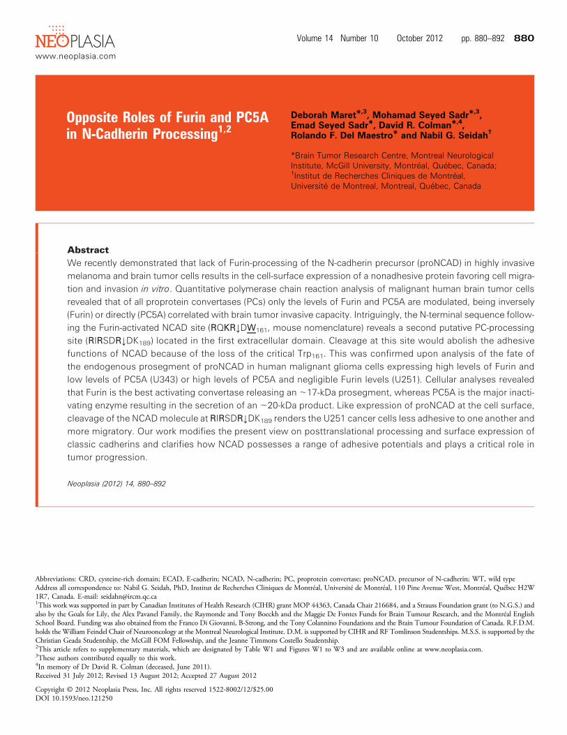
AbstractWe recently demonstrated that lack of Furin-processing of the N-cadherin precursor (proNCAD) in highly invasivemelanoma and brain tumor cells results in the cell-surface expression of a nonadhesive protein favoring cell migra-tion and invasion in vitro. Quantitative polymerase chain reaction analysis of malignant human brain tumor cellsrevealed that of all proprotein convertases (PCs) only the levels of Furin and PC5A are modulated, being inversely(Furin) or directly (PC5A) correlated with brain tumor invasive capacity. Intriguingly, the N-terminal sequence follow-ing the Furin-activated NCAD site (RQKR↓DW161, mouse nomenclature) reveals a second putative PC-processingsite (RIRSDR↓DK189) located in the first extracellular domain. Cleavage at this site would abolish the adhesivefunctions of NCAD because of the loss of the critical Trp161. This was confirmed upon analysis of the fate ofthe endogenous prosegment of proNCAD in human malignant glioma cells expressing high levels of Furin andlow levels of PC5A (U343) or high levels of PC5A and negligible Furin levels (U251). Cellular analyses revealedthat Furin is the best activating convertase releasing an ∼17-kDa prosegment, whereas PC5A is the major inacti-vating enzyme resulting in the secretion of an ∼20-kDa product. Like expression of proNCAD at the cell surface,cleavage of the NCADmolecule at RIRSDR↓DK189 renders the U251 cancer cells less adhesive to one another andmore migratory. Our work modifies the present view on posttranslational processing and surface expression ofclassic cadherins and clarifies how NCAD possesses a range of adhesive potentials and plays a critical role intumor progression.
Neoplasia (2012) 14, 880–892
Abbreviations: CRD, cysteine-rich domain; ECAD, E-cadherin; NCAD, N-cadherin; PC, proprotein convertase; proNCAD, precursor of N-cadherin; WT, wild typeAddress all correspondence to: Nabil G. Seidah, PhD, Institut de Recherches Cliniques de Montréal, Université de Montréal, 110 Pine Avenue West, Montréal, Québec H2W1R7, Canada. E-mail: [email protected] work was supported in part by Canadian Institutes of Health Research (CIHR) grant MOP 44363, Canada Chair 216684, and a Strauss Foundation grant (to N.G.S.) andalso by the Goals for Lily, the Alex Pavanel Family, the Raymonde and Tony Boeckh and the Maggie De Fontes Funds for Brain Tumour Research, and the Montréal EnglishSchool Board. Funding was also obtained from the Franco Di Giovanni, B-Strong, and the Tony Colannino Foundations and the Brain Tumour Foundation of Canada. R.F.D.M.holds the William Feindel Chair of Neurooncology at the Montreal Neurological Institute. D.M. is supported by CIHR and RF Tomlinson Studentships. M.S.S. is supported by theChristian Geada Studentship, the McGill FOM Fellowship, and the Jeanne Timmons Costello Studentship.2This article refers to supplementary materials, which are designated by Table W1 and Figures W1 to W3 and are available online at www.neoplasia.com.3These authors contributed equally to this work.4In memory of Dr David R. Colman (deceased, June 2011).Received 31 July 2012; Revised 13 August 2012; Accepted 27 August 2012
Copyright © 2012 Neoplasia Press, Inc. All rights reserved 1522-8002/12/$25.00DOI 10.1593/neo.121250
www.neoplasia.com
Volume 14 Number 10 October 2012 pp. 880–892 880
IntroductionOf the many processes that occur during tumor progression, invasionand the formation of secondary metastases are the most clinically rel-evant but the least well understood at the molecular level. Yet, it isnow well established that changes in surface expression of classes ofcell adhesion molecules correlate with malignant transformation[1,2]. Reduction of intercellular adhesive interactions of tumor cellsis necessary to promote detachment from the main tumor mass andto alter tumor-host cell interactions necessary for migration andinvasion [3].Classic cadherins are key cell adhesion molecules in epithelia
that mediate Ca2+-dependent intercellular interactions [4,5] andare strongly implicated in tumor development [6–8]. It has beenshown that a decrease in cadherin expression is linked to metastatictumor behavior [9–12]. Loss of E-cadherin (ECAD) expressionand/or function appears to be necessary for tumor metastasis [1]and correlates with high tumor grades and poor prognosis [13–15]. Contrary to ECAD, during the progression of several typesof carcinomas, an up-regulation of neural cadherin (NCAD; alsoknown as cadherin 2) expression has been shown to occur andthis correlates with increased tumor cell motility and metastasis[2,3,16,17]. In many cancers, a switch from ECAD to NCAD ac-companies a highly malignant phenotype [6,13,16,18,19]. How-ever, other tumors, such as primary gliomas, do not express ECADat any stage of development but upregulate NCAD compared to brainparenchyma [20].An important determinant of cadherin adhesive activity is the
proteolytic processing of its prosegment, which contains at its Cterminus a long and flexible linker region connecting it to the firstextracellular domain of NCAD [6]. The removal of the prosegmentis needed to expose a critical Trp at the second residue following thecleavage site (P2′ position) in mature NCAD. This Trp is function-ally implicated in the homophilic dimerization of cell-surface NCADresulting in efficient cell-cell adhesion [21] and the formation of acomplex between the cytosolic catenins and the cytoplasmic tail oftype 1 transmembrane cadherins [22,23]. The prosegment is composedof 138 amino acids (aa) and lacks the essential structural features re-quired for adhesion [21]. Following N -glycosylation in the ER andtrafficking to the Golgi, the prodomain is thought to be cleaved inthe trans-Golgi network (TGN) and/or at the cell surface by one ormore members of the proprotein convertase (PC) family. At the Cterminus of their prosegments, classic cadherins contain a consensuscleavage site for members of the PC family (RXKR↓) [24].The basic aa-specific mammalian subtilisin-like PCs are a family of
Ca2+-dependent endoproteases, responsible for the activation of pre-cursor proteins by cleavage at a consensus recognition site (R/K-(2X)-R/K↓; X = 0–4 aa). The PCs are PC1 (also known as PC1/3), PC2,Furin, PC4, PC5 [also known as PC5/6A and found in two isoforms,namely, a soluble PC5A (913 aa) and a longer (1860 aa) membrane-bound mostly intestinal PC5B isoform], PACE4, and PC7 [24,25].Whereas PC1 and PC2 are important in the neural and endocrinepathway, and PC4 only functions in germinal cells, Furin, PACE4,PC5A, and PC7 have a wide tissue distribution and proteolyticallyprocess precursors in the constitutive secretory pathway [25]. Furinhas catalytic activity primarily in the TGN, at the cell surface and/orendosomes [26], whereas in many instances PC5A and PACE4 exerttheir functions mostly at the cell surface [bound to heparin sulfateproteoglycans (HSPGs)] or extracellularly [25,27], and PC7 seems toact exclusively close to the cell surface [28]. Studies have demonstrated
that NCAD [29] and ECAD [30] can be cleaved by Furin to releasetheir prodomains and expose their critical Trp for adhesion.
What role do PCs play in tumor development? PCs are involved inthe processing and activation of key molecules in tumor progression,invasion, and metastasis, such as growth factors and membrane-typemetalloproteinases [31–34]. Studies have shown that Furin andPACE4 are overexpressed in tumor cell lines as well as in primaryhuman malignancies, and inhibition of these PCs correlates with de-creased tumorigenesis [31,34–38]. Conversely, cadherins, such asNCAD, are rendered adhesively active by Furin processing, resultingin tight cell-cell adhesion and suppression of tumor progression [29].However, some reports show an opposite effect of Furin and PACE4 inbreast cancer cell invasion [39], Furin in the growth of hepatocellularcarcinoma [40], and a protective effect of PC5 in intestinal tumori-genesis [41]. Thus, in some cancer models, Furin, PC5, and/or PACE4may act in similar or opposite fashion.
NCAD is also implicated in physiological invasion processes dur-ing embryonic development, including neural growth cone extension[42–45], gastrulation [42,46], and migration of neural crest cells overlarge distances [47,48]. It is thus conceivable that differentially pro-cessed cell-surface NCAD plays an important functional role in thesephysiological processes, as also supported by the presence of unprocessedprecursor of N-cadherin (proNCAD) on the surfaces of neurites in thedeveloping brain [49].
We recently showed that during malignant melanoma transforma-tion and in highly invasive glioma, e.g., U251 cells, in addition to ma-ture NCAD, significant amounts of nonadhesive uncleaved proNCADare present on the cell surface, promoting tumor cell migration andinvasion [29]. In contrast, in the less invasive U343 cells, most ofproNCAD was processed by endogenous Furin. The present studyextends our original findings and presents a novel role of PC5A incontrolling cell-cell adhesion and invasion through an atypical pro-cessing of proNCAD into a nonadhesive and pro-invasive form.
Materials and Methods
Cell Culture and TransfectionsHeLa cells were purchased from American Type Culture Collec-
tion (Manassas, VA). The human U343 malignant glioma cell linewas generously provided by A. Guha (University of Toronto, Toronto,Ontario), and the human U251 malignant glioma cell line was pro-vided by R. Bjervig (University of Bergen, Bergen, Norway). Cells werecultured in Dulbecco’s modified Eagle’s medium supplemented with10% FBS (Invitrogen, Burlington, Ontario) and maintained at 37°Cin a humidified atmosphere of 5% CO2. Lipofectamine Plus trans-fection reagent (Invitrogen) was used to transfect cells.
ConstructsWild-type (WT) NCAD-myc or mutant proNCAD-myc were
previously described [29]. V5-tagged Furin, PC5A, PC5A-ΔCRD,PACE4, PACE4-ΔCRD, and PC7 constructs were described else-where [27,50,51]. An NCAD myc-tagged mutated at S2 (NCAD-II)was generated using QuickChange II XL Site-DirectedMutagenesis Kit(Stratagene, La Jolla, CA), according to the manufacturer’s instructions.Mutagenesis primers are included in Table W1.
Antibodies and ReagentsThe following primary antibodies were used for Western immu-
noblot analysis and immunofluorescence: anti-proN [21], mouse
Neoplasia Vol. 14, No. 10, 2012 Opposite Roles of Furin and PC5A Maret et al. 881
monoclonal anti-myc (clone 9E10; Sigma-Aldrich, Oakville, ON),rabbit polyclonal anti-Furin and anti-PC5A (generated in-house),and fluorescent-conjugated secondary antibodies (Millipore, Billerica,MA). Fluorescence mounting media (DAKO, Burlington, ON) wasused to mount coverslips on glass slides.
Immunoblot analysisProtein samples were resolved on a 4% to 15% linear gradient sodium
dodecyl sulfate–polyacrylamide gel electrophoresis (SDS-PAGE; Bio-Rad, Mississauga, Ontario), transferred to nitrocellulose, blocked with5% milk protein, and incubated overnight with primary antibodies at4°C. Blots were then incubated with HRP-conjugated secondary anti-bodies (Jackson ImmunoResearch Laboratories, West Grove, PA), androutine washes were carried out. Blots were developed with the chemi-luminescence system (Pierce Biotechnology, Rockford, IL). Densito-metric analysis was carried out using the ImageJ software (NationalInstitutes of Health, Bethesda, MD). Bands were boxed and back-ground signal was subtracted from their relative intensities. Intensityvalues were normalized to reference values (loading control).
ImmunocytochemistryCells were plated onto poly-L-lysine–coated coverslips, fixed in 4%
paraformaldehyde, permeabilized in 0.3% Triton X-100 and phosphate-buffered saline (PBS), and blocked in 5% BSA, 5% goat serum, andPBS. Cells were then incubated for 1 hour in primary antibody (Ab)diluted in 1% BSA, 0.02% Triton X-100, and PBS, followed by a40-minute incubation in fluorescent-conjugated secondary anti-bodies. Three washes with PBS were performed before fixation, aswell as following each step. Coverslips were mounted and examinedby confocal laser microscopy using the Zeiss LSM 510 microscopewith the Zen image acquisition software and 60× oil immersionobjective. Images were acquired in the same plane of focus be-tween comparisons.
For live cell staining, cells were plated on coverslips with primaryAb diluted in medium without serum at 4°C for 1 hour. The cellswere washed with PBS and fixed in 3.7% paraformaldehyde. Follow-ing three washes with PBS, cells were incubated with fluorescent-conjugated secondary Ab diluted in 1% BSA, 0.02% Triton X-100,and PBS for 40 minutes at room temperature and washed, and cover-slips were mounted and analyzed as above.
Cell Aggregation AssaysAggregation assays were carried out as previously described [29].
Briefly, monolayer cultures were treated with 2 mM EDTA in PBS for5 minutes at 37°C. Cells were then washed gently in Hepes-bufferedCa2+-Mg2+–free Hank’s balanced salt solution supplemented with1 mM CaCl2 and 1% BSA for 30 minutes at 37°C to dissociate themonolayer into single cells while leaving cadherins intact on the cellsurface. Following cell dissociation, 5 × 10⁵ cells per well were trans-ferred to 24-well low-adherent dishes (VWR, Mississauga, Ontario)and brought up to a final volume of 0.5 ml of Hepes-buffered Ca2+-Mg2+–free Hank’s balanced salt solution containing 1% BSA with orwithout 1 mM Ca2+. The plates were rotated at 80 rpm at 37°C forobservation of aggregate formation during a 40-minute time course.At t = 0 minute, t = 20 minutes, and t = 40 minutes, 50 μl of the fixedaggregates was removed, placed on a slide, covered with a coverslip, andexamined by light microscopy. The number of single cells was countedand the percentage of single cells during the aggregation assay was de-termined by the index N t/N 0, where N t is the total number of single
cells after a certain incubation time, and N 0 is the total number ofsingle cells at the initiation of incubation.
Detection of NCAD Proteolytic ProductsN-terminal proNCAD segments were detected in the conditioned
medium of cells using the proN Ab. Cells were transiently transfectedwith the appropriate construct(s); the conditioned medium was col-lected 48 hours later and concentrated (Millipore Amicon Filters),and total protein concentration was determined using a Lowry assay(Bio-Rad). Conditioned medium (15 μg of protein) was resolved ona 4% to 15% SDS-PAGE (Bio-Rad), and cleavage products weredetected by Western immunoblot analysis as an ∼17-kDa bandcorresponding to cleavage at the S1 consensus site and an ∼20-kDaband representing cleavage at S2.
Wound-healing Migration AssaysTo assess two-dimensional migration of tumor cell lines, we seeded
3 × 10⁵ cells in six-well culture dishes. Before plating in these dishes,two parallel lines were drawn at the underside of the well with a marker,serving as fiducial marks for analysis of wound areas. The monolayerwas ∼100% confluent on the day of analysis. The growth mediumwas first aspirated and replaced by calcium-free PBS to prevent deathof cells at the edge of the wound by exposure to high calcium concen-trations. Monolayers were disrupted with a parallel scratch woundmade perpendicular to the marker lines with a fine pipette tip. Migra-tion into the wound was observed using phase contrast microscopy onan inverted microscope with 5× objective. Images of random areas ofthe wound were sampled for cell counting at regular time intervals ofareas flanking the intersections of the wound and the marker lines. Thenumber of cells that migrated into the wound was determined by mark-ing wound edges at t = 0 hour on the underside of the well and count-ing cells that migrated into the wound at specified time pointsappropriate for each cell line using Northern Eclipse software 6.0(EMPIX Imaging Inc., Mississauga, Ontario).
Small Interfering RNAPredesigned, small interfering RNA (siRNA) for Furin (#105594
and #112945), PC5A (#17520 and #144223), glyceraldehyde 3-phosphate dehydrogenase (GAPDH), and Cy3-labeled negativecontrol #1 were purchased from Ambion (Austin, TX). U343 andU251 cells were transfected with Furin siRNA (80 nM) and PC5AsiRNA (80 nM), respectively, using Lipofectamine plus reagent. Cellswere used in experiments 3 days after transfection. Furin-targetedsiRNA sequences have been described previously [29].
Reverse Transcriptase–Polymerase Chain ReactionReverse transcriptase–polymerase chain reaction (RT-PCR) was
carried out to determine the expression of PCs, and GAPDH was usedas a normalizing control. Real-time PCR was carried out to quantifyPC5A and Furin expression relative to hS14 expression, as previouslydescribed [10]. Primers are listed in Table W1.
Results
The PCs Furin and PC5A Are Differentially Expressed inGlioma Cells
Classic cadherins are synthesized as inactive precursors, which be-come functionally mature proteins upon posttranslational modifica-tions and cleavage. It has been shown that the PC Furin can cleave
882 Opposite Roles of Furin and PC5A Maret et al. Neoplasia Vol. 14, No. 10, 2012
human proECAD at RQRR↓DW156 [30], rendering the moleculefunctionally adhesive by exposing Trp156. Similarly, Furin also cleavesmouse proNCAD at RQRR↓DW161 [21,30] at the C-terminal end ofthe prodomain, thereby exposing the critical Trp161 (Figure 1B).In the present work, we concentrated on the various forms of
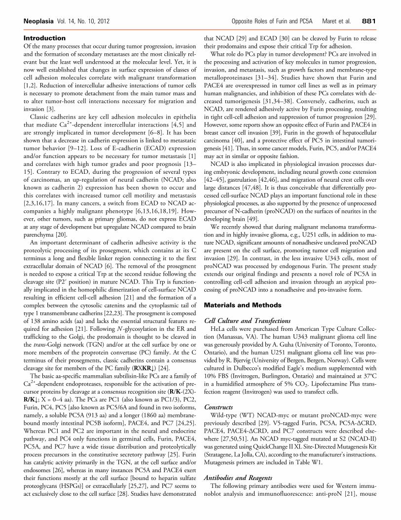
NCAD found in tumors. Accordingly, we evaluated the mRNAexpression levels of the relevant constitutive basic aa-specific PCs,namely, Furin, PC5A, PACE4, and PC7 in a panel of various tumorcells to determine whether differences in levels of these enzymesmight underlie the mechanism(s) leading to different forms of cell-surface NCAD. Because only 45% of the highly invasive glioma cells,compared to 70% of metastatic melanoma cells, expressed unprocessedproNCAD at the cell surface owing to the low expression of Furin [29],this suggested that there may be additional mechanisms associated withmalignant glioma cell invasion. RT-PCR analyses revealed similar levelsof PC7 in U343 and U251 cells, and expression of PACE4 was notdetected in either cell line (Figure 1A). Interestingly, the mRNA levelsof Furin were low in highly invasive U251 cells, relative to the lessinvasive U343 cells. In contrast, PC5 mRNA expression was high inU251 cells relative to U343 cells (Figure 1A). Quantitative RT-PCR(qRT-PCR) analyses showed that Furin is 36-fold more expressed thanPC5A in U343 cells, whereas PC5A is 4-fold more expressed thanFurin in U251 cells (Figure 1B). The relatively low Furin expressionin U251 cells could be a plausible explanation for the previously ob-served presence of proNCAD at the surface of these cells [29]. Thecontrasting expression of PC5A was intriguing, especially because itwas demonstrated that proECAD was processed in the Furin-deficientLoVo cells, indicating that another convertase could process cadherins[30], including proNCAD. Upon scrutiny of the mouse proNCADsequence, we identified a second putative S2 site at RIRSDR↓DK189
(conserved in human proNCAD) following the primary Furin-cleavableS1 site at RQKR↓DW161 (Figure 1B). Should cleavage occur at S2,the ensuing NCAD-ΔN28 product lacking the N-terminal 28 aa ofNCAD would have lost the critical Trp161 and likely be nonadhesive.Using an in-house–generated Ab against the prodomain of NCAD [29],we were surprised to find that the endogenous prodomain of NCADin U343 and U251 cells exhibits a different molecular size. Thus, inU343 and U251 cells, it predominantly migrates on SDS-PAGE asan ∼17-kDa or an ∼20-kDa protein, respectively (Figure 1C ), sug-gesting differential cleavage by endogenous proteases in these cells.To identify the cognate convertase at the S2 site (generating the∼20-kDa prodomain), we first showed that incubation of cells withthe general PC inhibitor decanoyl-RVKR-chloromethylketone (dec-RVKR-cmk) [25] completely abrogates both S1 and S2 cleavages, sug-gesting that both are accomplished by one or more PCs (Figure 1C ).To identify which PC is responsible for the generation of the
∼20-kDa prodomain following cleavage at S2, we expressed eachPC and proNCAD in HeLa cells (that lack endogenous PC5A[52]). As control, we showed that PC5A and Furin can similarly cleaveproPDGF-A [53] in these cells (not shown). The data showed thatof all PCs, PC5A is the only convertase capable of generating the∼20-kDa product from both endogenous and overexpressed proNCAD(Figure 1D), whereas Furin (Figure 1E) and PACE4 (Figure 1F) donot cleave proNCAD at the S2 site, and PC7 does not seem to cleaveat either site, as the level of the ∼17-kDa product does not changeupon expression of this enzyme (Figure 1G ).To unambiguously identify the important residues governing cleav-
age at the S2 site, we mutated the putative P1 residue Arg187 to Alagenerating the sequence to RIRSDA↓DK189. Co-expression of this
R187A mutant with PC5A in HeLa cells, lacking both endogenousNCAD [29] and PC5A expression [52], resulted in the generation ofthe ∼17-kDa product but not the ∼20-kDa product containing at itsC terminus the first 28 aa of mature NCAD, confirming that thePC5A-mediated production of NCAD-ΔN28 by cleavage at positionS2 is lost (Figure 1H ).
Cell-surface PC5A Cleaves NCAD at S2 in Gliomaand HeLa Cells
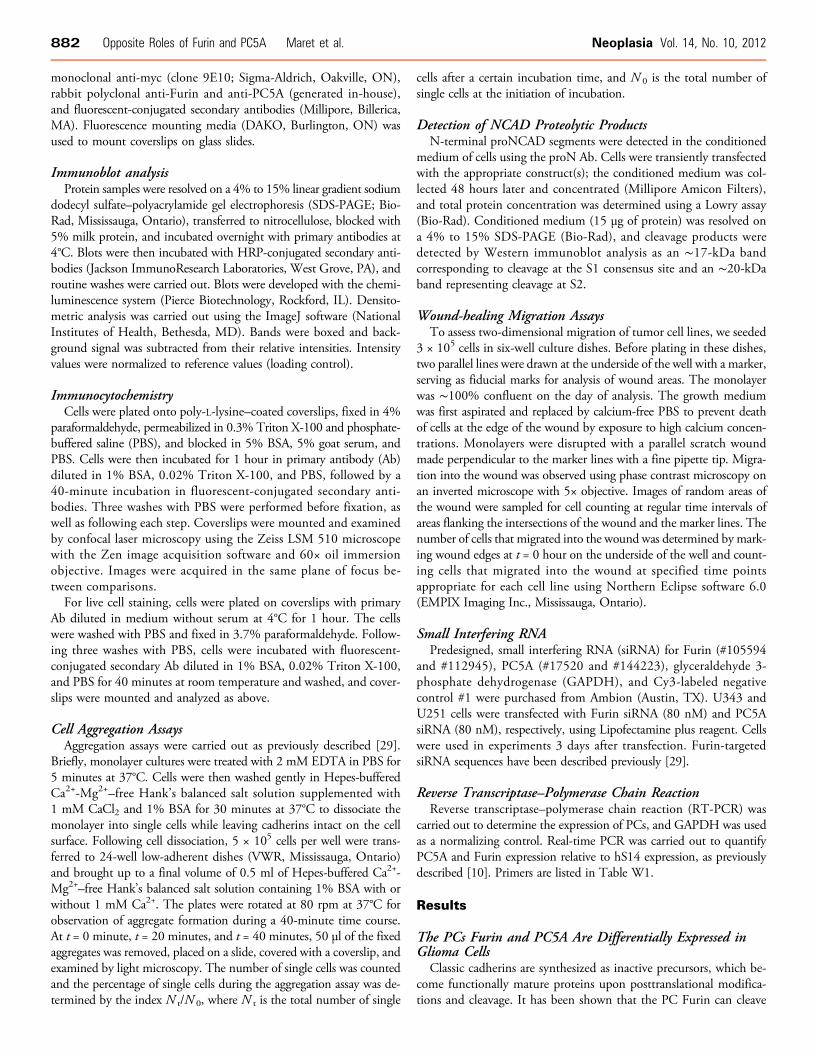
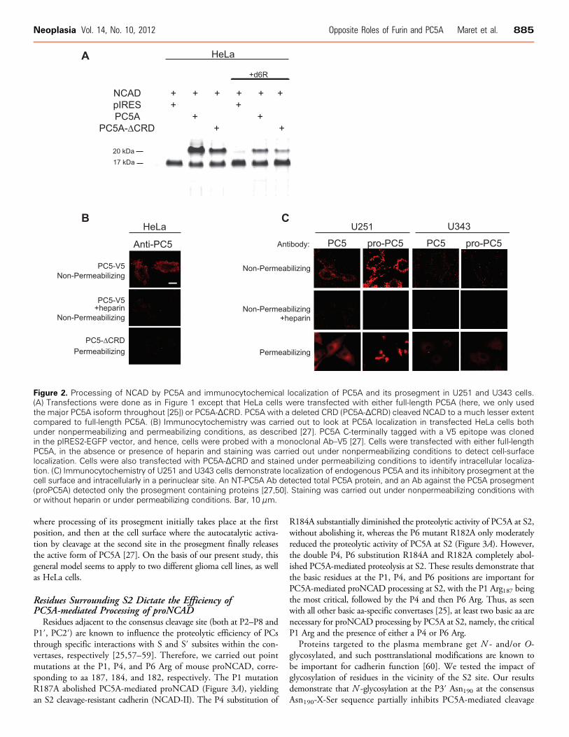
Proteolytic processing of proteins by PCs takes place in the TGN,cell surface, or endosomes [25,26]. To probe the subcellular location(s)where proNCAD processing by PC5A into NCAD-ΔN28 resulting inan ∼20-kDa prodomain takes place, we expressed in HeLa cells a trun-cated form of PC5A (PC5A-ΔCRD) lacking the C-terminal cysteine-rich domain (CRD) [27,50]. The results showed that co-expression ofproNCAD and PC5A-ΔCRD substantially reduced PC5A-mediatedproteolysis at S2 but did not abolish it (Figure 2A). This indicates thatin large part cleavage at S2 takes place at the plasma membrane, as sup-ported by previous studies showing that the CRD of PC5A mediatescell-surface anchoring of the enzyme through interactions with HSPGs[27,50]. To further support this conclusion, we demonstrated that in-cubations with the PC cell-surface inhibitor d6R [54,55] only partiallyabolished the proNCAD processing (Figure 2A), whereas dec-RVKR-cmk [54,55], a cell permeable PC inhibitor, completely blocked pro-cessing (Figure 1, C and D). Thus, proNCAD is processed by PC5Aboth at the cell surface and intracellularly, either at the TGN orfollowing endocytosis.
We then looked at the localization of PC5A in transfected HeLacells (Figure 2B). Herein, the protein localized to the cell surface, asassayed by immunofluorescence under nonpermeabilizing conditions(Figure 2B, upper panel); however upon heparin treatment, PC5Awas no longer located at the cell surface (Figure 2B, middle panel), be-fitting its cell-surface binding to HSPGs [27,50]. In addition, PC5A-ΔCRD did not localize to the cell surface of HeLa cells (Figure 2B,lower panel ), as also reported for COS-1 cells [27]. Thus, HSPG-boundPC5A localizes to the cell surface [27,50] where it presumably cleavesmost of proNCAD at the S2 site.
We next investigated the localization of endogenous PC5A byimmunofluorescence and detected substantial levels of the enzymeat the cell surface of U251 cells but very low levels at the surface ofU343 cells (Figure 2C , upper panel ). As we observed upon PC5Aoverexpression in HeLa cells (Figure 2B), endogenous PC5A immuno-reactivity was not detectable at the cell surface when heparin was addedto the medium of glioma cells (Figure 2C , middle panel ). Further-more, as previously reported in COS-1 cells [27], immunoreactivityof the prosegment of PC5A can also be detected at the cell surface ofU251 cells (but not U343 cells) with an Ab specific for the prosegmentof PC5A [50]. This suggests that a fraction of PC5A is found in aninactive form noncovalently bound to its inhibitory prosegment atthe surface of U251 cells, as reported for COS-1 cells [27]. Finally, un-der permeabilizing conditions, we note that in U251 cells endogenousimmunoreactive PC5A and its prosegment mostly localize to a peri-nuclear compartment, likely the TGN (Figure 2C , bottom panel ), sim-ilar to its intracellular localization in brain neurons [56]. We deducethat in this compartment a fraction of PC5A is still in an inactive formbound to its inhibitory prosegment. Thus, similar to COS-1 cells [27],we interpret the above data, combined with those in the literature de-scribing the zymogen activation of PC5A [25,27], to mean that auto-catalytic cleavage of proPC5A occurs in two steps, a first one in the ER
Neoplasia Vol. 14, No. 10, 2012 Opposite Roles of Furin and PC5A Maret et al. 883
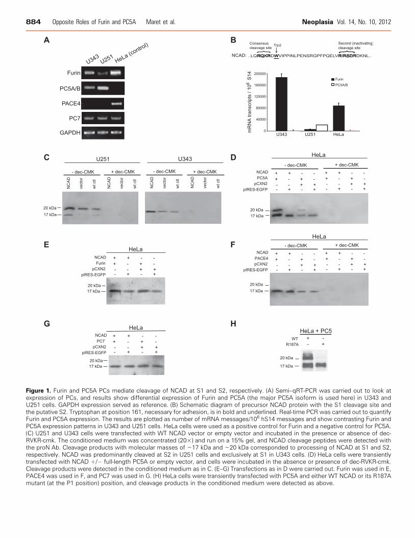
Figure 1. Furin and PC5A PCs mediate cleavage of NCAD at S1 and S2, respectively. (A) Semi–qRT-PCR was carried out to look atexpression of PCs, and results show differential expression of Furin and PC5A (the major PC5A isoform is used here) in U343 andU251 cells. GAPDH expression served as reference. (B) Schematic diagram of precursor NCAD protein with the S1 cleavage site andthe putative S2. Tryptophan at position 161, necessary for adhesion, is in bold and underlined. Real-time PCR was carried out to quantifyFurin and PC5A expression. The results are plotted as number of mRNA messages/106 hS14 messages and show contrasting Furin andPC5A expression patterns in U343 and U251 cells. HeLa cells were used as a positive control for Furin and a negative control for PC5A.(C) U251 and U343 cells were transfected with WT NCAD vector or empty vector and incubated in the presence or absence of dec-RVKR-cmk. The conditioned medium was concentrated (20×) and run on a 15% gel, and NCAD cleavage peptides were detected withthe proN Ab. Cleavage products with molecular masses of ∼17 kDa and ∼20 kDa corresponded to processing of NCAD at S1 and S2,respectively. NCAD was predominantly cleaved at S2 in U251 cells and exclusively at S1 in U343 cells. (D) HeLa cells were transientlytransfected with NCAD +/− full-length PC5A or empty vector, and cells were incubated in the absence or presence of dec-RVKR-cmk.Cleavage products were detected in the conditioned medium as in C. (E–G) Transfections as in D were carried out. Furin was used in E,PACE4 was used in F, and PC7 was used in G. (H) HeLa cells were transiently transfected with PC5A and either WT NCAD or its R187Amutant (at the P1 position) position, and cleavage products in the conditioned medium were detected as above.
884 Opposite Roles of Furin and PC5A Maret et al. Neoplasia Vol. 14, No. 10, 2012
where processing of its prosegment initially takes place at the firstposition, and then at the cell surface where the autocatalytic activa-tion by cleavage at the second site in the prosegment finally releasesthe active form of PC5A [27]. On the basis of our present study, thisgeneral model seems to apply to two different glioma cell lines, as wellas HeLa cells.
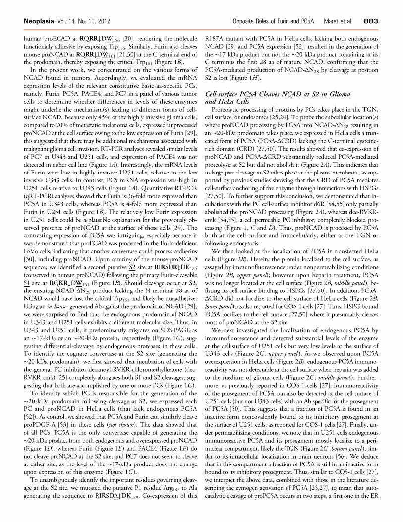
Residues Surrounding S2 Dictate the Efficiency ofPC5A-mediated Processing of proNCADResidues adjacent to the consensus cleavage site (both at P2–P8 and
P1′, PC2′) are known to influence the proteolytic efficiency of PCsthrough specific interactions with S and S′ subsites within the con-vertases, respectively [25,57–59]. Therefore, we carried out pointmutations at the P1, P4, and P6 Arg of mouse proNCAD, corre-sponding to aa 187, 184, and 182, respectively. The P1 mutationR187A abolished PC5A-mediated proNCAD (Figure 3A), yieldingan S2 cleavage-resistant cadherin (NCAD-II). The P4 substitution of
R184A substantially diminished the proteolytic activity of PC5A at S2,without abolishing it, whereas the P6 mutant R182A only moderatelyreduced the proteolytic activity of PC5A at S2 (Figure 3A). However,the double P4, P6 substitution R184A and R182A completely abol-ished PC5A-mediated proteolysis at S2. These results demonstrate thatthe basic residues at the P1, P4, and P6 positions are important forPC5A-mediated proNCAD processing at S2, with the P1 Arg187 beingthe most critical, followed by the P4 and then P6 Arg. Thus, as seenwith all other basic aa-specific convertases [25], at least two basic aa arenecessary for proNCAD processing by PC5A at S2, namely, the criticalP1 Arg and the presence of either a P4 or P6 Arg.
Proteins targeted to the plasma membrane get N - and/or O-glycosylated, and such posttranslational modifications are known tobe important for cadherin function [60]. We tested the impact ofglycosylation of residues in the vicinity of the S2 site. Our resultsdemonstrate that N -glycosylation at the P3′ Asn190 at the consensusAsn190-X-Ser sequence partially inhibits PC5A-mediated cleavage
Figure 2. Processing of NCAD by PC5A and immunocytochemical localization of PC5A and its prosegment in U251 and U343 cells.(A) Transfections were done as in Figure 1 except that HeLa cells were transfected with either full-length PC5A (here, we only usedthe major PC5A isoform throughout [25]) or PC5A-ΔCRD. PC5A with a deleted CRD (PC5A-ΔCRD) cleaved NCAD to a much lesser extentcompared to full-length PC5A. (B) Immunocytochemistry was carried out to look at PC5A localization in transfected HeLa cells bothunder nonpermeabilizing and permeabilizing conditions, as described [27]. PC5A C-terminally tagged with a V5 epitope was clonedin the pIRES2-EGFP vector, and hence, cells were probed with a monoclonal Ab–V5 [27]. Cells were transfected with either full-lengthPC5A, in the absence or presence of heparin and staining was carried out under nonpermeabilizing conditions to detect cell-surfacelocalization. Cells were also transfected with PC5A-ΔCRD and stained under permeabilizing conditions to identify intracellular localiza-tion. (C) Immunocytochemistry of U251 and U343 cells demonstrate localization of endogenous PC5A and its inhibitory prosegment at thecell surface and intracellularly in a perinuclear site. An NT-PC5A Ab detected total PC5A protein, and an Ab against the PC5A prosegment(proPC5A) detected only the prosegment containing proteins [27,50]. Staining was carried out under nonpermeabilizing conditions withor without heparin or under permeabilizing conditions. Bar, 10 μm.
Neoplasia Vol. 14, No. 10, 2012 Opposite Roles of Furin and PC5A Maret et al. 885
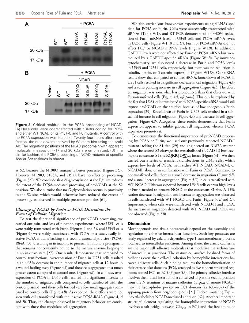
at S2, because the N190Q mutant is better processed (Figure 3C ).However, N120Q, S185A, and S192A have no effect on processing(Figure 3C). We conclude that N -glycosylation at the P3′ site reducesthe extent of the PC5A-mediated processing of proNCAD at the S2position. We also surmise that no O-glycosylation occurs in proximityto the S2 site, which would otherwise have reduced the extent ofprocessing, as observed in multiple precursor proteins [61].
Cleavage of NCAD by Furin or PC5A Determines theExtent of Cellular Migration
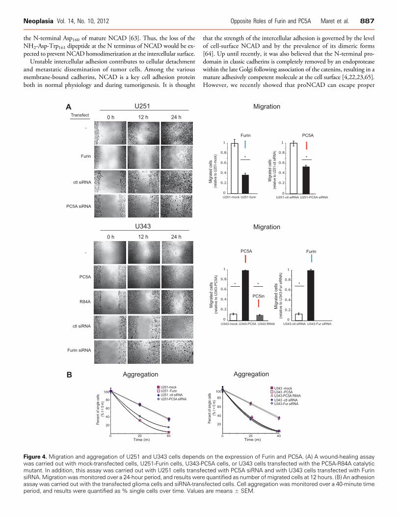
To test the functional significance of proNCAD processing, wecarried out gain- and loss-of-function experiments, where U251 cellswere stably transfected with Furin (Figures 4 and 5), and U343 cells(Figure 4) were stably transfected with PC5A or a catalytically in-active PC5A mutant lacking the second autocatalytic site (PC5A-R84A; [50]), resulting in its inability to process its inhibitory prosegmentthat remains noncovalently bound to the mature enzyme keeping itin an inactive state [27]. Our results demonstrate that compared tocontrol transfections, overexpression of Furin in U251 cells resultedin an ∼65% decrease in the number of migrated cells at 12 hours ina wound-healing assay (Figure 4A) and these cells aggregated to a muchgreater extent compared to control ones (Figure 4B). In contrast, over-expression of PC5A in U343 cells resulted in a significant increase inthe number of migrated cells compared to cells transfected with thecontrol plasmid, and these cells formed very few small aggregates com-pared to control cells (Figure 4B). As expected, these effects were notseen with cells transfected with the inactive PC5A-R84A (Figure 4, Aand B). Thus, the changes observed in migratory behavior are consis-tent with those that modulate cell aggregation.
We also carried out knockdown experiments using siRNAs spe-cific for PC5A or Furin. Cells were successfully transfected withsiRNAs (Table W1), and RT-PCR demonstrated an ∼80% reduc-tion of Furin mRNA levels in U343 cells and PC5A mRNA levelsin U251 cells (Figure W1, B and C ). Furin or PC5A siRNAs did notaffect PC7 or NCAD mRNA levels (Figure W1B). In addition,GAPDH levels were not affected by Furin or PC5A siRNA but werereduced by a GAPDH-specific siRNA (Figure W1B). By immuno-cytochemistry, we also noted a decrease in Furin and PC5A levelsin U343 and U251 cells, respectively, but there was no reduction intubulin, nestin, or β-catenin expression (Figure W1D). Our siRNAresults show that compared to control siRNA, knockdown of PC5A inU251 cells resulted in a significant decrease in cell migration (Figure 4A)and a corresponding increase in cell aggregation (Figure 4B). The effecton migration was somewhat less pronounced than that observed withFurin-transfected cells (Figure 4A, left panel). This can be explained bythe fact that U251 cells transfected with PC5A-specific siRNAwould stillexpress proNCAD on their surface because of low endogenous Furinexpression [29]. Knockdown of Furin in U343 cells resulted in a sub-stantial increase in cell migration (Figure 4A) and decrease in cell aggre-gation (Figure 4B). Altogether, these results demonstrate that Furinexpression appears to inhibit glioma cell migration, whereas PC5Aexpression promotes it.
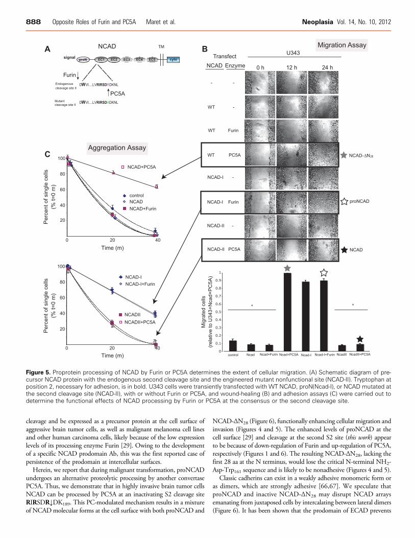
To demonstrate the functional importance of proNCAD process-ing by PC5A or Furin, we used our previously reported NCAD-Imutant lacking the S1 site [29] and engineered an R187A mutantwhere the second S2 cleavage site was abolished (NCAD-II) but leav-ing the consensus S1 site RQKR↓DW161 intact (Figure 5A). We thencarried out a series of transient transfections in U343 cells, whichexpress low levels of PC5A, with either WT NCAD, NCAD-I, orNCAD-II, alone or in combination with Furin or PC5A. Compared tonontransfected cells, there is a small decrease in migration (Figure 5B)and a small increase in aggregation (Figure 5C) in cells transfected withWT NCAD. This was expected because U343 cells express high levelsof Furin needed to process NCAD at the consensus S1 site. A 15%further decrease in migration and increase in aggregation was observedin cells transfected with WT NCAD and Furin (Figure 5, B and C).Importantly, when cells were transfected with NCAD-II and PC5A,the increase in migration detected with WT NCAD and PC5A wasnot observed (Figure 5B).
DiscussionMorphogenesis and tissue homeostasis depend on the assembly andregulation of cohesive intercellular junctions. Such key processes arefinely regulated by calcium-dependent type 1 transmembrane proteinslocalized to intercellular junctions. Among these, the classic cadherinsare the major cell adhesive molecules that modulate the architectureof intercellular junctions. The mature cell-surface localized forms ofcadherins exert their cell-cell cohesion by homophilic interactions be-tween adjacent cells. Such binding requires the homodimerization oftheir extracellular domains (ECs), arranged as five tandem structural seg-ments named EC1 to EC5 (Figure 5A). The primary adhesive interfaceinvolves the mutual insertion of a conserved Trp at the second positionfrom the N terminus of mature cadherins (Trp161 of mouse NCAD)into the hydrophobic pocket on EC1 domain (aa 160–267) of theapposing protein from neighboring cells [21]. Indeed, mutating Trp161into Ala abolishes NCAD-mediated adhesion [62]. Another importantstructural element regulating the homophilic interaction of NCADinvolves a salt bridge between Glu248 in EC1 and the free amine of
Figure 3. Critical residues in the PC5A processing of NCAD.(A) HeLa cells were co-transfected with cDNAs coding for PC5Aand either WT NCAD or its P1, P4, and P6 mutants. A control withno PC5A expression was included. Twenty-four hours after trans-fection, the media were analyzed by Western blot using the proNAb. The migration positions of the NCAD prodomain with apparentmolecular masses of ∼17 and 20 kDa are emphasized. (B) In asimilar fashion, the PC5A processing of NCAD mutants at specificAsn or Ser residues is shown.
886 Opposite Roles of Furin and PC5A Maret et al. Neoplasia Vol. 14, No. 10, 2012
the N-terminal Asp160 of mature NCAD [63]. Thus, the loss of theNH2-Asp-Trp161 dipeptide at the N terminus of NCAD would be ex-pected to prevent NCAD homodimerization at the intercellular surface.Unstable intercellular adhesion contributes to cellular detachment
and metastatic dissemination of tumor cells. Among the variousmembrane-bound cadherins, NCAD is a key cell adhesion proteinboth in normal physiology and during tumorigenesis. It is thought
that the strength of the intercellular adhesion is governed by the levelof cell-surface NCAD and by the prevalence of its dimeric forms[64]. Up until recently, it was also believed that the N-terminal pro-domain in classic cadherins is completely removed by an endoproteasewithin the late Golgi following association of the catenins, resulting in amature adhesively competent molecule at the cell surface [4,22,23,65].However, we recently showed that proNCAD can escape proper
Figure 4. Migration and aggregation of U251 and U343 cells depends on the expression of Furin and PC5A. (A) A wound-healing assaywas carried out with mock-transfected cells, U251-Furin cells, U343-PC5A cells, or U343 cells transfected with the PC5A-R84A catalyticmutant. In addition, this assay was carried out with U251 cells transfected with PC5A siRNA and with U343 cells transfected with FurinsiRNA.Migrationwasmonitored over a 24-hour period, and results were quantified as number ofmigrated cells at 12 hours. (B) An adhesionassay was carried out with the transfected glioma cells and siRNA-transfected cells. Cell aggregation was monitored over a 40-minute timeperiod, and results were quantified as % single cells over time. Values are means ± SEM.
Neoplasia Vol. 14, No. 10, 2012 Opposite Roles of Furin and PC5A Maret et al. 887
cleavage and be expressed as a precursor protein at the cell surface ofaggressive brain tumor cells, as well as malignant melanoma cell linesand other human carcinoma cells, likely because of the low expressionlevels of its processing enzyme Furin [29]. Owing to the developmentof a specific NCAD prodomain Ab, this was the first reported case ofpersistence of the prodomain at intercellular surfaces.
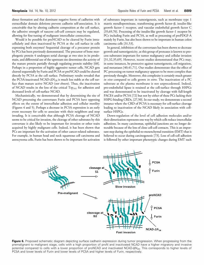
Herein, we report that during malignant transformation, proNCADundergoes an alternative proteolytic processing by another convertasePC5A. Thus, we demonstrate that in highly invasive brain tumor cellsNCAD can be processed by PC5A at an inactivating S2 cleavage siteRIRSDR↓DK189. This PC-modulated mechanism results in a mixtureof NCADmolecular forms at the cell surface with both proNCAD and
NCAD-ΔN28 (Figure 6), functionally enhancing cellular migration andinvasion (Figures 4 and 5). The enhanced levels of proNCAD at thecell surface [29] and cleavage at the second S2 site (this work) appearto be because of down-regulation of Furin and up-regulation of PC5A,respectively (Figures 1 and 6). The resulting NCAD-ΔN28, lacking thefirst 28 aa at the N terminus, would lose the critical N-terminal NH2-Asp-Trp161 sequence and is likely to be nonadhesive (Figures 4 and 5).
Classic cadherins can exist in a weakly adhesive monomeric form oras dimers, which are strongly adhesive [66,67]. We speculate thatproNCAD and inactive NCAD-ΔN28 may disrupt NCAD arraysemanating from juxtaposed cells by intercalating between lateral dimers(Figure 6). It has been shown that the prodomain of ECAD prevents
Figure 5. Proprotein processing of NCAD by Furin or PC5A determines the extent of cellular migration. (A) Schematic diagram of pre-cursor NCAD protein with the endogenous second cleavage site and the engineered mutant nonfunctional site (NCAD-II). Tryptophan atposition 2, necessary for adhesion, is in bold. U343 cells were transiently transfected with WT NCAD, proN(Ncad-I), or NCAD mutated atthe second cleavage site (NCAD-II), with or without Furin or PC5A, and wound-healing (B) and adhesion assays (C) were carried out todetermine the functional effects of NCAD processing by Furin or PC5A at the consensus or the second cleavage site.
888 Opposite Roles of Furin and PC5A Maret et al. Neoplasia Vol. 14, No. 10, 2012
dimer formation and that dominant negative forms of cadherins withextracellular domain deletions prevent cadherin self-association. It isconceivable that by altering cadherin composition at the cell surface,the adhesive strength of nascent cell-cell contacts may be regulated,allowing for fine-tuning of malignant intercellular connections.Would it be possible for proNCAD to be cleaved by Furin intra-
cellularly and then inactivated by PC5A at the cell surface in cellsexpressing both enzymes? Sequential cleavage of a precursor proteinby PCs has been previously demonstrated. The precursor of bone mor-phogenic protein 4 undergoes serial cleavage at two sites in its prodo-main, and differential use of the upstream site determines the activity ofthe mature protein partially through regulating protein stability [68].Perhaps in a proportion of highly aggressive tumor cells, NCAD getscleaved sequentially by Furin and PC5A or proNCAD could be cleaveddirectly by PC5A at the cell surface. Preliminary results revealed thatthe PC5A-inactivated NCAD-ΔN28 is much less stable at the cell sur-face than mature active NCAD (not shown). Thus, the inactivationof NCAD results in the loss of the critical Trp161 for adhesion anddecreased levels of cell-surface NCAD.Mechanistically, we demonstrated that by determining the site of
NCAD processing the convertases Furin and PC5A have opposingeffects on the extent of intercellular adhesion and cellular motility(Figures 4 and 5). Perhaps a decrease in PC5A expression is an earlyevent necessary for cells to associate with their neighbors and stopinvading. It is conceivable that although PC5A cleavage of NCADseems to be critical for invasion, the cleavage of other substrates by thisconvertase is also likely to be important for invasion or other traitsacquired by highly malignant cells. Indeed, it has been shown thatPCs are important for the activation of other cancer-related substrates.For example, in human head and neck squamous cell carcinoma andastrocytoma cells, Furin has been shown to be important for activation
of substrates important in tumorigenesis, such as membrane type 1matrix metalloproteinase, transforming growth factor–β, insulin-likegrowth factor–1 receptor, and vascular endothelial growth factor C[35,69,70]. Processing of the insulin-like growth factor–1 receptor byPCs including Furin and PC5A, as well as processing of proPDGF-Amainly by Furin, has also been shown to be important in human coloncarcinoma cells [31,53].
In general, inhibition of the convertases has been shown to decreasegrowth and tumorigenicity, as this group of proteases is known to pro-cess substrates important for tumor malignancy and cellular invasion[31,32,35,69]. However, recent studies demonstrated that PCs may,in some instances, be protective against tumorigenesis, cell migration,and metastases [40,41,71]. Our studies demonstrate that the effect ofPC processing on tumor malignancy appears to be more complex thanpreviously thought. Moreover, this complexity is certainly much greaterin vivo compared to cells grown in vitro. The inactivation of a PCsubstrate at the plasma membrane is not unprecedented. Indeed,pro-endothelial lipase is retained at the cell-surface through HSPGsand was demonstrated to be inactivated by cleavage with full-lengthPACE4 and/or PC5A [72] but not by either of these PCs lacking theirHSPG-binding CRDs, [27,50]. In our study, we demonstrate a secondinstance where the CRD of PC5A is necessary for cell-surface cleavageleading to inactivation of the NCAD likely in association with cell-surface HSPGs.
Down-regulation of the level of cell adhesion molecules and/ortheir dimerization represents one way by which cells reduce intercellularadhesions. In many carcinomas, epithelial junctions are no longer de-tectable because of the loss of close cell-cell contacts. This is an impor-tant step during the epithelial-to-mesenchymal transition (EMT) that isbelieved to occur during carcinogenesis [73]. Loss of cell-cell adhesionis followed by other important phenotypic changes during EMT such
Figure 6. Proposed schematic diagram depicting surface cadherin expression during tumor progression. When progressing from thepremalignant to malignant stage, cells with a high proportion of proN and inactivated NCAD have a higher migratory and invasivepotential compared to cells with a lower proportion of proNCAD and inactivated NCAD-ΔN28. This corresponds to higher levels ofPC5A and lower levels of Furin and lower levels of PC5A and higher levels of Furin, respectively.
Neoplasia Vol. 14, No. 10, 2012 Opposite Roles of Furin and PC5A Maret et al. 889
as loss of cell polarity and the acquisition of migratory and invasiveproperties. The loss of ECAD is considered to be a critical event inEMT, as ECAD repressors are capable of functioning as full EMTinducers in different cell types [74]. This is accompanied by an up-regulation of NCAD in many types of carcinomas and the switch fromECAD to NCAD correlates with induced cellular motility [2,3,16].It is believed that because of disruption of adhesion junctions, loss ofECAD allows malignant cells to detach from the epithelium and invadehost tissue, whereas up-regulation of NCAD expression facilitates inva-sion of tumor cells by mediating adhesion of malignant cells to stromalor endothelial cells rather than epithelial cells [75].
Alternatively, as we have demonstrated, intercellular adhesioncould be modulated by surface expression of a nonadhesive proNCAD[29], or NCAD-ΔN28 (this work). Thus, our results should lead to therevision of the current model of cadherin posttranslational processingand surface expression [3,16,18,76,77]. We propose that in later stagesof tumor progression nonadhesive surface proNCAD and functionallyinactivated NCAD-ΔN28 determine the degree of cell invasiveness andmetastasis and shed light on the stages of malignancy during tumor pro-gression. Consequently, the switch from ECAD to NCAD duringEMT is in fact a switch to a mixture of NCAD molecules where onlya certain proportion is functionally adhesive. In brain tumor cells,which do not undergo an ECAD-to-NCAD shift, the switch frommature NCAD to nonadhesive NCAD-ΔN28 molecules mediatesdetachment from the main tumor mass and migration over extensivedistances in our in vitro assay (Figures 4 and 5). Preliminary data inan intracranial xenograft mouse model show that prevention of theformation of NCAD-ΔN28 results in a less aggressive phenotypecompared to WT (in preparation).
We speculate that differential expression of PCs may be a commonmechanism in many types of tumors to regulate cellular motility andperhaps other malignant traits and is perhaps central in developmentalprograms. In addition, precursor forms of other types of cadherins atthe cell surface may play a similar role in tumor cell motility. Accord-ingly, alignment of the processing sites of various cell adhesion mole-cules (Figure W2) revealed that the seminal observation reported heremay be more general than previously appreciated. Indeed, the Furindibasic cleavage site and the alternative PC5A-mediated monobasicprocessing site characterized herein for proNCAD are found to beconserved in a number of cadherin family members including ECAD,P-cadherin, and cadherin-11 that play pivotal roles in cancer develop-ment and progression [76,78,79]. It remains to be defined if PC5Aalso mediates these inactivation processes and whether such a mecha-nism regulates the functions of these adhesion molecules both physiolog-ically and/or pathologically. In that context, mining the ONCOMINEcancer gene expression database (http://www.oncomine.org) revealedthat PC5A expression was significantly reduced in most tumors [41]but was actually upregulated in 3 of 10 tumor types including pancre-atic ductal adenocarcinoma, glioblastoma multiforme, and anaplasticoligoastrocytoma/oligodendroglioma (Figure W3). On the basis ofour present work, it is possible that in these latter types of tumorsPC5A would also promote cancer metastasis.
Malignant primary brain tumors are extremely complex and hetero-geneous microenvironments, characterized by subpopulations of highlyinvasive cells that infiltrate the normal brain parenchyma, often sig-nificant distances from the primary tumor mass [80]. Unlike mostcarcinomas, these tumors do not enter blood vessels and form metas-tases in distant organs. Instead, fatality is because of “micro-metastasis”within the brain, resulting in destruction of regions essential for survival
of the patient [81]. Uncontrollable invasion leads to failure of treatmentmodalities such as radiotherapy and chemotherapy and recurrencefollowing radical resection.
The mechanism proposed herein sheds light on putative noveltreatment strategies with the potential to attenuate extensive infiltra-tion of the brain parenchyma by malignant tumor cells and reducethe frequency of fatal recurrences following tumor resection. Further-more, in cancers such as malignant melanoma and malignant glialtumors [29], characterization of the cell-surface forms of NCADmay serve as a prognostic tool for staging and progression of the dis-ease. Our work may potentially lead to treatment strategies to controlthe metastatic spread of malignant brain carcinomas, through thedevelopment of specific PC5 inhibitors.
References[1] Birchmeier W (1994). Molecular aspects of the loss of cell adhesion and gain of
invasiveness in carcinomas. Princess Takamatsu Symp 24, 214–232.[2] Suyama K, Shapiro I, Guttman M, and Hazan RB (2002). A signaling pathway
leading to metastasis is controlled by N-cadherin and the FGF receptor. CancerCell 2, 301–314.
[3] Nieman MT, Prudoff RS, Johnson KR, and Wheelock MJ (1999). N-cadherinpromotes motility in human breast cancer cells regardless of their E-cadherinexpression. J Cell Biol 147, 631–644.
[4] Ozawa M and Kemler R (1990). Correct proteolytic cleavage is required for thecell adhesive function of uvomorulin. J Cell Biol 111, 1645–1650.
[5] Takeichi M (1988). The cadherins: cell-cell adhesion molecules controllinganimal morphogenesis. Development 102, 639–655.
[6] Christofori G (2006). New signals from the invasive front. Nature 441, 444–450.[7] Hsu MY, Meier FE, Nesbit M, Hsu JY, Van Belle BP, Elder DE, and Herlyn M
(2000). E-cadherin expression in melanoma cells restores keratinocyte-mediatedgrowth control and down-regulates expression of invasion-related adhesion recep-tors. Am J Pathol 156, 1515–1525.
[8] Hazan RB, Qiao R, Keren R, Badano I, and Suyama K (2004). Cadherin switchin tumor progression. Ann N Y Acad Sci 1014, 155–163.
[9] Frixen UH, Behrens J, Sachs M, Eberle G, Voss B, Warda A, Lochner D, andBirchmeier W (1991). E-cadherin-mediated cell-cell adhesion prevents invasive-ness of human carcinoma cells. J Cell Biol 113, 173–185.
[10] Vleminckx K, Vakaet L Jr, Mareel M, Fiers W, and van Roy RF (1991).Genetic manipulation of E-cadherin expression by epithelial tumor cells revealsan invasion suppressor role. Cell 66, 107–119.
[11] Takeichi M (1993). Cadherins in cancer: implications for invasion and metastasis.Curr Opin Cell Biol 5, 806–811.
[12] Birchmeier W, Behrens J, Weidner KM, Frixen UH, and Schipper J (1991).Dominant and recessive genes involved in tumor cell invasion. Curr Opin CellBiol 3, 832–840.
[13] Perl AK, Wilgenbus P, Dahl U, Semb H, and Christofori G (1998). A causalrole for E-cadherin in the transition from adenoma to carcinoma. Nature 392,190–193.
[14] Hirohashi S (1998). Inactivation of the E-cadherin-mediated cell adhesionsystem in human cancers. Am J Pathol 153, 333–339.
[15] Christofori G and Semb H (1999). The role of the cell-adhesion moleculeE-cadherin as a tumour-suppressor gene. Trends Biochem Sci 24, 73–76.
[16] Islam S, Carey TE, Wolf GT, Wheelock MJ, and Johnson KR (1996). Ex-pression of N-cadherin by human squamous carcinoma cells induces a scatteredfibroblastic phenotype with disrupted cell-cell adhesion. J Cell Biol 135,1643–1654.
[17] Hazan RB, Kang L, Whooley BP, and Borgen PI (1997). N-cadherin promotesadhesion between invasive breast cancer cells and the stroma. Cell Adhes Commun4, 399–411.
[18] Hazan RB, Phillips GR, Qiao RF, Norton L, and Aaronson SA (2000). Exog-enous expression of N-cadherin in breast cancer cells induces cell migration,invasion, and metastasis. J Cell Biol 148, 779–790.
[19] Li G and Herlyn M (2000). Dynamics of intercellular communication duringmelanoma development. Mol Med Today 6, 163–169.
[20] Asano K, Duntsch CD, Zhou Q, Weimar JD, Bordelon D, Robertson JH, andPourmotabbed T (2004). Correlation of N-cadherin expression in high gradegliomas with tissue invasion. J Neurooncol 70, 3–15.
890 Opposite Roles of Furin and PC5A Maret et al. Neoplasia Vol. 14, No. 10, 2012
[21] Koch AW, Farooq A, Shan W, Zeng L, Colman DR, and Zhou MM (2004).Structure of the neural (N-) cadherin prodomain reveals a cadherin extracellulardomain-like fold without adhesive characteristics. Structure 12, 793–805.
[22] Wahl JK III, Kim YJ, Cullen JM, Johnson KR, and Wheelock MJ (2003).N-cadherin-catenin complexes form prior to cleavage of the proregion andtransport to the plasma membrane. J Biol Chem 278, 17269–17276.
[23] Shore EM and Nelson WJ (1991). Biosynthesis of the cell adhesion moleculeuvomorulin (E-cadherin) in Madin-Darby canine kidney epithelial cells. J BiolChem 266, 19672–19680.
[24] Seidah NG and Chretien M (1999). Proprotein and prohormone convertases: afamily of subtilases generating diverse bioactive polypeptides. Brain Res 848,45–62.
[25] Seidah NG and Prat A (2012). The biology and therapeutic targeting of theproprotein convertases. Nat Rev Drug Discov 11, 367–383.
[26] Thomas G (2002). Furin at the cutting edge: from protein traffic to embryo-genesis and disease. Nat Rev Mol Cell Biol 3, 753–766.
[27] Mayer G, Hamelin J, Asselin MC, Pasquato A, Marcinkiewicz E, Tang M,Tabibzadeh S, and Seidah NG (2008). The regulated cell surface zymogenactivation of the proprotein convertase PC5A directs the processing of its secre-tory substrates. J Biol Chem 283, 2373–2384.
[28] Rousselet E, Benjannet S, Marcinkiewicz E, Asselin MC, Lazure C, and SeidahNG (2011). Proprotein convertase PC7 enhances the activation of the EGFreceptor pathway through processing of the EGF precursor. J Biol Chem 286,9185–9195.
[29] Maret D, Gruzglin E, Sadr MS, Siu V, Shan W, Koch AW, Seidah NG,Del Maestro RF, and Colman DR (2010). Surface expression of precursorN-cadherin promotes tumor cell invasion. Neoplasia 12, 1066–1080.
[30] Posthaus H, Dubois CM, Laprise MH, Grondin F, Suter MM, and Muller E(1998). Proprotein cleavage of E-cadherin by furin in baculovirus over-expressionsystem: potential role of other convertases in mammalian cells. FEBS Lett 438,306–310.
[31] Khatib AM, Siegfried G, Prat A, Luis J, Chretien M, Metrakos P, and SeidahNG (2001). Inhibition of proprotein convertases is associated with loss ofgrowth and tumorigenicity of HT-29 human colon carcinoma cells: importanceof insulin-like growth factor-1 (IGF-1) receptor processing in IGF-1-mediatedfunctions. J Biol Chem 276, 30686–30693.
[32] Khatib AM, Siegfried G, Chretien M, Metrakos P, and Seidah NG (2002).Proprotein convertases in tumor progression and malignancy: novel targets incancer therapy. Am J Pathol 160, 1921–1935.
[33] Bassi DE, Fu J, Lopez DC, and Klein-Szanto AJ (2005). Proprotein convertases:“master switches” in the regulation of tumor growth and progression. MolCarcinog 44, 151–161.
[34] Coppola JM, Bhojani MS, Ross BD, and Rehemtulla A (2008). A small-molecule furin inhibitor inhibits cancer cell motility and invasiveness. Neoplasia10, 363–370.
[35] Bassi DE, Lopez DC, Mahloogi H, Zucker S, Thomas G, and Klein-Szanto AJ(2001). Furin inhibition results in absent or decreased invasiveness and tumor-igenicity of human cancer cells. Proc Natl Acad Sci USA 98, 10326–10331.
[36] D’Anjou F, Routhier S, Perreault JP, Latil A, Bonnel D, Fournier I, Salzet M,and Day R (2011). Molecular validation of PACE4 as a target in prostatecancer. Transl Oncol 4, 157–172.
[37] Scamuffa N, Siegfried G, Bontemps Y, Ma L, Basak A, Cherel G, Calvo F,Seidah NG, and Khatib AM (2008). Selective inhibition of proprotein conver-tases represses the metastatic potential of human colorectal tumor cells. J ClinInvest 118, 352–363.
[38] Bassi DE, Zhang J, Cenna J, Litwin S, Cukierman E, and Klein-Szanto AJ(2010). Proprotein convertase inhibition results in decreased skin cell prolifer-ation, tumorigenesis, and metastasis. Neoplasia 12, 516–526.
[39] Lapierre M, Siegfried G, Scamuffa N, Bontemps Y, Calvo F, Seidah NG, andKhatib AM (2007). Opposing function of the proprotein convertases furin andPACE4 on breast cancer cells’ malignant phenotypes: role of tissue inhibitors ofmetalloproteinase-1. Cancer Res 67, 9030–9034.
[40] Huang Y-H, Lin K-H, Liao CH, Lai M-W, Tseng Y-H, and Yeh C (2012).Furin overexpression suppresses tumor growth and predicts a better postopera-tive disease-free survival in hepatocellular carcinoma. PLoS One 7, e40738.
[41] Sun X, Essalmani R, Seidah NG, and Prat A (2009). The proprotein convertasePC5/6 is protective against intestinal tumorigenesis: in vivo mouse model. MolCancer 8, 73.
[42] Gumbiner BM (2005). Regulation of cadherin-mediated adhesion in morpho-genesis. Nat Rev Mol Cell Biol 6, 622–634.
[43] Matsunaga M, Hatta K, Nagafuchi A, and Takeichi M (1988). Guidance ofoptic nerve fibres by N-cadherin adhesion molecules. Nature 334, 62–64.
[44] Kiryushko D, Berezin V, and Bock E (2004). Regulators of neurite outgrowth:role of cell adhesion molecules. Ann N Y Acad Sci 1014, 140–154.
[45] Skaper SD (2005). Neuronal growth-promoting and inhibitory cues in neuro-protection and neuroregeneration. Ann N Y Acad Sci 1053, 376–385.
[46] Hatta K and Takeichi M (1986). Expression of N-cadherin adhesion moleculesassociated with early morphogenetic events in chick development. Nature 320,447–449.
[47] Derycke LD and Bracke ME (2004). N-cadherin in the spotlight of cell-celladhesion, differentiation, embryogenesis, invasion and signalling. Int J Dev Biol48, 463–476.
[48] Tucker RP (2004). Neural crest cells: a model for invasive behavior. Int JBiochem Cell Biol 36, 173–177.
[49] Reines A, Bernier LP, McAdam R, Belkaid W, Shan W, Koch AW, Seguela P,Colman DR, and Dhaunchak AS (2012). N-cadherin prodomain processingregulates synaptogenesis. J Neurosci 32, 6323–6334.
[50] Nour N, Mayer G, Mort JS, Salvas A, Mbikay M, Morrison CJ, Overall CM,and Seidah NG (2005). The cysteine-rich domain of the secreted proproteinconvertases PC5A and PACE4 functions as a cell surface anchor and interactswith tissue inhibitors of metalloproteinases. Mol Biol Cell 16, 5215–5226.
[51] Rousselet E, Benjannet S, Hamelin J, Canuel M, and Seidah NG (2011). Theproprotein convertase PC7: unique zymogen activation and trafficking pathways.J Biol Chem 286, 2728–2738.
[52] Essalmani R, Hamelin J, Marcinkiewicz J, Chamberland A, Mbikay M,Chretien M, Seidah NG, and Prat A (2006). Deletion of the gene encodingproprotein convertase 5/6 causes early embryonic lethality in the mouse. MolCell Biol 26, 354–361.
[53] Siegfried G, Khatib AM, Benjannet S, Chretien M, and Seidah NG (2003). Theproteolytic processing of pro-platelet-derived growth factor-A at RRKR86 bymembers of the proprotein convertase family is functionally correlated to platelet-derived growth factor-A-induced functions and tumorigenicity. Cancer Res 63,1458–1463.
[54] KacprzakMM, Peinado JR, ThanME, Appel J, Henrich S, Lipkind G, HoughtenRA, Bode W, and Lindberg I (2004). Inhibition of furin by polyarginine-containing peptides: nanomolar inhibition by nona-D-arginine. J Biol Chem279, 36788–36794.
[55] Susan-Resiga D, Essalmani R, Hamelin J, Asselin MC, Benjannet S, ChamberlandA, Day R, Szumska D, Constam D, Bhattacharya S, et al. (2011). Furin is themajor processing enzyme of the cardiac-specific growth factor bone morphogeneticprotein 10. J Biol Chem 286, 22785–22794.
[56] Villeneuve P, Seidah NG, and Beaudet A (1999). Immunohistochemical distribu-tion of the prohormone convertase PC5-A in rat brain. Neuroscience 92, 641–654.
[57] Henrich S, Lindberg I, Bode W, and Than ME (2005). Proprotein convertasemodels based on the crystal structures of furin and kexin: explanation of theirspecificity. J Mol Biol 345, 211–227.
[58] Henrich S, Cameron A, Bourenkov GP, Kiefersauer R, Huber R, Lindberg I,Bode W, and Than ME (2003). The crystal structure of the proprotein processingproteinase furin explains its stringent specificity. Nat Struct Biol 10, 520–526.
[59] Komiyama T, VanderLugt B, Fugere M, Day R, Kaufman RJ, and Fuller RS(2003). Optimization of protease-inhibitor interactions by randomizing adven-titious contacts. Proc Natl Acad Sci USA 100, 8205–8210.
[60] Guo HB, Johnson H, Randolph M, and Pierce M (2009). Regulation of homo-typic cell-cell adhesion by branched N -glycosylation of N-cadherin extracellularEC2 and EC3 domains. J Biol Chem 284, 34986–34997.
[61] Gram Schjoldager KT, Vester-Christensen MB, Goth CK, Petersen TN,Brunak S, Bennett EP, Levery SB, and Clausen H (2011). A systematic studyof site-specific GalNAc-type O-glycosylation modulating proprotein convertaseprocessing. J Biol Chem 286, 40122–40132.
[62] Leckband D and Prakasam A (2006). Mechanism and dynamics of cadherinadhesion. Annu Rev Biomed Eng 8, 259–287.
[63] Harrison OJ, Bahna F, Katsamba PS, Jin X, Brasch J, Vendome J, Ahlsen G,Carroll KJ, Price SR, Honig B, et al. (2010). Two-step adhesive binding byclassical cadherins. Nat Struct Mol Biol 17, 348–357.
[64] Tanaka H, Shan W, Phillips GR, Arndt K, Bozdagi O, Shapiro L, HuntleyGW, Benson DL, and Colman DR (2000). Molecular modification of N-cadherinin response to synaptic activity. Neuron 25, 93–107.
[65] Ozawa M (2002). Lateral dimerization of the E-cadherin extracellular do-main is necessary but not sufficient for adhesive activity. J Biol Chem 277,19600–19608.
Neoplasia Vol. 14, No. 10, 2012 Opposite Roles of Furin and PC5A Maret et al. 891
[66] Shan W, Yagita Y, Wang Z, Koch A, Fex SA, Gruzglin E, Pedraza L, andColman DR (2004). The minimal essential unit for cadherin-mediated intercellularadhesion comprises extracellular domains 1 and 2. J Biol Chem 279, 55914–55923.
[67] Boggon TJ, Murray J, Chappuis-Flament S, Wong E, Gumbiner BM, andShapiro L (2002). C-cadherin ectodomain structure and implications for celladhesion mechanisms. Science 296, 1308–1313.
[68] Cui Y, Hackenmiller R, Berg L, Jean F, Nakayama T, Thomas G, and ChristianJL (2001). The activity and signaling range of mature BMP-4 is regulated bysequential cleavage at two sites within the prodomain of the precursor. GenesDev 15, 2797–2802.
[69] Lopez DC, Bassi DE, Zucker S, Seidah NG, and Klein-Szanto AJ (2005).Human carcinoma cell growth and invasiveness is impaired by the propeptideof the ubiquitous proprotein convertase furin. Cancer Res 65, 4162–4171.
[70] Mercapide J, Lopez DC, Bassi DE, Castresana JS, Thomas G, and Klein-SzantoAJ (2002). Inhibition of furin-mediated processing results in suppression ofastrocytoma cell growth and invasiveness. Clin Cancer Res 8, 1740–1746.
[71] Nejjari M, Berthet V, Rigot V, Laforest S, Jacquier MF, Seidah NG, Remy L,Bruyneel E, Scoazec JY, Marvaldi J, et al. (2004). Inhibition of proproteinconvertases enhances cell migration and metastases development of humancolon carcinoma cells in a rat model. Am J Pathol 164, 1925–1933.
[72] Jin W, Fuki IV, Seidah NG, Benjannet S, Glick JM, and Rader DJ (2005).Proprotein convertases are responsible for proteolysis and inactivation of endo-thelial lipase. J Biol Chem 280, 36551–36559.
[73] Thiery JP, Acloque H, Huang RY, and NietoMA (2009). Epithelial-mesenchymaltransitions in development and disease. Cell 139, 871–890.
[74] Peinado H, Olmeda D, and Cano A (2007). Snail, Zeb and bHLH factorsin tumour progression: an alliance against the epithelial phenotype? Nat RevCancer 7, 415–428.
[75] Qi J, Chen N, Wang J, and Siu CH (2005). Transendothelial migration ofmelanoma cells involves N-cadherin-mediated adhesion and activation of thebeta-catenin signaling pathway. Mol Biol Cell 16, 4386–4397.
[76] Tomita K, van Leenders BA, van Leenders GJ, Ruijter ET, Jansen CF,Bussemakers MJ, and Schalken JA (2000). Cadherin switching in humanprostate cancer progression. Cancer Res 60, 3650–3654.
[77] Li G, Satyamoorthy K, and Herlyn M (2001). N-cadherin-mediated inter-cellular interactions promote survival and migration of melanoma cells. CancerRes 61, 3819–3825.
[78] Bussemakers MJ, Van Bokhoven BA, Tomita K, Jansen CF, and Schalken JA(2000). Complex cadherin expression in human prostate cancer cells. Int JCancer 85, 446–450.
[79] Nakajima G, Patino-Garcia A, Bruheim S, Xi Y, San JM, Lecanda F,Sierrasesumaga L, Muller C, Fodstad O, and Ju J (2008). CDH11 expressionis associated with survival in patients with osteosarcoma. Cancer GenomicsProteomics 5, 37–42.
[80] Bigner DD, Brown MT, Friedman AH, Coleman RE, Akabani G, FriedmanHS, Thorstad WL, McLendon RE, Bigner SH, Zhao XG, et al. (1998). Iodine-131-labeled antitenascin monoclonal antibody 81C6 treatment of patients withrecurrent malignant gliomas: phase I trial results. J Clin Oncol 16, 2202–2212.
[81] Burger PC (1990). Morphologic correlates in gliomas: where do we stand?Monogr Pathol 32, 16–29.
892 Opposite Roles of Furin and PC5A Maret et al. Neoplasia Vol. 14, No. 10, 2012
_-FLA]_tbl1",5,"place_anchor">_-FLA]_tbl1",5,"pla-ce_anchor">
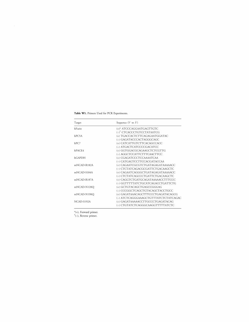
Table W1. Primers Used for PCR Experiments.
Target Sequence (5′ to 3′)
hFurin (+)* ATCCCAGGAATGAGTTGTC(−)† CTCACCCTGTCCTATAATCG
hPC5A (+) TGACCACTCTTCAGAGAATGGATAC(−) GAGATACCCACTAGGGCAGC
hPC7 (+) CATCATTGTCTTCACAGCCACC(−) ATGACTCATCCCCGACATCC
hPACE4 (+) GGTGGACGCAGAAGCTCTCGTTG(−) AGGCTCCATTCTTTCAACTTCC
hGAPDH (+) CGAGATCCCTCCAAAATCAA(−) CATGAGTCCTTCCACGATACCAA
mNCAD-R182A (+) CAGAATCGCGTCTGATAGAGATAAAAACC(−) CTCTATCAGACGCGATTCTGACAAGCTC
mNCAD-S184A (+) CAGAATCAGGGCTGATAGAGATAAAAACC(−) CTCTATCAGCCCTGATTCTGACAAGCTC
mNCAD-R187A (+) CAGGTCTGATGCAGATAAAAACCTTTCCC(−) GGTTTTTATCTGCATCAGACCTGATTCTG
mNCAD-N120Q (+) GCTGTACAGCTGAGCCGGGAG(−) CCCGGCTCAGCTGTACAGCTACCTGCC
mNCAD-N190Q (+) GAGATAAACAGCTTTCCCTGAGATACAGCG(−) ATCTCAGGGAAAGCTGTTTATCTCTATCAGAC
NCAD-S192A (+) GAGATAAAAACCTTGCCCTGAGATACAG(−) CTGTATCTCAGGGCAAGGTTTTTATCTC
*(+), Forward primer.†(−), Reverse primer.
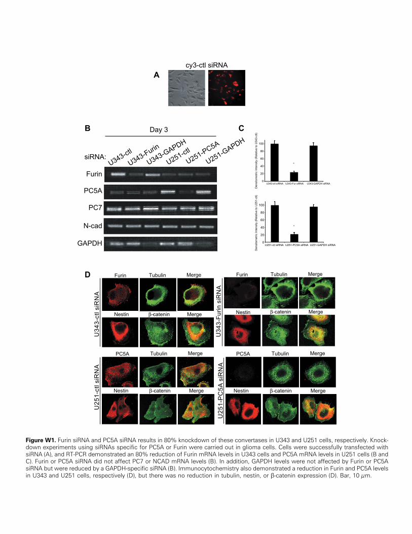
Figure W1. Furin siRNA and PC5A siRNA results in 80% knockdown of these convertases in U343 and U251 cells, respectively. Knock-down experiments using siRNAs specific for PC5A or Furin were carried out in glioma cells. Cells were successfully transfected withsiRNA (A), and RT-PCR demonstrated an 80% reduction of Furin mRNA levels in U343 cells and PC5A mRNA levels in U251 cells (B andC). Furin or PC5A siRNA did not affect PC7 or NCAD mRNA levels (B). In addition, GAPDH levels were not affected by Furin or PC5AsiRNA but were reduced by a GAPDH-specific siRNA (B). Immunocytochemistry also demonstrated a reduction in Furin and PC5A levelsin U343 and U251 cells, respectively (D), but there was no reduction in tubulin, nestin, or β-catenin expression (D). Bar, 10 μm.
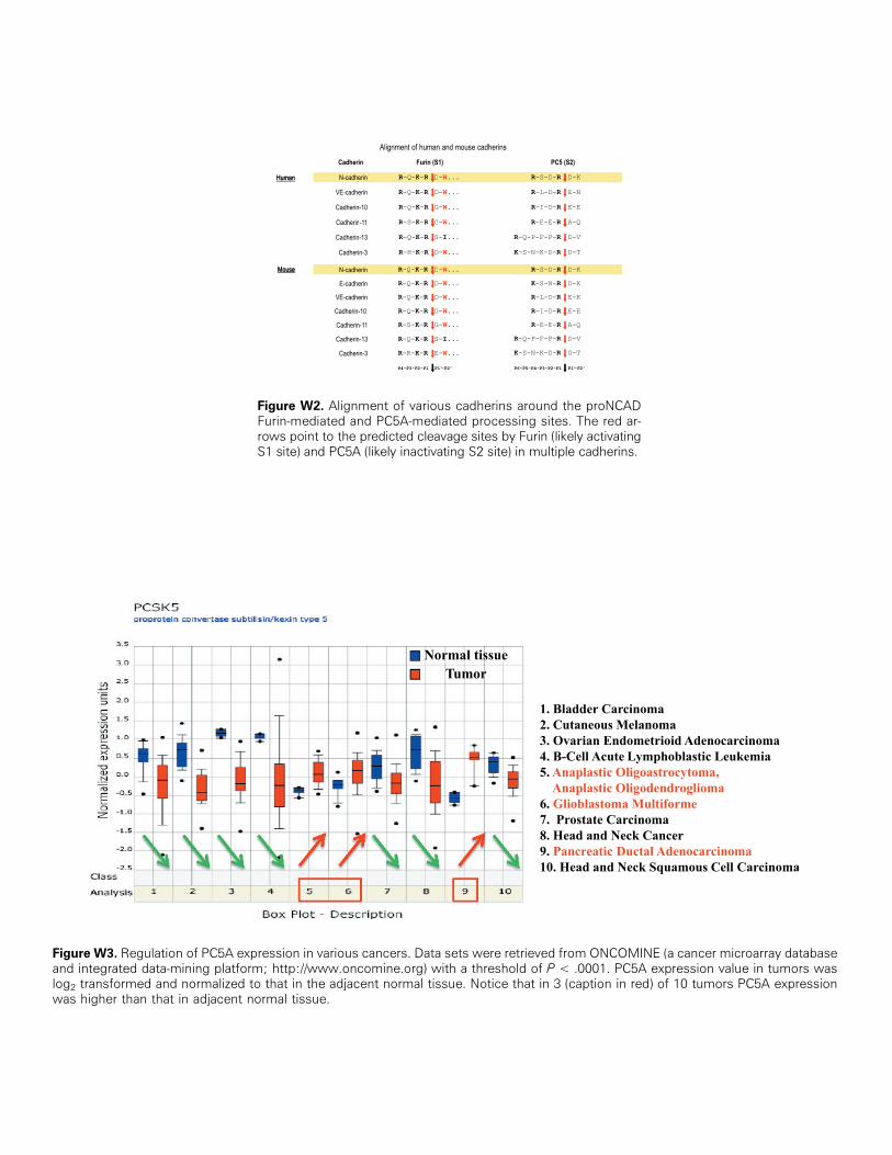
Figure W2. Alignment of various cadherins around the proNCADFurin-mediated and PC5A-mediated processing sites. The red ar-rows point to the predicted cleavage sites by Furin (likely activatingS1 site) and PC5A (likely inactivating S2 site) in multiple cadherins.
Figure W3. Regulation of PC5A expression in various cancers. Data sets were retrieved from ONCOMINE (a cancer microarray databaseand integrated data-mining platform; http://www.oncomine.org) with a threshold of P < .0001. PC5A expression value in tumors waslog2 transformed and normalized to that in the adjacent normal tissue. Notice that in 3 (caption in red) of 10 tumors PC5A expressionwas higher than that in adjacent normal tissue.
Recommended