10.1111/j.1469-0691.2008.02191.x
Seroprevalence survey of equine anaplasmosis in Franceand in sub-Saharan AfricaL. Maurizi1, J.-L. Marie2, C. Courtin3, S. Gorsane4, D. Chal5 and B. Davoust6
1Secteur Veterinaire de Rennes, France, 2Secteur Veterinaire de Marseille, France, 3SecteurVeterinaire de Metz, France, 4Secteur Veterinaire de Chalons-en-Champagne, France, 5SecteurVeterinaire de Strasbourg, France and 6Direction Regionale du Service de Sante des Armees de Toulon,France
Equine anaplasmosis is a vector-borne diseasetransmitted by ticks belonging to the Ixodes genus,especially I. ricinus in metropolitan France. Thecausative agent is a bacterium of the Anaplasma-taceae family: Anaplasma phagocytophilum [1]. Theclinical presentation ranges from asymptomatic,where horses are infected but do not showoutward signs of disease, to others that presentwith signs consistent with a febrile illness (fever,lethargy, anorexia), icterus, ataxia and oedema ofmembers. The first clinical case was described inthe USA and then in Europe (predominately inFrance in 2000) [2]. The aspects in veterinarypublic health are important: human cases wererecognised in 1994 in the USA and in Europe in1995 [3]. The epidemiology of equine anaplasmo-sis is quite unknown: the reservoir has not beenidentified and the cases’ repartition has to bedetailed.
The goal of this study is to assess the preva-lence of Anaplasma in equines in France andAfrica using a serological method and to detecteventual risk factors.
Blood samples were obtained on 570 horses in13 sites in metropolitan France (408), in FrenchGuyana (49) and in five African countries (IvoryCoast, Chad, Gabon, Congo (DR) and Djibouti)(113). On each site, if the number of horses wasless than 30 all horses were tested, otherwise only30 subjects were selected.
The blood samples were tested using an ELISADot-Blot method (SNAP� 4Dx, IDEXX Laborato-ries Inc., Westbrook, ME, USA), allowing detec-tion of both Ig M and Ig G. The percentages werecompared using the chi-squared test (a = 5%).
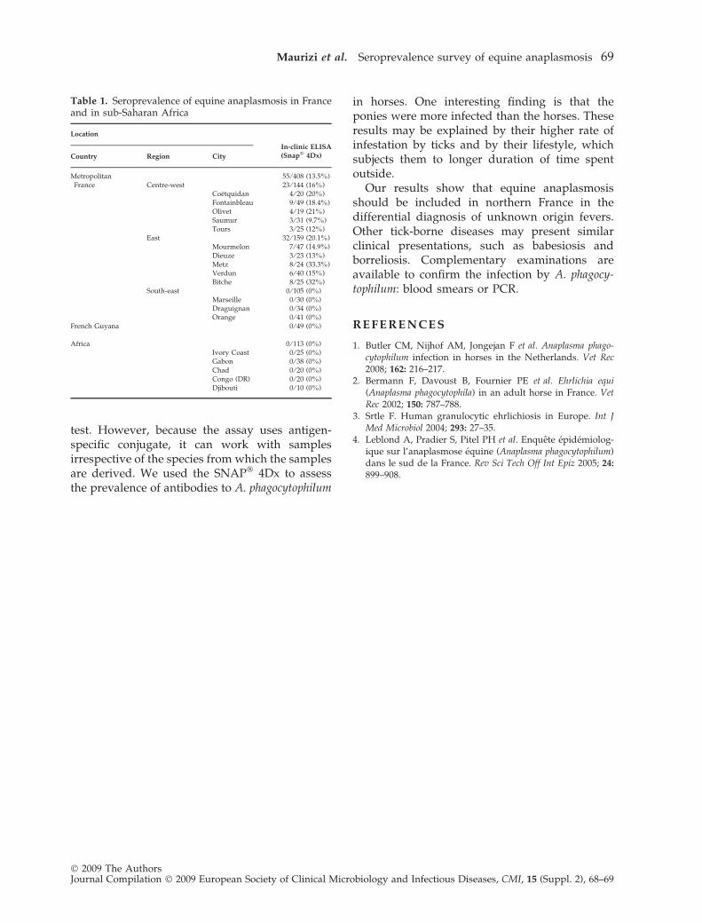
The results are shown in Table 1. None of thehorses tested from Africa, French Guyana or inthe southern France group had antibodies toA. phagocytophilum. The seroprevalences in thecentre-western and eastern regions in France were16.0% and 20.1%, respectively. Among the north-ern France group (included the centre-westernand eastern France groups), 23.1% of females and15.8% of males tested positive; 24.5% of poniesand 16.8% of horses; 14.3% of 0–5 year oldsubjects, 18.6% of 6–10 year old ones, 17.7% of11–15 year old ones, 21.4% of 16–20 year old onesand 14.3% of 21–25 year old ones. The prevalenc-es in Africa, French Guyana and metropolitanFrance were statistically different (p< 0.01). Inmetropolitan France, a significant differenceappeared between the northern and the southerngroups (p< 0.00001). The difference between thecentre-western and eastern groups wasn’t statis-tically significant. Among the northern group, norisk factors were identified (sex, pony vs. horseand ⁄ or age). At the site of Metz, the ponies hadhigher infection rate than the adult horses(p < 0.0001).
The vector of equine anaplasmosis in metro-politan France is a hard tick, Ixodes ricinus. Itspreferred environment is humid areas nearwoods or prairies. The ticks don’t constitute thereservoir of the disease. This reservoir has notbeen determined. Our study confirms that thepositive horses are only detected in the preferen-tial areas of the vector (northern France). Thisfinding is in contrast to other studies that haveshown the existence of the pathogen in the south(11.3% in Leblond et al. in 2005) [4]. The differ-ences may be explained by the poor specificity ofthe serological method and the fact that differentgeographic regions in southern France wereevaluated.
The commercial kit used in this study isrecommended by the manufacturer as a canine
Corresponding author and reprint requests: B. Davoust,Direction Regionale du Service de Sante des Armees deToulon, BP 80, 83800 Toulon Armees, FranceE-mail: [email protected]
No conflicts of interest declared.
� 2009 The AuthorsJournal Compilation � 2009 European Society of Clinical Microbiology and Infectious Diseases, CMI, 15 (Suppl. 2), 68–69
test. However, because the assay uses antigen-specific conjugate, it can work with samplesirrespective of the species from which the samplesare derived. We used the SNAP� 4Dx to assessthe prevalence of antibodies to A. phagocytophilum
in horses. One interesting finding is that theponies were more infected than the horses. Theseresults may be explained by their higher rate ofinfestation by ticks and by their lifestyle, whichsubjects them to longer duration of time spentoutside.
Our results show that equine anaplasmosisshould be included in northern France in thedifferential diagnosis of unknown origin fevers.Other tick-borne diseases may present similarclinical presentations, such as babesiosis andborreliosis. Complementary examinations areavailable to confirm the infection by A. phagocy-tophilum: blood smears or PCR.
R E F E R E N C E S
1. Butler CM, Nijhof AM, Jongejan F et al. Anaplasma phago-cytophilum infection in horses in the Netherlands. Vet Rec2008; 162: 216–217.
2. Bermann F, Davoust B, Fournier PE et al. Ehrlichia equi(Anaplasma phagocytophila) in an adult horse in France. VetRec 2002; 150: 787–788.
3. Srtle F. Human granulocytic ehrlichiosis in Europe. Int JMed Microbiol 2004; 293: 27–35.
4. Leblond A, Pradier S, Pitel PH et al. Enquete epidemiolog-ique sur l’anaplasmose equine (Anaplasma phagocytophilum)dans le sud de la France. Rev Sci Tech Off Int Epiz 2005; 24:
899–908.
Table 1. Seroprevalence of equine anaplasmosis in Franceand in sub-Saharan Africa
Location
In-clinic ELISA(Snap� 4Dx)Country Region City
MetropolitanFrance
55 ⁄ 408 (13.5%)Centre-west 23 ⁄ 144 (16%)
Coetquidan 4 ⁄ 20 (20%)Fontainbleau 9 ⁄ 49 (18.4%)Olivet 4 ⁄ 19 (21%)Saumur 3 ⁄ 31 (9.7%)Tours 3 ⁄ 25 (12%)
East 32 ⁄ 159 (20.1%)Mourmelon 7 ⁄ 47 (14.9%)Dieuze 3 ⁄ 23 (13%)Metz 8 ⁄ 24 (33.3%)Verdun 6 ⁄ 40 (15%)Bitche 8 ⁄ 25 (32%)
South-east 0 ⁄ 105 (0%)Marseille 0 ⁄ 30 (0%)Draguignan 0 ⁄ 34 (0%)Orange 0 ⁄ 41 (0%)
French Guyana 0 ⁄ 49 (0%)
Africa 0 ⁄ 113 (0%)Ivory Coast 0 ⁄ 25 (0%)Gabon 0 ⁄ 38 (0%)Chad 0 ⁄ 20 (0%)Congo (DR) 0 ⁄ 20 (0%)Djibouti 0 ⁄ 10 (0%)
Maurizi et al. Seroprevalence survey of equine anaplasmosis 69
� 2009 The AuthorsJournal Compilation � 2009 European Society of Clinical Microbiology and Infectious Diseases, CMI, 15 (Suppl. 2), 68–69
Recommended